
Quiz
Bio 211 Final Exam
Este quiz tem tipos de pergunta que abrem no player de sempre.
Perguntas do quiz (82)
1Fill in the blank for each function with its corresponding match.
- [blank_start]Schwann Cell[blank_end]: myelin-producing neuroglia of the peripheral nervous system
- [blank_start]Neuroglia[blank_end]: fill spaces, provide structure, produce myelin
- [blank_start]Oligodendrocytes[blank_end]: form the myelin sheath in the brain and spinal cord
- [blank_start]Astrocytes[blank_end]: found between blood vessels and neurons. Fills scar tissue in brain, form BBB (blood brain barrier)- ASchwann Cell
- BNeuroglia
- COligodendrocytes
- DAstrocytes
2How is the RMP established and maintained?
- ANa+ moves out and K+ moves in, ATPase maintains RMP
- BThe ATPase releases MG2+ which allows for Ca+ to flow through the NMDA
- CMore AMPA channels bind to the cell membrane
- DK+ moves out, Na+ moves in, ATPase maintains RMP
3Which of the following is true of the Goldman equation?
- ANeeded when a single ion is crossing over the membrane
- BNeeded when more than one ion is crossing over the membrane
4Which of the following is true of the Nernst equation?
- ANeeded when more than one ion is crossing over the membrane
- BNeeded when one ion is crossing over the membrane
5Which of the following variable's change when new conditions are presented?
- AGraded potential
- BRMP potential
- CIontropic potential
- DmM values
- EAction potential
- FEPSP
- GIPSP
6The process of starting/stopping an AP:
[blank_start]Gated[blank_end] Na+ and K+ channels are closed, only [blank_start]leak[blank_end] channels are open. [blank_start]RMP[blank_end] is maintained and you need threshold before [blank_start]depolarization[blank_end] occurs in order for an [blank_start]AP[blank_end] to occur.- AGated
- Bleak
- CRMP
- Ddepolarization
- EAP
7Match the term with its definition in respect to action potentials.
- [blank_start]Depolarization[blank_end]: membrane becomes more positive away from the RMP
- [blank_start]Re-polarization[blank_end]: membrane becomes more negative away from the RMP
- [blank_start]Hyper-polarization[blank_end]: membrane becomes even more negative than the RMP- ADepolarization
- BRe-polarization
- CHyper-polarization
8The role of the Na/K ATPase is to reset the chemical conditions in the RMP. After repolarization occurs the pump restores electrical conditions.
- AVerdadeiro
- BFalso
9Label the diagram with is respective phases.
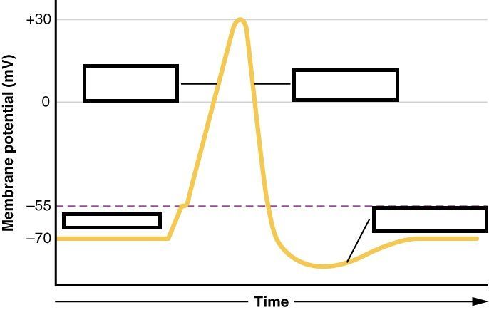
- Adepolarization
- Bresting state
- Chyperpolarization
- Drepolarization
10Fill in the blank to explain how one neuron signals to another once an AP reaches the axon terminal in the pre-synaptic neuron.
An AP in the axon terminal causes [blank_start]voltage gated[blank_end] Ca+ channels to [blank_start]open[blank_end] and enter the cell. Ca+ inside the cell causes the [blank_start]synaptic vesicle[blank_end] to [blank_start]fuse[blank_end] with the membrane. Each has a certain amount of neurotransmitter released into the cleft. Neurotransmitter [blank_start]diffuses[blank_end] across the cleft. Neurotransmitter [blank_start]binds[blank_end] to receptors on the post synaptic neuron where it can act.- Avoltage gated
- Bopen
- Csynaptic vesicle
- Dfuse
- Ediffuses
- Fbinds
11Which of the following is true of Iontropic NT receptors?
- AMonamines
- BAA
- CNeuropeptides
- DACh
- Eallows ions to flow through central pores
- Fchanges inside the cell
- GEPSP/IPSP
- Husually involves G protein
- Ifast/brief period
- Jslow/long period
12EPSP: leads to [blank_start]depolarization[blank_end] of the neuron. [blank_start]Excited[blank_end] the post synaptic neuron
IPSP: leads to [blank_start]hyper-polarization[blank_end] of the neuron. [blank_start]Inhibits[blank_end] the post synaptic neuron- Adepolarization
- Bhyper-polarization
- CExcited
- DInhibits
13[blank_start]Spatial summation[blank_end]: simultaneous EPSP in different location - produces AP, EPSP & IPSP in different location - reduces chance of AP
[blank_start]Temporal summation[blank_end]: rapid repeat of EPSP in same location - produces AP- ASpatial summation
- BTemporal summation
14[blank_start]Myelinated[blank_end]: AP is faster, contains [blank_start]nodes of ranvier[blank_end]: voltage gated Na+ channels where AP is generated.
[blank_start]Un-myelinated[blank_end]: AP is slower, contains [blank_start]leak channels[blank_end]: less flow of ions, difficult to get incoming Na+ charges to move.- AMyelinated
- BUn-myelinated
- Cnodes of ranvier
- Dleak channels
15If the Na/K ATPase stopped working the membrane would be more negative which would be harder to start an AP.
- AVerdadeiro
- BFalso
16The brains ability to form or change synaptic connections is known as _____________.
- ALong term potentiation
- BSynaptic Pruning
- CNeuroplasticity
- DMyelination
17Early LTP: [blank_start]AMPA[blank_end] receptor becomes [blank_start]excitatory[blank_end] which leads to an increase of [blank_start]Na+[blank_end] which then depolarizes the post synaptic cell. The EPSP that has been started releases the [blank_start]Mg+[blank_end] from the [blank_start]NMDA[blank_end] receptor and allows [blank_start]Ca+[blank_end] to enter.
Late LTP: Begins when [blank_start]sustained[blank_end] activation from early LTP leads to protein synthesis. The post synaptic changes increase dendritic spines and increase AMPA receptors.- AAMPA
- Bexcitatory
- CNa+
- DMg+
- ENMDA
- FCa+
- Gsustained
18Match the term with its corresponding scenario:
[blank_start]Ca2+ levels[blank_end]: a decrease in this could cause a neuron to not pass through the NMDA receptors resulting in no LTP.
[blank_start]AMPA receptors[blank_end]: if there are none of these in early LTP then there will not be any LTP at all but if the release of NMDA occurs there none to make the membrane more permeable; causing late LTP to occur slower or not at all.
[blank_start]Lack of threshold[blank_end]: this would result in Na+ not flowing through the cell therefore no LTP would be initiated. Mg2+ would possibly block the NMDA receptor and no late LTP would occur.
[blank_start]Excess Mg2+[blank_end]: this would cause for more Na+ to flow in causing depolarization of the cell faster
[blank_start]Lack of Na+[blank_end]: there would be no EPSP, no Mg2+ release, no LTP- ACa2+ levels
- BAMPA receptors
- CLack of threshold
- DExcess Mg2+
- ELack of Na+
19The [blank_start]photo-receptor[blank_end] cell receives light signals and is located in the [blank_start]retina[blank_end].
- Aphoto-receptor
- Bbi polar
- Cganglionic
- Dretinal
- Eretina
- Ffovea
- Gmacula
- Hprimary visual cortex
20The rods are used for [blank_start]dim[blank_end] light and [blank_start]high[blank_end] sensitivity.
The cones are used for [blank_start]bright[blank_end] light and [blank_start]low[blank_end] sensitivity.- Adim
- Bhigh
- Cbright
- Dlow
21The macula contains the fovea.
- AVerdadeiro
- BFalso
22The fovea has a low density of photoreceptors.
- AVerdadeiro
- BFalso
23What photo-receptors are found in the periphery?
- ACones
- BRods
24Which ion is released when signaling photo-receptors?
- ANa+
- BK+
- CCa+
- DCl-
25Pathway of light from eye to brain.
[blank_start]Light[blank_end] -> [blank_start]Retina[blank_end] -> [blank_start]Photoreceptor[blank_end] -> [blank_start]Bi Polar Cell[blank_end] -> [blank_start]Ganglionic Cell[blank_end] -> [blank_start]Optic Nerve[blank_end] -> [blank_start]Optic Chiasm[blank_end] -> [blank_start]LGN[blank_end] -> [blank_start]V1[blank_end]- ALight
- BRetina
- CPhotoreceptor
- DBi Polar Cell
- EGanglionic Cell
- FOptic Nerve
- GOptic Chiasm
- HLGN
- IV1
26What axons make up the optic nerve?
- ABi polar cell
- BPhotoreceptor
- CGanglionic Cell
- DNerve endings
27The optic chiasm is the crossing over point for vision.
- AVerdadeiro
- BFalso
28Label the pathway from the outer to inner ear.
[blank_start]External meatus[blank_end] -> [blank_start]Tympanic Membrane[blank_end] -> [blank_start]Malleus[blank_end] -> [blank_start]Incus[blank_end] -> [blank_start]Stapes[blank_end] -> [blank_start]Oval Window[blank_end] -> [blank_start]Inner Ear[blank_end]- AExternal meatus
- BTympanic Membrane
- CMalleus
- DIncus
- EStapes
- FOval Window
- GInner Ear
29Top tube: [blank_start]scala vestibule[blank_end]: connects to the oval window
Middle tube: [blank_start]scala media[blank_end]: holds organ of corti - hair cells that change waves to electric signals - sits on basilar membrane and covered by tectorial membrane
Bottom tube: [blank_start]scala tympani[blank_end]: connects to round window- Ascala vestibule
- Bscala media
- Cscala tympani
30The stereocilia depolarizes in response to sound waves.
- AVerdadeiro
- BFalso
31Sound waves come in and bend the stereocilia which allow for Na+ to flow in.
- AVerdadeiro
- BFalso
32The [blank_start]endolymph[blank_end] is high in potassium.
- Aendolymph
- Bperilymph
33Stereocilia is stuck in the [blank_start]tectorial membrane[blank_end] and allows for tugging when sound waves pass through. The organ of corti sits on the [blank_start]basilar membrane[blank_end].
- Atectorial membrane
- Bbasilar membrane
34The [blank_start]depolarization[blank_end] of stereocilia causes them to release [blank_start]glutamate[blank_end] onto the [blank_start]spiral ganglion[blank_end] cells of the cochlear nerve.
- Adepolarization
- Bhyperpolarization
- Crepolarization
- Dcrossing over
- Eglutamate
- Fpotassium
- Gsodium
- Hcalcium
- Ispiral ganglion
- Jhair
- Kodorant
- Lreceptor
35Cilia repolarize as K+ leaves the endolymph.
- AVerdadeiro
- BFalso
36Fill in the blanks for the pathway of sound from the cochlea to the brain.
[blank_start]Hair Cell[blank_end] -> [blank_start]Spiral Ganglion Axon[blank_end] -> [blank_start]Cochlear Nerve[blank_end] -> [blank_start]Cochlear Nuclei in the medulla[blank_end] -> [blank_start]Superior Olive in the pons[blank_end] -> [blank_start]Inferior Colliculus in the midbrain[blank_end] -> [blank_start]Medial Geniculate Nucleus in the thalamu[blank_end]s -> [blank_start]A1 in the superior temporal lobe[blank_end].- AHair Cell
- BSpiral Ganglion Axon
- CCochlear Nerve
- DCochlear Nuclei in the medulla
- ESuperior Olive in the pons
- FInferior Colliculus in the midbrain
- GMedial Geniculate Nucleus in the thalamu
- HA1 in the superior temporal lobe
37Where is the crossing over point for the auditory system?
- Amedulla
- Bmidbrain
- Cpons
- Dthalamus
38The primary auditory cortex is located in the Superior temporal lobe.
- AVerdadeiro
- BFalso
39[blank_start]Pitch[blank_end] is based on frequency.
High pitch: [blank_start]base of cochlea[blank_end]
Low pitch: [blank_start]apex of cochlea[blank_end]
[blank_start]Loudness[blank_end] is based on intensity
Increased [blank_start]amplitude[blank_end] of waves: louder sounds- APitch
- Bbase of cochlea
- Capex of cochlea
- DLoudness
- Eamplitude
40Explain the way in which odor molecules start an AP in sensory neurons.
[blank_start]Odorant particles bind to receptors[blank_end] on the cilia -> [blank_start]transduction begins[blank_end] -> [blank_start]signals the G Protein[blank_end] -> [blank_start]cAMP production[blank_end] -> [blank_start]cAMP binds to open ion channels[blank_end] -> [blank_start]allow Na+ and Ca2+ to enter[blank_end] -> [blank_start]depolarization[blank_end]- AOdorant particles bind to receptors
- Btransduction begins
- Csignals the G Protein
- DcAMP production
- EcAMP binds to open ion channels
- Fallow Na+ and Ca2+ to enter
- Gdepolarization
41Trace the path of olfaction signals from the nose to brain
[blank_start]Mucus[blank_end] -> [blank_start]olfactory cells[blank_end] -> [blank_start]dendrite[blank_end] -> [blank_start]olfactory receptor[blank_end] -> [blank_start]basal cell[blank_end] -> [blank_start]axon[blank_end] -> [blank_start]olfactory gland[blank_end] -> [blank_start]olfactory nerve[blank_end] -> [blank_start]cribiform plate[blank_end] -> [blank_start]glomeruli[blank_end] -> [blank_start]mitral cell[blank_end] -> [blank_start]olfactory tract[blank_end]- AMucus
- Bolfactory cells
- Cdendrite
- Dolfactory receptor
- Ebasal cell
- Faxon
- Golfactory gland
- Holfactory nerve
- Icribiform plate
- Jglomeruli
- Kmitral cell
- Lolfactory tract
42How are different odors interpreted?
- A100 different receptor types
- B500 different receptor types
- C350 different receptor types
- Dunknown amount of receptor types
43Which cells make up the olfactory nerve?
- Amitral cell
- Bbasal cell
- Cepithelial cell
- Dgustatory cell
44Which of the following have pertain to taste buds?
- AGustatory cells
- BEpithelial cells
- CBasal cells
- DGanglionic cells
- EOlfactory cells
45Gustatory cells are ciliated and bind to dissolved chemicals.
- AVerdadeiro
- BFalso
46Match the NT to its tastant:
T1R1: [blank_start]umami/MSG[blank_end]
T1R2: [blank_start]sweet[blank_end]
T1R3: [blank_start]sweet/umami[blank_end]
T2: [blank_start]bitter[blank_end]
ENaC: [blank_start]salty[blank_end]
PKD2L: [blank_start]sour[blank_end]- Aumami/MSG
- Bsweet
- Csweet/umami
- Dbitter
- Esalty
- Fsour
47[blank_start]T1R1 & T2[blank_end]: binds G protein -> increase IP3 Ca2+ -> open TRP channels -> depol.
[blank_start]ENaC & PKD2L[blank_end]: enters through membrane Na+ channels -> depol.- AT1R1 & T2
- BENaC & PKD2L
48Fill in the blanks for the path of taste bud to brain:
[blank_start]Cranial nerves[blank_end] -> [blank_start]medulla oblongata[blank_end] -> [blank_start]Pons (medulla)[blank_end] -> [blank_start]VPN thalamus[blank_end] -> [blank_start]Primary Gustatory Cortex (insula)[blank_end] -> [blank_start]Hypothalamus (limbic system)[blank_end]- ACranial nerves
- Bmedulla oblongata
- CPons (medulla)
- DVPN thalamus
- EPrimary Gustatory Cortex (insula)
- FHypothalamus (limbic system)
49The direction in which sensory information travels is from spinal cord to periphery
- AVerdadeiro
- BFalso
50Fill in the blanks to show the path of sensory information from mechanoreceptors to S1 using the MLP:
[blank_start]Primary afferent[blank_end] -> [blank_start]medulla[blank_end] -> [blank_start]secondary afferent[blank_end] -> [blank_start]thalamus[blank_end] (where it crosses over) -> [blank_start]VPN[blank_end] -> [blank_start]tertiary afferent[blank_end] -> [blank_start]S1[blank_end] (parietal lobe)- APrimary afferent
- Bmedulla
- Csecondary afferent
- Dthalamus
- EVPN
- Ftertiary afferent
- GS1
51Fill in the blank for the sensory information for nociceptors to S1 using the spinothalamic tract.
-[blank_start]Spinal cord[blank_end] -> [blank_start]cross at ventral route[blank_end] -> [blank_start]medulla[blank_end]- ASpinal cord
- Bcross at ventral route
- Cmedulla
52In the medial lemniscal pathway crossing over occurs in the [blank_start]thalamus[blank_end] and in the spinothalamic pathway crossing over occurs in the [blank_start]spinal cord[blank_end].
- Athalamus
- Bmedulla
- Cspinal cord
- Dpons
- Espinal cord
- Fthalamus
- Gmedulla
- Hpons
53[blank_start]A delta[blank_end]: myelinated, fast conduction, skin pressure/touch vibration
[blank_start]A beta[blank_end]: myelinated, sharp pain, cool/cold info
[blank_start]C fibers[blank_end]: unmyelinated, slow conduction, pain, warm/hot information- AC fibers
- BA delta
- CA beta
54Match each mechano-receptor with the sensory information they detect:
[blank_start]Merkel Cell[blank_end]: mostly in fingertips, feel touch
[blank_start]Meisnner Corpuscle[blank_end]: respond to low freq. vibrations, brushing across skin, slippage of objects in hands
[blank_start]Paccinian Corpuscle[blank_end]: poor spatial resolution, respond to high freq. vibration, use of tools- AMerkel Cell
- BMeisnner Corpuscle
- CPaccinian Corpuscle
55S1 is in the parietal lobe and is somatotopically organized
- AVerdadeiro
- BFalso
562 point discrimination is the regions far apart in the body synapse in the cortex close together.
- AVerdadeiro
- BFalso
57Both Substance P and Glutamate ________ interneurons. - Secondary afferents in the spinal cord.
- Ainhibit
- Bexcite
- Chyperpolarize
- Deliminate
58How do endogeous opiods block incoming nociceptor signals in order to reduce the perception of pain?
They all bind to opiod ligand -> release inhibitory [blank_start]NT[blank_end] on nociceptor neuron terminals in spinal cord and either block [blank_start]Ca2+[blank_end] channels or open [blank_start]K+[blank_end] channels.- ANT
- BCa2+
- CK+
59Match each NT to its interneuron.
Mu: [blank_start]endorphin[blank_end]
Delta: [blank_start]enkephalin[blank_end]
Kappa: [blank_start]dynorphin[blank_end]- Aendorphin
- Benkephalin
- Cdynorphin
60What structures below are a part of the NMJ? (CATA)
- Aaxon terminal of a neuron
- BACh
- CSarcolemma
- Dsynaptic cleft
61During muscle contraction:
- Athin filaments allow thick filaments to slide on top
- Bthin and thick filaments slide past each other
- Cthin and thick filaments begin to convulse
62Myosin heads bind to actin during muscle contraction
- AVerdadeiro
- BFalso
63Match each term with their definition:
- [blank_start]Sarcolemma[blank_end]: plasma membrane
- [blank_start]Sarcoplasmic Reticulum[blank_end]: smooth ER; stores Ca2+ has voltage gated channels
- [blank_start]Muscle Fiber[blank_end]: thing filaments, myosin attaches, think filaments binding site for actin and ATP.- ASarcolemma
- BSarcoplasmic Reticulum
- CMuscle Fiber
64The [blank_start]direct[blank_end] pathway signals inhibit some basal ganglia structures which lead to movement. The [blank_start]indirect[blank_end] pathway signals excite some basal ganglia structures which inhibit movement.
- Adirect
- Bindirect
- Cindirect
- Ddirect
65M1 is not organized somatotopically.
- AVerdadeiro
- BFalso
66The size of the area determines the movement in M1.
- AVerdadeiro
- BFalso
67Fill in the blanks with the pathway for motor movement from the brain to the muscle:
[blank_start]M1[blank_end] -> [blank_start]pons[blank_end] -> [blank_start]medulla[blank_end] -> [blank_start]spinal cord[blank_end] -> [blank_start]muscle[blank_end]- AM1
- Bpons
- Cmedulla
- Dspinal cord
- Emuscle
68Where do the motor neurons decussate?
- Athalamus
- Bventral horn
- Cdorsal horn
- Dmedulla
- Epons
69The UMN synapses on the LMN at the __________________
- Adorsal horn
- Bventral horn
70Dorsal Horn: [blank_start]sensory neuron[blank_end]
Ventral Horn: [blank_start]upper motor neuron[blank_end]
Dorsal Route: [blank_start]sensory pathway[blank_end]
Ventral Route: [blank_start]motor pathway[blank_end]
[blank_start]Spinal Nerve[blank_end]: mixed between sensory & motor neurons
[blank_start]Left[blank_end] side of spinal cord: motor/descending ([blank_start]efferent[blank_end]) pathway
[blank_start]Right[blank_end] side of spinal cord: sensory/ascending ([blank_start]afferent[blank_end]) pathway- Asensory neuron
- Bupper motor neuron
- Cupper motor neuron
- Dsensory neuron
- Esensory pathway
- Fmotor pathway
- Gmotor pathway
- Hsensory pathway
- ISpinal Nerve
- JDorsal Nerve
- KAutonomic Nerve
- LSpinothalamic Nerve
- MLeft
- NRight
- Oefferent
- Pafferent
- QRight
- RLeft
- Safferent
- Tefferent
71Sensory neuron connect motor neurons through interneurons.
- AVerdadeiro
- BFalso
72[blank_start]Flexor[blank_end] reflex causes automatic withdrawal of threatened body parts and uses simple reflex arc.
[blank_start]Stretch[blank_end] reflex maintains posture and adjusts.- AFlexor
- BStretch
- CStretch
- DFlexor
73Proprioceptors _________________.
- Adetect stretch and fire AP - causes muscle contraction
- Bdetect stretch and contains AP - constricting muscle contraction
- Cmaintain homeostasis
- Dcarry signals from outer motor neurons to inner
74Proprioception is ...
- Athe constriction of limbs
- BThe reflex of limbs
- CThe sensation of limbs
- DThe involuntary movement of limbs
75[blank_start]Autonomic[blank_end] NS: involuntary, impulses go to [blank_start]cardiac[blank_end] muscles
[blank_start]Somatic[blank_end] NS: voluntary, impulses go to [blank_start]skeletal[blank_end] muscles- AAutonomic
- BSomatic
- CSomatic
- DAutonomic
- Ecardiac
- Fskeletal
- Gskeletal
- Hcardiac
76Check all that apply to the Parasympathetic Nervous System.
- AConstricts airways
- BDecrease heart rate
- CIncrease digestion
- DIncrease Urine Output
- EDecrease Blood Pressure
- FIncrease heart rate
- GDecrease digestion
- HDecrease Urine Output
- IDialtate Airways
- JIncrease Blood Pressure
77Receptor Types:
- [blank_start]Nicotinic[blank_end]: stimulatory, found on skeletal muscles at the neuromuscular juntion, all postganglionic neurons.
- [blank_start]Muscarinic[blank_end]: inhibitory/excitatory, found on all effector cells.- ANicotinic
- BMuscarinic
- CMuscarinic
- DNicotinic
78What Neurotransmitter is used for signaling?
- Sympathetic: [blank_start]NE[blank_end]
- Parasympathetic: [blank_start]ACh[blank_end]- ANE
- BACh
79What type of fibers are used for in the SNS?
- Acholinergic
- Bmuscarinic
- Cadrenergic
- Dnicotinic
80What type of fiber is used in the PNS?
- Aadrenergic
- Bcholinergic
- Cmuscarinic
- Dnicotinic
81The Central Nervous System is controlled by the Autonomic Nervous System.
- AVerdadeiro
- BFalso
82Is the ANS controlled unconsciously by the CNS?
- AYes
- BNo
Informações
- Descrição
- Spring 2017 - Final Exam - Dr. Jones
- Autor
- Toni Demoff
- Idioma
- English
- Visualizações
- 69
- Etiquetas
- Avaliação
- Ainda sem avaliações Avalie este quiz