
Flashcards
BTEC Applied Science Unit 1- Biology

Flashcards do conjunto (61)
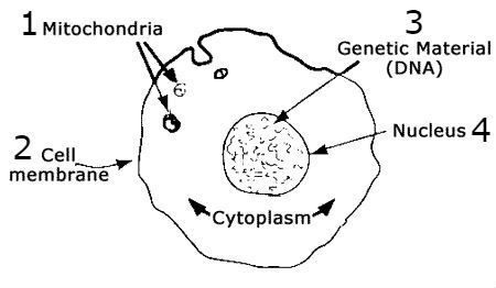
- 1What are organelles?Structures within a cell e.g. nucleus, chloroplast, vacuole
Features seen under a light microscope - 2What is the ultra-structure?The detail inside a cell revealed by an electron microscope
- 3What is cell theory?Cells are the basic unit of life.
Cells are the fundamental unit of structure, function and organisation in all living organisms.
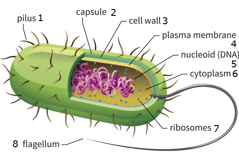
Only developed from existing cells. - 4What are the features of Prokaryotic cells?Pilli
Flagellum
Coil of DNA
No membrane bound organelles
70S Ribosome
Small size 0.1-10 um - 5What are the features of Eukaryotic cells?Nucleus
Membrane bound organelles
80S Ribosomes
Bigger size 10-100 um - 6What organelles are in Prokaryotes? (Bacteria) (9)Plasmid
Slime Capsule
Pilli
Cell wall
Cell membrane
Nucleoid
Cytoplasm
70S ribosome
Flagellum - 7What organelles are in Animal cells? (16)Vesicles
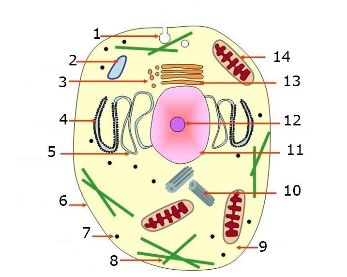
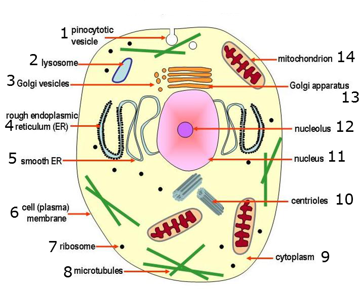
Golgi apparatus
80S ribosomes
Rough endoplasmic reticulum
Nucleolus
Nucleus
Nuclear pore
Nuclear membrane
Mitochondria
Cytoplasm
Cell membrane
Lysosome
Smooth endoplasmic reticulum
Small vacuole
Centrioles
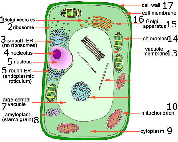
Vesicles - 8What organelles are in Plant cells? (17)Tonoplast
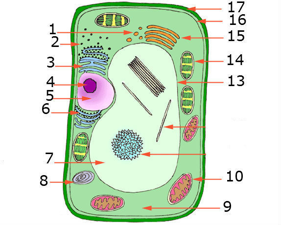
Vacuole
Amyloplast
Plasmodesmata
Smooth endoplasmic reticulum
Cell wall
Cell membrane
Mitochondria
Chloroplast
Nuclear Membrane
Nucleus
Nucleolus
Nuclear pore
Rough endoplasmic reticulum
80S ribosome
Golgi apparatus
Secretary vesicles - 9What is Microscopy?Organelle recognition
- 10What is a Micrograph?A photograph from an electron microscope
- 11What is Resolution?The ability to distinguish between two points
- 12What radiation source does a Light microscope use?Light
- 13What radiation source does an Electron microscope use?Electrons
- 14What magnification does a Light microscope have?Up to 1000x
(up to one thousand times) - 15What magnification does an Electron microscope have?Up to 1000,000x
(up to one million times) - 16What resolution does a Light microscope have?0.2 um
(micrometres) - 17What resolution does an Electron microscope have?50 pm
(????) - 18Are Light microscopes in colour?Yes
- 19Are Electron microscopes in colour?No- Black and white
- 20—
- 21—
- 22—
- 23What are microscopes used for?Tells us about how the structure relates to function so we understand how cells work.
Cytology- studying whether tissue samples are diseased or healthy. - 24What is magnification?How many times bigger the image appears compared to the actual object.
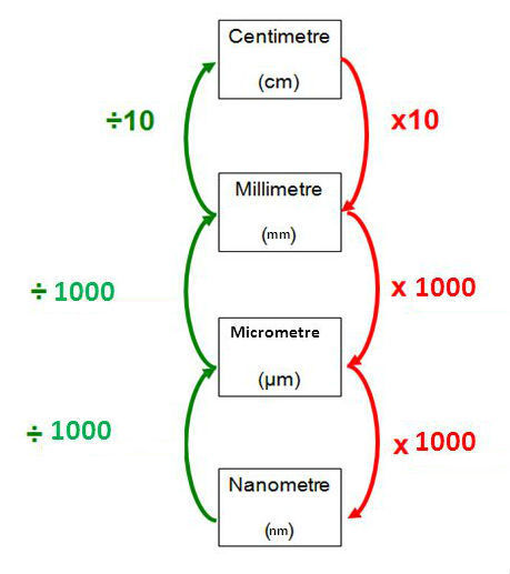
- 25How do you convert between cm, mm, um and nm?
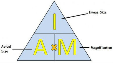
- 26What is the formula to work out Image/ Magnification/ Actual?
- 27What is the method of working out the actual size of a magnified image?Measure the image size using a ruler (mm)
Convert mm to micrometres (um)
Divide this number by the given magnification - 28What is the structure of gram negative bacteria?Gram negative bacteria has an additional membrane that allows them to control reaches the inner cell enabling them to remove threats in the space between the membranes before it reaches the cell.
- 29What is the structure of gram positive bacteria?Gram positive bacteria has thick peptidoglycan layer that absorbs antibiotics and cleaning products easily.
- 30What colour result will there be for gram-positive bacteria?Purple
- 31What colour result will there be for gram-negative bacteria?Pink
- 32What is tissue?A collection of similar cells that perform a common role e.g. epithelial
- 33What are organs?A collection of tissues working together to perform a particular function e.g. the liver
- 34What are organ systems?Several organs working together to perform an over all life function e.g. digestive system
- 35What is cell differentiation and specialisation?When multicellular organisms consist of many cells, these cells specialise to perform a role.
- 36How do cells differentiate?Change in the number of a particular organelle.
Changes in shape of cell.
Changes in the contents of the cell.
(Differentiation can include all three types of change in a cell) - 37Where do unspecialised cells come from?Bone marrow
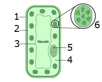
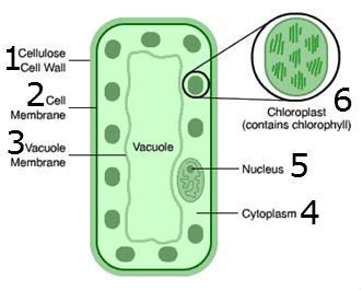
- 38What is the structure of Palisade Mesophyll Cells?Packed with chloroplasts containing chlorophyll which absorbs sunlight.
Regular columnar shape closely packed together forming a layer for maximum sunlight absorption.
Mitochondria that produce ATP to support photosynthesis.
Thin walled cells for rapid diffusion of gasses.
Clear cell walls to absorb maximum sunlight.
Large vacuole to maintain turgor pressure. - 39—
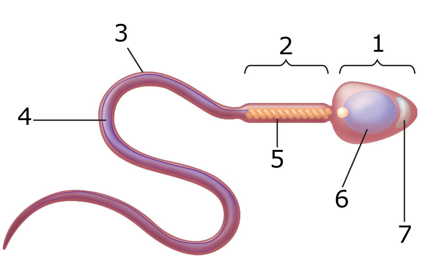
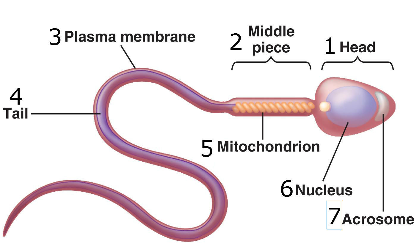
- 40What is the structure of Sperm?Nucleoid=haploid, 23 chromosomes.
Streamline head and body to reduce resistance.
head contains acrosomes which contain hydrolytic enzymes to digest the egg cell membrane for penetration of the sperm head (nucleus).
Flagellum (tail)to propel the sperm to the egg.
Mitochondria to generate ATP for the movement of flagellum. - 41—
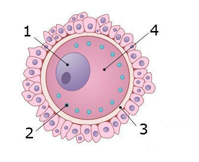
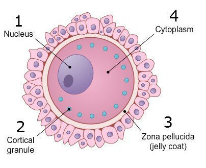
- 42What is the structure of an egg cell?Many mitochondria for cell division after fertilisation.
Haploid nucleus containing 23 chromosomes.
Cytoplasm containing nutrients and fat for embryo development.
Zona pellucida protects the cell and restricts sperm entry.
Cortical granules harden the zona pellucida to prevent multiple fertilisation. - 43—
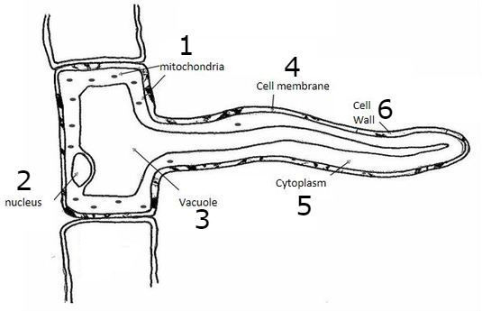
- 44What is the structure of a root hair cell?Large surface area for absorption of water by osmosis.
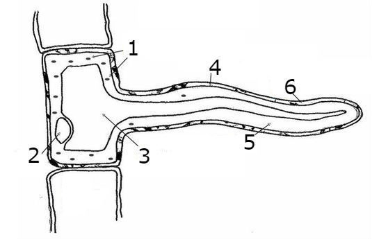
Some minerals are transported down a concentration gradient from the soil by facilitated diffusion through channel proteins.
Thin permeable cell walls reduce diffusion distance.
Large numbers of mitochondria providing ATP for active transport of minerals from soil. - 45—
- 46What is the structure of a white blood cell?Flexible shape allows engulfing of pathogens (phagocytosis).
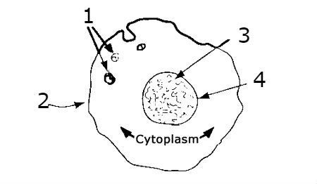
Multi-loved nucleus allows flexibility.
Contains lysosomes to break down pathogens.
Contains large amounts of RER for protein synthesis (proteins used to make antibodies).
Granular appearance- granules containing hydrolytic enzymes.
Many receptors on the membrane for antigens to attach. - 47—
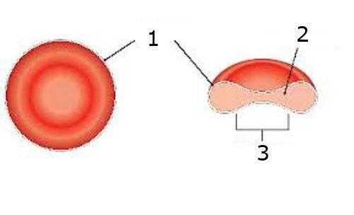
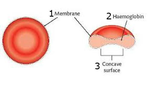
- 48What is the structure of a red blood cell?Contains haemoglobin proteins to bind oxygen.
Thin outer membrane for short diffusion distance.
Bi-concaved surface increases surface area for diffusion- allows more heamoglobin to be packed arround the edges.
Flexible membrane to squeeze through narrow capillaries.
No nucleus so there is more room for haemoglobin. - 49—
- 50What type of tissue is this?Squamous epithelial tissue

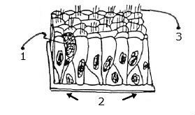
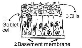
- 51What type of tissue is this?Columnar epithelial tissue
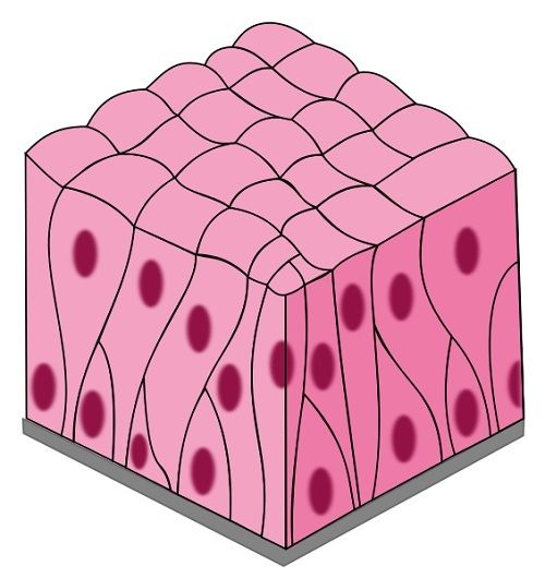
- 52—
- 53What is Squamous Epithelial Tissue?Located in the lungs and lines alveoli.
Single layer of thin cells that fit tightly together- large surface area for diffusion of gasses.
They allow diffusion and filtration (CO2 and O2 exchange).
One cell thick- short diffusion pathway.
Good blood supply- maintains concentration gradient. - 54What is COPD?Chronic Obstructive Pulmonary Disease
- 55What affect does smoking have on cilia and lung tissue?It slows the movement of cilia.
Mucus is not moved efficiently.
It causese blockages in the airways.
Persistent cough to expel mucus. - 56What is the function of goblet cells?Goblet cells are specialised as gland cells that synthesise and secrete mucus.
- 57What is endothelial tissue?Cells that line blood vessels and the heart, they are one cell thick and form a barrier between blood and tissue.
- 58What is the structure and function of endothelial tissue?One cell thick providing short diffusion pathways.
Movement of substances e.g. digestion products into capillaries, blood plasma and tissue fluid in and out of blood capillaries. - 59What risk factors can cause damage to endothelium?Carbon-monoxide
Lack of exercise
Smoking
High blood pressure- lumen is narrowed
Too much salt
High cholesterol - 60What is atherosclerosis?1- Deposits of fatty substances under the endothelium lining of arteries.
2- The fatty deposits build up and break through endothelial lining forming plaque in lume.
3- This reduces the size of the lumen and restricts blood flow. - 61Which tissue contain cilia and goblet cells?Columnar epithelial tissue
Informações
- Descrição
- Flashcards Including- -Cell structure - Organelles in Prokayotes and Eukaryotes (incl. Plant and Animal cells) -Microscopy -Magnification calculations -Gram staining -Cell specialisation -Epithelial and Endothelial tissue
- Criado por
- Megan Whitehead · fev 2018
- Atualizado
- 12 mar 2018
- Visualizações
- 9.298
- Avaliação