20491865
Description
Quiz by Charlotte Jakes, updated more than 1 year ago
|
|
Created by Charlotte Jakes
over 4 years ago
|
|
Question 1
Question
What name is given to the cells that secrete ground substance and collagen to form the rigid gel of cartilage?
Answer
-
Chondroblasts
-
Epiblasts
-
Hypoblasts
-
Fibroblasts
Question 2
Question
Cartilage has its own blood and nerve supply.
Answer
- True
- False
Question 3
Question
Why must cartilage be relatively thin?
Answer
-
It doesn't have its own blood supply so relies on diffusion for nutrients
-
It needs to be flexible
-
It needs to be able to be compressed
-
It needs to allow blood vessels to penetrate it
Question 4
Question
When deformed, [blank_start]proteoglycans[blank_end] in the cartilage 'give up' fluid. When the force is removed, fluid is 'drawn back in'. This provides a mass movement of [blank_start]nutrient[blank_end] supply and [blank_start]metabolite[blank_end] removal.
Answer
-
proteoglycans
-
nutrient
-
metabolite
Question 5
Question
Bones provide a store of calcium and which other element?
Answer
-
Phosphorus
-
Sodium
-
Potassium
-
Nitrogen
Question 6
Question
Label this image of a long bone to show the different parts.
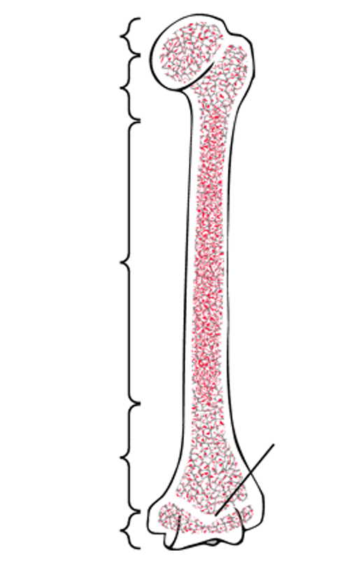
Answer
-
Proximal epiphysis
-
Distal epiphysis
-
Metaphysis
-
Diaphysis
-
Metaphysis
-
Epiphyseal plate
Question 7
Question
What name is given to the line that forms after the epiphysial plate stops producing bone (i.e. when growth stops)?
Answer
-
Epiphyseal line
-
Metaphyseal line
-
Epiphyseal shunt
-
Epiphyseal foramen
Question 8
Question
There are three types of cells in bone.
[blank_start]Osteoblasts[blank_end] form new bone via the production of osteoid. They also control the deposition of calcium and other minerals.
[blank_start]Osteocytes[blank_end] are formed when an osteoblast become embedded in the matrix they secrete. These can send out 'branches' to connect to other cells of their kind.
[blank_start]Osteoclasts[blank_end] dissolve bone to prep for repair and replacement. They are usually formed from the fusion of multiple cells, meaning they are multinucleate.
Answer
-
Osteoblasts
-
Osteocytes
-
Osteoclasts
Question 9
Question
What name is given to the bones of the head and trunk?
Answer
-
Axial skeleton
-
Appendicular skeleton
-
Trunk skeleton
-
Spinal skeleton
Question 10
Question
What name is given to the bones that support the appendages (i.e. the limbs and the pelvis)?
Answer
-
Axial skeleton
-
Appendicular skeleton
-
Appendegal skeleton
-
Limbic skeleton
Question 11
Question
Label this image with the classifications of each type of bone.
Answer
-
Flat bone
-
Long bone
-
Sesamoid bone
-
Short bone
-
Irregular bone
Question 12
Question
Which type of ossification occurs whereby the bone develops by replacing hyaline cartilage in the embryo?
Answer
-
Endochronal ossification
-
Intramembranous ossification
-
Compact ossification
-
Mesenchymal ossification
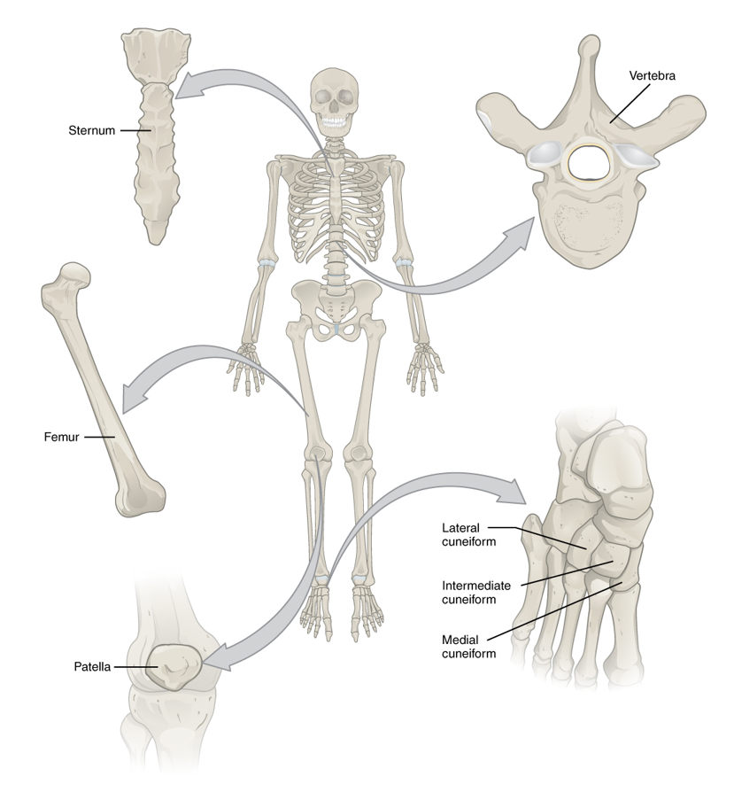
Question 13
Question
Endochronal ossification is the origin of all bones below the skull except the...
Answer
-
Clavicle
-
Sternum
-
Patella
-
Humerus
Question 14
Question
Fill in the blanks below to describe the process of endochronal ossification.
1. A bone [blank_start]collar[blank_end] forms around the [blank_start]hyaline[blank_end] cartilage model. This occurs when the perichondreum is vascularised, supplying nutrients which allow the [blank_start]mesenchymal[blank_end] cells to differentiate into [blank_start]osteoblasts[blank_end] which gather at the diaphysis wall.
2. [blank_start]Chondrocytes[blank_end] within the central cavity enlarge, causing the matrix to [blank_start]calcify[blank_end]. This matrix is then impermeable to nutrients causing the cells within to [blank_start]die[blank_end]. This forms a cavity supported by the bone collar.
3. The [blank_start]periosteal bud[blank_end] invades the cavity. forming spongy bone.
4. [blank_start]Osteoblasts[blank_end] break down new spongy bone to form the [blank_start]medullary[blank_end] cavity. The [blank_start]secondary[blank_end] ossification centres appear at the [blank_start]epiphyses[blank_end].
5. Once ossification of the epiphyses is complete, hyaline cartilage remains only at the epihyseal [blank_start]plates[blank_end] and the [blank_start]articular[blank_end] surfaces.
Answer
-
collar
-
hyaline
-
mesenchymal
-
osteoblasts
-
Chondrocytes
-
calcify
-
die
-
periosteal bud
-
Osteoblasts
-
medullary
-
secondary
-
epiphyses
-
plates
-
articular
Question 15
Question
What name is given to the connective tissue containing arteries, veins, lymphatics and nerves that invades the central cavity of a developing bone to deliver osteogenic cells?
Answer
-
Periosteal bud
-
Periosteal cord
-
Paraosteal bud
-
Osteogenic cord
Question 16
Question
Intramembranous ossification forms mainly flat bones such as the cranial bones and the clavicle.
Answer
- True
- False
Question 17
Question
Fill in the blanks to describe the process of intramembranous ossification.
1. [blank_start]Mesenchymal[blank_end] cells aggregate and differntiate into [blank_start]osteoblasts[blank_end], forming the [blank_start]ossification[blank_end] centre. The osteoblasts begin to secrete [blank_start]osteoid[blank_end] inwards towards this centre.
2. [blank_start]Peripheral[blank_end] msecnhymal cells continue to differentiate. Osteoblasts become trapped in the central space and differentiate into [blank_start]osteocytes[blank_end].
3. The ostoid [blank_start]calcifies[blank_end] and hardens to form the hardened bone matrix.
4. Osteoid continues to be deposited randomly around embryonic [blank_start]blood vessels[blank_end] to form the trabeculae.
5. The remaining mesenchymal cells around the central bone differentiate into the [blank_start]periosteum[blank_end]. [blank_start]Lamellar[blank_end] bone replaces spongy bone at the outer edges of the trabecular bone in compact layers.
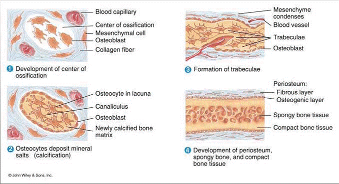
Answer
-
Mesenchymal
-
osteoblasts
-
ossification
-
osteoid
-
Peripheral
-
osteocytes
-
calcifies
-
blood vessels
-
periosteum
-
Lamellar
Question 18
Question
What is an osteon?
Answer
-
The functional unit of compact bone
-
A cell that secretes osteoid
-
The central canal between bone cells through which arterial supply travels
-
Networks of gaps between cells which route nutrients to bone cells
Question 19
Question
Osteons are aligned in the same direction in compact bone.
Answer
- True
- False
Question 20
Question
Label this diagram to show the different histological structures within bone.
Image:
Osteon (binary/octet-stream)
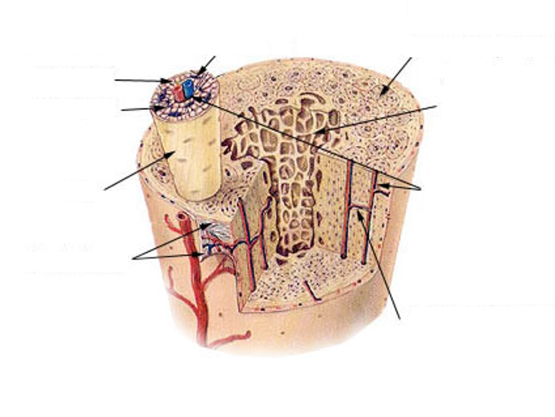
Answer
-
Osteon
-
Ostein
-
Canaliculi
-
Lamellae
-
Lacunae
-
Trabeculae
-
Harversian canal
-
Volkmann's canal
Question 21
Question
An osteon forms a cylindrical structure with a central cavity. In the centre is the [blank_start]Haversian[blank_end] canal. This contains [blank_start]blood vessels[blank_end] and nerve fibres to supply the bone. Surrounding the Haversian canal are concentric layers of compact matrix known as [blank_start]lamellae[blank_end]. These lamella are connected by [blank_start]canaliculi[blank_end] and contain spaces known as [blank_start]lacunae[blank_end] which are occupied by [blank_start]osteocytes[blank_end].
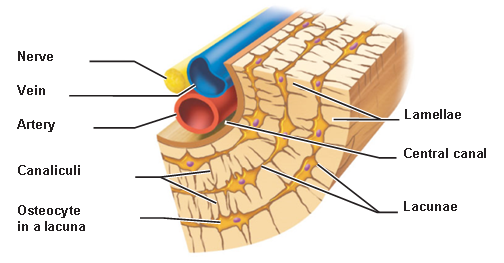
Answer
-
Haversian
-
blood vessels
-
lamellae
-
canaliculi
-
lacunae
-
osteocytes
Question 22
Question
What name is given to the canals which connect adjacent osteons via their Haversian canals and connect these Haversian canals to the periosteum?
Answer
-
Volkmann's canals
-
Transverse canal
-
Canaliculi
-
Osteoid canals
Question 23
Question
What is the periosteum?
Answer
-
Dense fibrous layer around the entire surface of the bone to which tendons attach
-
Dense fibrous layer around the bone except at the articular surfaces to which tendons attach
-
The cartilage found at the articular surfaces of joints
-
The functional unit of compact bone
Question 24
Question
How does trabecular bone help withstand pressure?
Answer
-
Diverts exerted forces onto compact bone
-
Diverts exerted forces into bone marrow
-
Is more dense in cells and osteoid than compact bone
-
It has fewer blood vessels than compact bone
Question 25
Question
Fill in the blanks to describe the inner structure of trabecular bone.
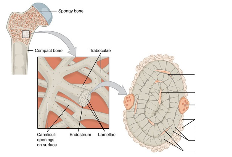
Answer
-
Osteocyte
-
Lacunae
-
Osteoclast
-
Osteoblasts
-
Canaliculi
-
Lamellae
Question 26
Question
What classification name is given to joints where two bones are united by collagen?
Answer
-
Collagenous joints
-
Fibrous joints
-
Suture joint
-
Syndesmosal joints
Question 27
Question
Which type of fibrous joint is seen only in the skull whereby the cranial bones are joined by interdigitations and Sharpey's fibres?
Answer
-
Sutures
-
Syndesmoses
-
Gomphoses
Question 28
Question
Sharpey's fibres join two cranial bones together at a suture joint and consist of bundles of what type of collagen?
Answer
-
Type I
-
Type II
-
Type IV
-
Type VII
Question 29
Question
What type of fibrous joint is formed when an interosseus ligament joins two bones, often seen in the lower arm and leg between adjacent long bones?
Answer
-
Suture joints
-
Syndesmoses
-
Gomphoses
-
Synovial joints
Question 30
Question
What type of fibrous joint binds the teeth to bony sockets in the maxilla mandible consisting of tough ligaments?
Answer
-
Suture joints
-
Syndesmoses
-
Gomphoses
-
Dental joints
Question 31
Question
Cartilaginous joints occur where two bones are united by cartilage.
Answer
- True
- False
Question 32
Question
A symphysis is a [blank_start]cartilginous[blank_end] joint where two adjacent bones join. The apposing surfaces of the two bones are covered in [blank_start]cartilage[blank_end] but are separated by an intervening disc of [blank_start]fibrocartilage[blank_end]. This contains numerous bundles of thick [blank_start]collagen[blank_end] fibres to provide resistance against shock. Symphysis are found between the spinal vertebrae and between the [blank_start]pubic[blank_end] bones of the pelvis.
Answer
-
cartilginous
-
fibrocartilage
-
collagen
-
cartilage
-
pubic
Question 33
Question
A synchrondrosis is a [blank_start]cartilaginous[blank_end] joint that develops between bones of [blank_start]endochronal[blank_end] origin. A solid plate of [blank_start]hyaline[blank_end] cartilage occurs between the two apposing surfaces forming the epiphyseal [blank_start]plate[blank_end] and is seen between ribs and the [blank_start]sternum[blank_end].
Answer
-
cartilaginous
-
endochronal
-
hyaline
-
plate
-
sternum
Question 34
Question
What name is given to a joint where two ends of bone are united by a fibrous capsule even though they don't technically make contact with one another?
Answer
-
Synovial joint
-
Synchrondoses
-
Symphysis
-
Gel joint
Question 35
Question
Fill in the blanks on this diagram to label the 6 different types of synovial joint.
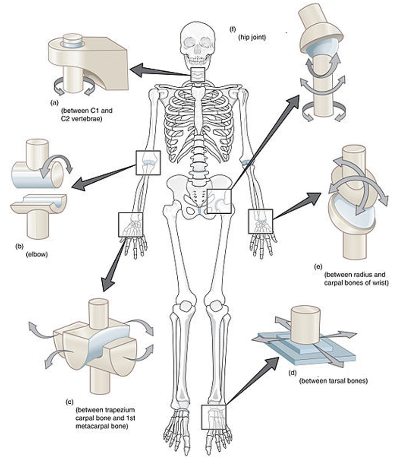
Answer
-
Ball and socket joint
-
Pivot joint
-
Saddle joint
-
Hinge joint
-
Condyloid joint
-
Plane joint
Question 36
Question
What name is given to the sacs filled with synovial fluid that occur where structures move in tight apposition to prevent bone-bone contact?
Answer
-
Bursa
-
Articular capsule
-
Osteoid sac
-
Synovial sac
Question 37
Question
The synovial membrane lines the capsule but not the articular surface.
Answer
- True
- False
Question 38
Question
Fill in the blanks to label the synovial joint shown here.
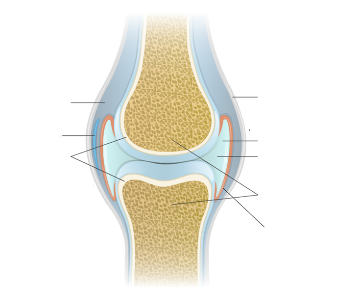
Answer
-
Synovial cavity
-
Ligament
-
Bursa
-
Articular cartilage
-
Bones
-
Synovial membrane
-
Synovial fluid
-
Joint cavity
Question 39
Question
The viscosity of the synovial fluid is constant.
Answer
- True
- False
Question 40
Question
What happens to the hyaline cartilage on the articular surface of a bone at a synovial joint when force is exerted on the bone?
Answer
-
Deforms to ensure all parts of bone are in contact to spread the load of the force
-
Transmits force into the centre of the cartilage at one single point
-
Becomes rigid to prevent deformation which would be painful
-
Decreases the viscosity of the synovial fluid
Question 41
Question
Which part of the synovial joint has a rich capillary network and allows both secretion and absorption of the synovial fluid?
Answer
-
Articular cartilage
-
Ligament
-
Epiphyseal plate
-
Synovial membrane
Question 42
Question
[blank_start]Concentric[blank_end] contraction is muscle contraction that increases tension on a muscle as it shortens to generate force.
[blank_start]Eccentric[blank_end] contraction is muscle contraction occurring as it lengthens under load.
[blank_start]Isometric[blank_end] contraction is contraction that does not change the length of the muscle as it holds a steady load.
Answer
-
Concentric
-
Eccentric
-
Isometric
Question 43
Question
A [blank_start]uniaxial[blank_end] joint can move about one axis of movement.
A [blank_start]biaxial[blank_end] joint can move about two axes of movement perpendicular to one another.
A [blank_start]multiaxial[blank_end] joint can move about three or more axes of movement.
A [blank_start]non-axial[blank_end] joint can move in all directions.
Answer
-
uniaxial
-
biaxial
-
multiaxial
-
non-axial
Want to create your own Quizzes for free with GoConqr? Learn more.