
Quiz
Control of heart rate
This quiz has question types that open in the usual player.
Questions in this quiz (6)
1With each heartbeat, a pulse of blood is ejected. This can be felt passing along the arteries; easily felt at the wrist ([blank_start]radial artery[blank_end]) or at the neck ([blank_start]carotid artery[blank_end]). Resting heart rate between 60 and 100 bpm. It's slightly higher in [blank_start]females[blank_end]. The differences in resting heart rate are caused by many factors. Our hearts differ in size, due to differences in [blank_start]body size[blank_end] and [blank_start]genetic[blank_end] factors. A larger heart will usually have a [blank_start]lower[blank_end] resting heart rate. It will expel more blood with each beat as so, other things being equal, doesn't have to beat as [blank_start]frequently[blank_end] to circulate the same volume of blood around the body. [blank_start]Endurance training[blank_end] produces a lower resting heart rate, largely due to an increase in the [blank_start]size[blank_end] of the heart, resulting from the thickening of the heart muscle walls.
- Aradial artery
- Bcarotid artery
- Cfemales
- Dbody size
- Egenetic
- Flower
- Gfrequently
- HEndurance training
- Isize
2What does myogenic mean?
- AThe heart muscle can contract without external nervous stimulation
- BThe heart muscle can contract without conscious thought
3Contraction of cardiac muscle is initiated by small changes in the [blank_start]electrical charge[blank_end] of cardiac muscle cells. When these cells have a slight positive charge on the outside, they're [blank_start]polarised[blank_end]. When this charge is reversed, they're [blank_start]depolarised[blank_end]. A change in polarity spreads like a wave from cell to cell and causes the cells to [blank_start]contract[blank_end]. Depolarisation starts at the [blank_start]sinoatrial node (SAN)[blank_end]. This is a small area of specialised muscle fibres located in the wall of the [blank_start]right[blank_end] atrium, beneath the opening to the [blank_start]superior[blank_end] vena cava. The SAN is aka the pacemaker. The SAN generates an electrical impulse; this spreads across the right and left atria, causing them to contract at [blank_start]the same time[blank_end]. The impulse also travels to some specialised cells called the [blank_start]atrioventricular node (AVN)[blank_end]. From here, the impulse is conducted to the ventricles after a delay of about [blank_start]0.13[blank_end] seconds.
- Aelectrical charge
- Bconcentration gradient
- Cpolarised
- Ddepolarised
- Edepolarised
- Fpolarised
- Gcontract
- Hrelax
- Isinoatrial node (SAN)
- Jatrioventricular node (AVN)
- Katrioventricular node (AVN)
- Lsinoatrial node (SAN)
- Msuperior
- Ninferior
- Oright
- Pleft
- Qthe same time
- Rdifferent times
- S0.13
- T1
4Why is the delay important?
- AIt ensures the atria have finished contracting and that the ventricles have filled with blood before they contract
- BIt gives the heart a chance to compose itself before contracting
5After the delay, the signal reaches the [blank_start]Purkyne fibres[blank_end]. These are large, specialised muscle fibres that conduct impulses rapidly to the [blank_start]apex[blank_end] (tip) of the ventricles. There are right and left bundles of fibres, and they're collectively called the [blank_start]bundle of His[blank_end]. The Purkyne fibres continue around each ventricle and divide into smaller branches that penetrate the ventricular muscle. These branches carry the impulse to the inner cells of the ventricles and from here it spreads through the entire ventricle walls. The first ventricular cells to be depolarised are at the apex of the heart, so that contraction begins at this point and travels upwards towards the atria. This produces a [blank_start]wave[blank_end] of contraction moving up the ventricles, pushing the blood into the [blank_start]aorta[blank_end] and the pulmonary artery.
- APurkyne fibres
- Bapex
- Cbundle of His
- Dwave
- Eaorta
6Label the diagram:
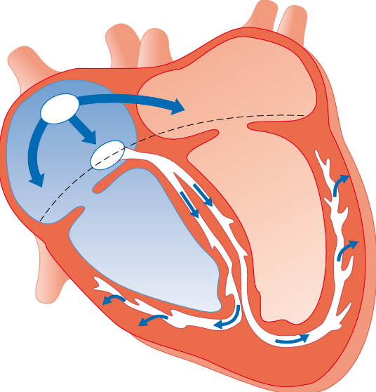
- Asinoatrial node (SAN)
- Batrioventricular node (AVN)
- Cnon-conducting layer in heart wall
- DPurkyne fibres in bundle of His
- EApex of heart
Information
- Description
- 6 questions
- Author
- Ella Middlemiss
- Language
- English
- Views
- 3
- Tags
- Rating
- No ratings yet Rate this quiz