
null
null
17927364
9. Specialised Cells: B cells
Description
No tags specified

Slide Set by Chloe Cavarretta, updated more than 1 year ago
More
Less

|
Created by Chloe Cavarretta
over 7 years ago
|
|
null
Resource summary
Slide 1
Learning Outcomes: L18
The mechanism of B cell activation
Importance of coreceptors for signalling and costimulatory molecules for amplification
The functions of FcϒRIIB and shutting down of the B cell response
The basic organisation of germinal centres and the roles of the various cells
Follicular dendritic cells
Follicular T helper cells; IL-21, Bcl-6
Somatic hypermutation and AID
Antibody class switching and AID
The different types of antibodies/immunoglobulins in our bodies and their role in the immune response
Primary versus secondary responses
Opsonisation, neutralisation, complement fixation
Link to allergy, asthma and how microbes can promote such types of responses
Slide 2
Activation of B Cells
B cells recognise antigen via BCR
Lock and Key approach
The part of antigen that BCR/Ab recognise= epitope of antigen
Continuous: Ab recognises sequence on antigen that is linear
Discontinuous: Forms the shape that Ab recognises when molecule folds
Important for experiments:
If Ab recognises linear epitope: ELISA is fine as whole protein in plate, western blot fine (protein denatured on gel)
If Ab recognises discontinous epitope: western blot not fine, -ve result may not mean antigen not their but Ab may no longer recognise
Slide 3
Activation of B Cells
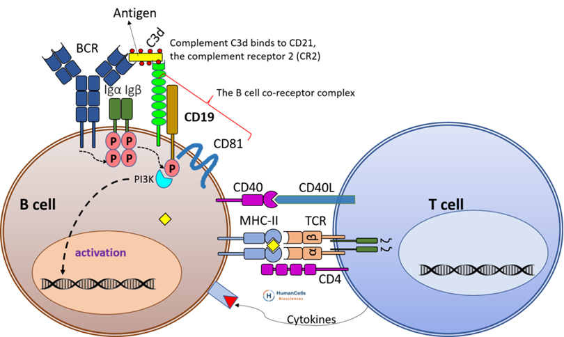
Like TCR, BCR signals through co-receptor, signal potentiated via co-stimulatory molecules- receptor alone has no capacity to signal:
Co-receptors Igα and Igβ have tyrosine kinase activity
Antigen binds BCR, conformational change in intracellular part, Igα and Igβ phosphorylated (activated) + phosphorylate costimulatory molecules
CD19/CD81 form a co-receptor complex that provides costimulatory amplification of signals through the BCR
C3d complement bound to pathogen also helps target pathogen to BCR, B cell has receptors for complement (CR2/CD21)
CD4+ T cell help activation + differentiation of B cells into plasma cells
Slide 4
Slide 5
B Cell Response No Longer Needed
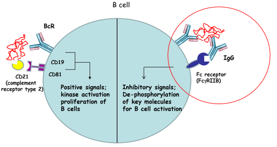
When B response not need, B cell shut down by signals through the negative regulator- FcϒRIIB (Fc receptor):
Positive signals:
Kinase activation proliferation of B cells
CD19, CD81 act as costimulatory molecules for B cell activation
Negative signals:
Fc gamma receptor looks for excess IgG
Dephosphorylation of key molecules for B cell activation (like CTLA4)
Mutations in FcϒRIIB linked to autoimmune production of Ab's to host cell- cant shut down B cell response efficiently
Slide 6
B Cell Response No Longer Needed
Slide 7
Activation In Secondary Lymphoid Tissues
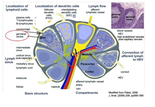
Highly organised structures where cells are compartmentalised:
Inside B cell follicle is germinal centre
B cells in outer follicle, upon antigen encounter move to germinal centre: activation, proliferation and differentiation
Germinal centre= less dense, only contains B cells with BCR triggered by antigen
Slide 8
Germinal Centre
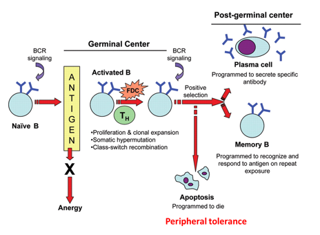
Critical for development of B cell responses:
Within germinal centre have interactions between activated B cell, follicular DC and Th cell
B cell proliferation
Somatic hypermutation of B cell
Class switch
Once these are achieved B cell has different fates: Memory cell, Plasma cell, Apoptosis (check point at germinal centre for autoreactivity- dies unlike in BM get receptor editing)
Slide 9
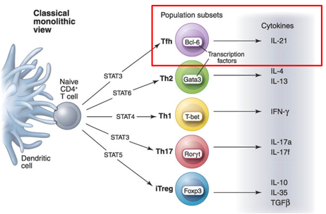
GC: CD4+ Follicular Th cell (Tfh)
Central to formation and propagation of germinal centre, resides in germinal centre tells B cell to do the 3 functions
TF for differentiation into Tfh cell is Bcl-6 and it makes cytokine IL-21
Slide 10
GC: Function
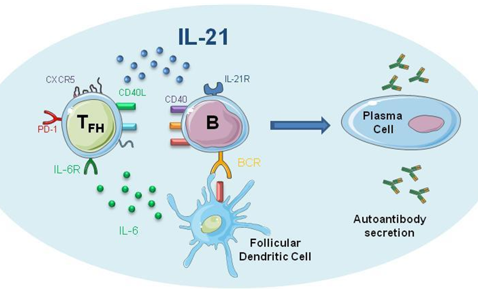
IL-21 is criticial mediator in germinal centre function:
Tfh express chemokine receptor CXCR5 , responsive to CXCL13 produced by GC to recruit and keep B cell and Tfh cell there
IL-21:
Helps stromal cells of GC to maintain themselves
Signals back to Tfh to keep alive
Signal into B cell for proliferation)
Tfh continually expresses antigens on its surface, that B cell responds to, to ensure B cell performs the 3 function
Slide 11
GC: Function
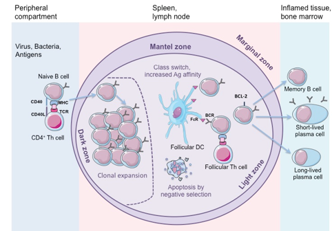
Infection occurs in outer B cell follicle, interaction with conventional Th cell, activated and moves into GC
In dark zone= clonal expansion occurs
Moves into light zone= interacts with follicular DC and Th cell and receives signals to transform into:
Memory B cell
Short-lived plasma cell that secretes Abs
Moves into BM and remains as long-lived plasma cell, secreting small amounts of Ab to that pathogen (protective immunity)
Slide 12
Somatic Hypermutation (SHM) by AID
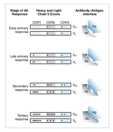
Advanced Induced Deaminase (AID) is active in GC to facilitate SHM
First encounter of R with antigen= not good fit but still triggers initial activation
Mutations in Hv region enable a tighter fit (stronger interaction= better signal)- secondary response= memory cells
Each subsequent encounter 'hones' the antigen binding site further
B cells that have undergone SHM will have their BCRs re-screened to determine if the mutations lead to autoreactive BCR (B cell dies if so)
Slide 13
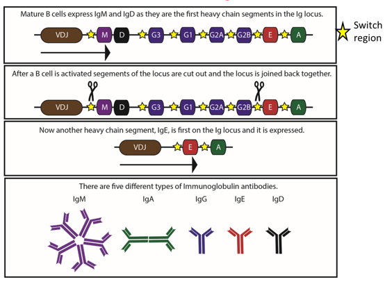
Class Switching by AID
Need specific Ab for specific response- remove Fc region of IgM and replace with different Ab Fc region
Upstream every Ab Fc region is a switch region (except igD so igM expressing cells also express igD)
AID causes switch regions to form haripin loop that brings together Fc constant regions to change
Cleave out intermediate regions
Brings new Fc region to the VDJ so specifcity of Ab stays same
Slide 14
Maturation of Humoral Response
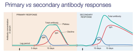
Maturation of humoral response characterised by Ig with increased affinity for its cognate antigen and switching of Ig class:
Primary response- takes few days to start making Ab's- need B cell activation, CD4 T help, functioning in GC to class switch
Initially get IgM- replaced by IgG after 15 days
Short lived, low-grade Ab production
Secondary infection- immune system stimulates IgG B cell to secrete Ab because antigen binding site better fit
Slide 15
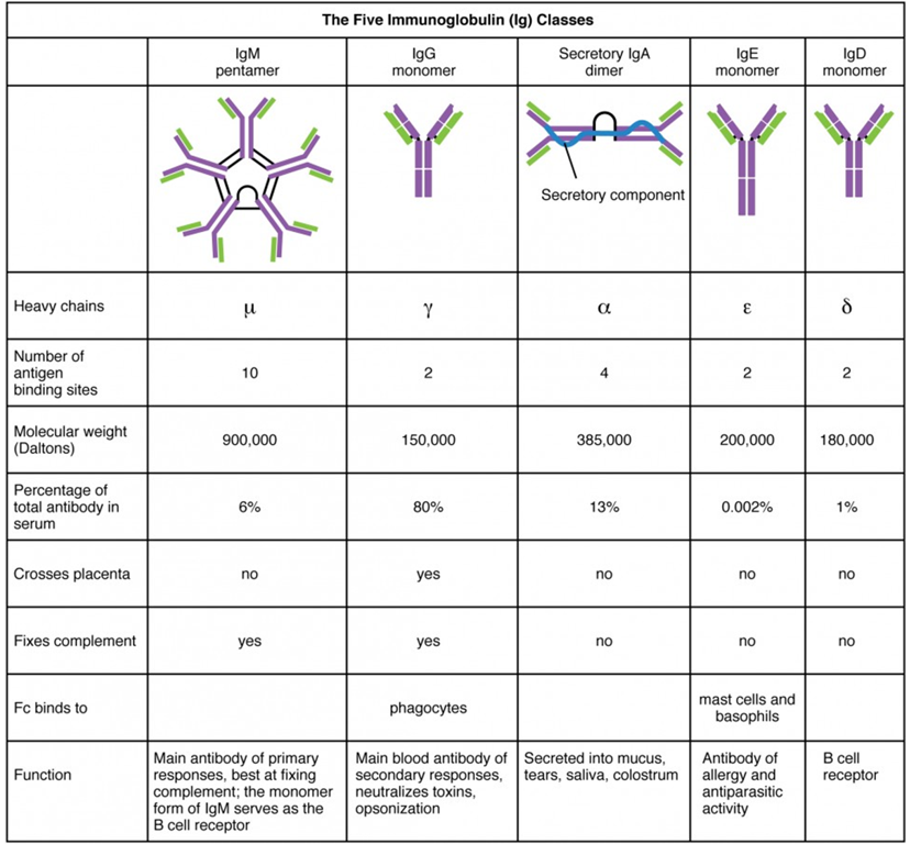
IgM
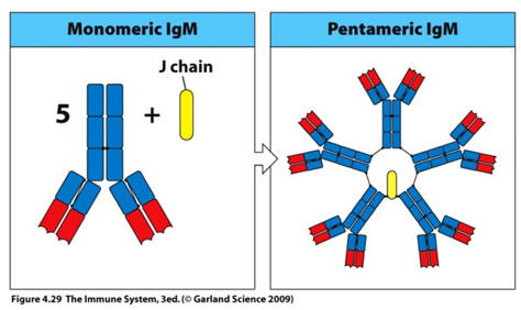
Forms pentameric str held together by a protein called J chain
5 weak binding sites, 2 per arm= 10 antigen binding sites
Ensures pathogens dont escape primary response
Low affinity
Key role= protection during immune response but 10 binding sites compromise low affinity
First Ig produced during initial immune response
Slide 16
IgD
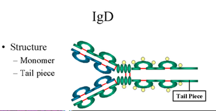
Found in serum- unknown function
Usually found co-expressed with IgM on surface of unactivated B cells
Acts as receptor
Slide 17
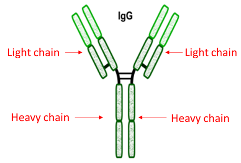
IgG
High affinity
Key role= memory immune response
Class switched in primary and secondary response
Occurs as disulphide linkage of 2 heavy chains and 2 light
Most common Ig in serum
Generated following initial infection due to class switching of IgM
Can cross placenta (receptors for Fc regions of IgG on placenta)
Exceptional at activating C' and ADCC
Slide 18
IgA
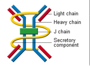
Exists as dimer: two monomeric Ig units held together by J chain
Present in serum and secretory fluids, most abundant in secretory fluids (tears, colostrum, saliva, intestinal tract
Monomer in serum
Dimer in mucosal tissue
Secretory component (yellow chain) makes IgA well suited to reside in mucosa (wraps around dimer, protects J chain, stops dimer doesn't falling apart)
Neutralises toxins (in blood stream)
Slide 19
IgE
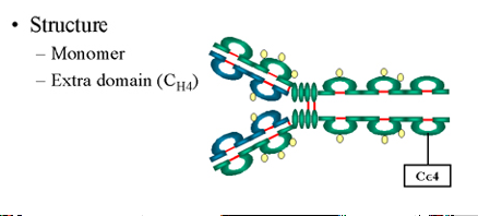
Least common Ig in serum
Additional constant region domain
Binds to specialised Fc epsilon receptors on mast cells, basophils and eosinophils
Strongly involved in allergic reactions and clearance of parasitic infections
Slide 20
IgE Roles: Deal with Parasitic Infections
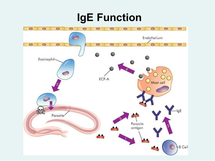
Enriched in mucosal tissues and associated with mast cells (innate) so are well placed to deal with parasites
Parasitic antigen picked up by B cell, CD4 T cell help class switch to IgE
Mast cells have receptors to bind IgE Fc region: parasitic antigen bound by IgE, bound by mast cell receptor
Triggers mast cell to secrete eosinophil chemoattractant factor
Eosinophils have toxic granules
Slide 21
Allergic Reactions
Susceptibility to allergic reactions influences by your genes:
Allergy= immune response to something harmless
Individuals predisposed to developing allergies= atopic individuals that have higher circulating levels of IgE than non-atopic individuals
Allergies are inherited (not always) and strong link to maternal side of family
Slide 22
Allergic Reactions: Asthma
Narrower airways- can't take in as much air
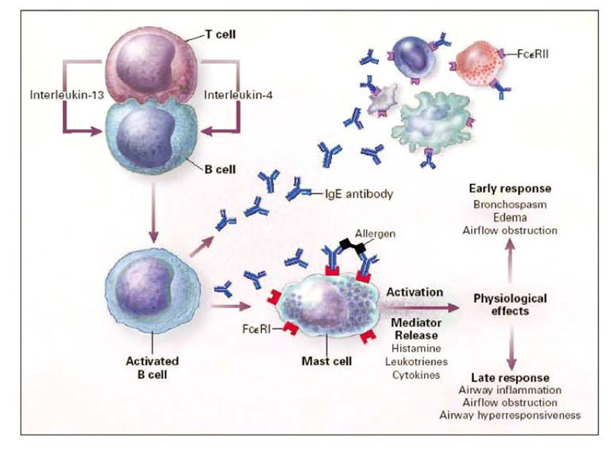
IgE secreting B cell activates mast cell to release inflammatory mediators
IgE binds mast cell causing secretion of histamines, leuktorienes, cytokines (inflammatory mediators) from mast cell
Secretory molecules affect airways by:
Early response- bronchospasms, swelling and airflow obstruction
Late response= airway inflammatiion, obstruction and hyperresponsiveness
Slide 23
Allergic Reactions: Asthma
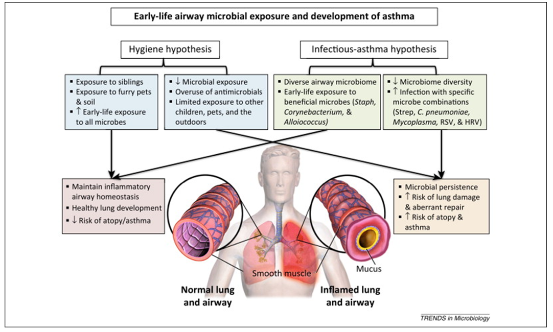
Microbes linked to asthma development:
Up until toddler, immune system predominantly Th2 biased, becomes more Th1 with age
Children at risk of asthma/allergies retain high Th2 environment
Theories Th1 vs Th2 biased immunity:
Hygiene hypothesis: Children with siblings and pets have lower risk of allergies/asthma
Siblings pick up infections as they grow, when a baby is exposed to wide range of infections, need Th1 response, shapes immune system to Th1
Infection-asthma hypothesis: Have own microbiome (gut bacteria)
Infection when young, with beneficial microbes= shapes immune system to be less responsive to allergens
Infection when young, with specific microbe combinations= more likely develop allergies and asthma
Slide 24
Allergic Reactions: Asthma
Slide 25
Summary Ab Classes
Want to create your own Slides for free with GoConqr? Learn more.
Similar
null