
Notizen
Microscopy
Inhalt der Notizen (2)
Seite 1
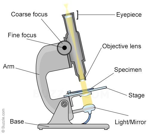
Light microscope:
Use a number of lenses to produce an image that can be viewed directly at the eyepiece.
Light passes through a bulb under the stage, through a condenser lens, then through the specimen.
This beam of light is focused through the objective lens, then through the eyepiece lens.
To view specimens at different magnifications, light microscopes have a number of objective lenses that can be rotated into position.
They usually have four objective lenses, x4, x10, x40 and x100. The x100 is an oil immersion lens.
The eyepiece lens magnifies the image again, this is usually x10.
Total magnification is given by the objective magnification times the eyepiece magnification.
Advantages and limitations:
Magnification - most light microscopes are capable of magnification up to a maximum of x1500
Resolution - The maximum resolving power using light is 200nm. This is due to the wavelength of light. Two objects can only be seen separately if light waves can pass between them.
Specimens - Living organisms such as Euglena and Daphnia can be viewed using a light microscope. You can also view thin sections of larger plants and animals and smear preparations of blood or cheek cells.
Preparations of specimens:
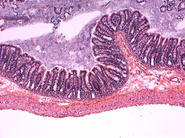
Staining - Coloured stains are chemicals that bind to chemicals on or in the specimen. This allows the specimen to be seen. Some stains bind to specific cell structures. Acetic orcein stains DNA dark red. Gentian violet stains bacterial cell walls.
Sectioning - Specimens are embedded in wax. Thin sections are then cut without distorting the structure of the specimen. This is especially useful for making sections of soft tissue such as brain.
Electron microscopes:
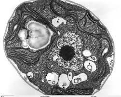
Transmission Electron Microscope (TEM)
The electron beam passes through a very thin prepared sample.
Electrons pass through the denser parts of the sample less easily, so giving some contrast.
The final image produced is two dimensional
The magnification possible with a TEM is x500 000
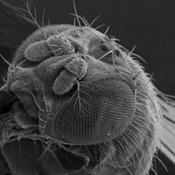
Scanning Electron Microscope (SEM)
The electron beam is directed onto a sample. The electrons don't pass through the specimen.
They are bounced off the sample.
The final image produced is a 3D view of the surface of the sample.
The magnification possible with an SEM is about x100 000
Advantages:
The resolution is 0.1nm
This means that electron microscopes can be used to produce detailed image of cell structure (organelles)
The SEM produces 3D images that can reveal the detail of contours and cellular or tissue arrangements; this is not possible with a light microscope.
Limitations:
Electron beams are deflected by the molecules in air, so samples have to be placed in a vacuum.
Electron microscopes are extremely expensive.
Preparing samples and using an electron microscope both require a high degree of skill and training.
Preparation of samples:
Fix the specimen in glutaraldehyde to make the tissue firm
Dehydrate it to replace the water with ethanol
Embed the dehydrated tissue in a solid resin
Cut very thin slices using a diamond knife
Stain it using lead salts to scatter electrons differently - this gives it contrast.
Mount it on a copper grid
Place the specimen on the grid in a vacuum in the microscope.
Laser Scanning Confocal microscope:
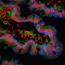
In laser scanning confocal microscopy, a laser is used to scan the sample. This allows us to see a very precise layer of the sample. This means that it has a higher resolution than conventional light microscopy.
Advantages:
Can view living cells like light microscopy
Has a higher resolution than light microscopy
Can produce 3D images of cells
We can target specific proteins and structures within the cell and watch them move.
Seite 2
Eyepiece Graticule
can measure Mic
Informationen
- Beschreibung
- AS - Level Biology (2.1.1 Cell Structure) Notiz am Microscopy, erstellt von 10egordon am 26/03/2016.
- Erstellt von
- 10egordon
- Aufrufe
- 2
- Bewertung