
Quiz
Pathophysiology revision - SECTION II - Respiratory Systems
Dieses Quiz enthält Fragetypen, die sich im gewohnten Player öffnen.
Fragen in diesem Quiz (56)
1Label the areas of the respiratory CONDUCTING zones as appropriate:
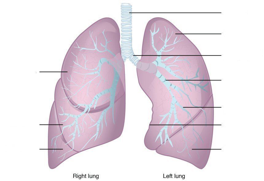
- ATrachea
- BSuperior Lobe
- CMain (primary) bronchus
- DLobar (secondary) bronchus
- ESegmental (tertiary) bronchus
- FCardiac notch
- GInferior lobe
- HMiddle lobe
- ITrachea
- JSuperior lobe
- KMain (primary) bronchus
- LLobar (secondary) bronchus
- MSegmental (tertiary) bronchus
- NCardiac notch
- OInferior lobe
- PMiddle lobe
- QTrachea
- RSuperior lobe
- SMain (primary) bronchus
- TLobar (secondary) bronchus
- USegmental (tertiary) bronchus
- VCardiac notch
- WInferior lobe
- XMiddle lobe
- YTrachea
- ZSuperior lobe
- AAMain (primary) bronchus
- ABLobar (secondary) bronchus
- ACSegmental (tertiary) bronchus
- ADCardiac notch
- AEInferior lobe
- AFMiddle lobe
- AGTrachea
- AHSuperior lobe
- AIMain (primary) bronchus
- AJLobar (secondary) bronchus
- AKSegmental (tertiary) bronchus
- ALCardiac notch
- AMInferior lobe
- ANMiddle lobe
- AOTrachea
- APSuperior lobe
- AQMain (primary) bronchus
- ARLobar (secondary) bronchus
- ASSegmental (tertiary) bronchus
- ATCardiac notch
- AUInferior lobe
- AVMiddle lobe
- AWTrachea
- AXSuperior lobe
- AYMain (primary) bronchus
- AZLobar (secondary) bronchus
- BASegmental (tertiary) bronchus
- BBCardiac notch
- BCInferior lobe
- BDMiddle lobe
- BETrachea
- BFSuperior lobe
- BGMain (primary) bronchus
- BHLobar (secondary) bronchus
- BISegmental (tertiary) bronchus
- BJCardiac notch
- BKInferior lobe
- BLMiddle lobe
- BMTrachea
- BNSuperior lobe
- BOMain (primary) bronchus
- BPLobar (secondary) bronchus
- BQSegmental (tertiary) bronchus
- BRCardiac notch
- BSInferior lobe
- BTMiddle lobe
- BUTrachea
- BVSuperior lobe
- BWMain (primary) bronchus
- BXLobar (secondary) bronchus
- BYSegmantal (tertiary) bronchus
- BZCardiac notch
- CAInferior lobe
- CBMiddle lobe
2The passage of air into the lungs is governed by pressure differences caused by changes in [blank_start]lung volume[blank_end]. The [blank_start]thoracic diaphragm[blank_end] is the principle muscle of respiration which is innervated by the phrenic nerves derived from the ventral rami of cervical segments 3 – 5.
- Alung volume
- Blung capacity
- Clung residue
- Dlung resilience
- Ethoracic diaphragm
- Flumbar diaphram
- Gcervical diaphram
- Hadregeneric diaphragm
3When the diaphragm [blank_start]contracts[blank_end], the vertical diameter of the thoracic cavity increases and air moves into the lungs. This relationship between volume and pressure is represented in [blank_start]Boyle’s Law[blank_end]
- Acontracts
- Brelaxes
- Cinverts
- DBoyle’s Law
- ECharles' Law
- FHenry's Law
- GStarling's Law
4[blank_start]Muscles of inspiration[blank_end]: external intercostals, sternocleidomasoids, scalei, anterior serrate, trapezius, posterior neck muscles.
[blank_start]Muscles of expiration[blank_end]: internal intercostals, posterior inferior serrate, abdominal muscles.
[blank_start]Normal conditions[blank_end]: diaphragm and intercostals- AMuscles of inspiration
- BMuscles of expiration
- CNormal conditions
- DMuscles of expiration
- EMuscles of inspiration
- FNormal conditions
- GNormal conditions
- HMuscles of inspiration
- IMuscles of expiration
5In a patient with a pneumothorax, the lung collapses when the [blank_start]intraplural space[blank_end] is violated and the [blank_start]negative[blank_end] pressure between the lung and chest wall is no longer maintained
- Aintraplural space
- Bextraplural space
- Cintercostal space
- Dmyocardial space
- Enegative
- Fpositive
- Ginerrant
- Hbackforce
6Patients with Asthma have narrowed air passages due to smooth muscle contraction, mucus plugging and inflammation. This leads to an [blank_start]increase[blank_end] in residual volume, a [blank_start]decrease[blank_end] in vital capacity and a [blank_start]decrease[blank_end] in expiratory reserve volume
- Aincrease
- Bdecrease
- Cdecrease
- Dincrease
- Edecrease
- Fincrease
7In patients with emphysema, lung tissue is destroyed and weakened airway walls collapse during [blank_start]expiration[blank_end]. Because of chronic lung tissue inflammation and swelling, the airway walls have decreased recoil and elasticity. Thus, inspiritory capacity and expiratory reserve volume [blank_start]decease[blank_end] while reserve volume [blank_start]increases[blank_end]
- Aexpiration
- Binspiration
- Cdecease
- Dincrease
- Eincreases
- Fdecreases
- Gremains the same
8According to [blank_start]La Place’s[blank_end] Law, the surface tension of smaller alveoli is [blank_start]greater[blank_end] than that of larger alveoli. [blank_start]Surfactant[blank_end] is a substance produced by type II alveolar cells which decreases surface tension of the alveoli and is an important component to think about when dealing with [blank_start]pre-mature babies[blank_end]
- ALa Place’s
- BBoyle's
- CCharles'
- DHenry's
- EStarling's
- Fgreater
- Glesser
- Hthe same
- ISurfactant
- JLubriol
- KProtasine
- LPluratol
- Mpre-mature babies
- Nobese patients
- Ogeriatrics
- Pteenagers
9Damage to the capillary membrane, heart failure and valvular disease can all lead to [blank_start]pulmonary odeama[blank_end]. This is due to an imbalance in the normal forces at the alveolar capillary membrane, causing fluid to accumulate in the pulmonary interstitial space and alveoli. This is best illustrated by [blank_start]Starling’s Law[blank_end]
- Apulmonary odeama
- BEmphysema
- CAsthma
- DBronchitis
- EStarling’s Law
- FHenry's Law
- GCharles's Law
- HLa Place's Law
10Excess fluid accumulating within the pulmonary interstitial space is usually taken up by the [blank_start]lymphatic system[blank_end]
- Alymphatic system
- Bartieral system
- Cpulmonary system
- Dendocrine system
11Airway diameter and secretions are primarily controlled by the [blank_start]autonomic nervous system[blank_end]. [blank_start]Beta-2[blank_end] receptors function to dilate the bronchioles when activated by adrenergic agonists such as epinephrine and norepinephrine. Stimulation of these receptors also [blank_start]decreases[blank_end] the secretions of mucus and serous glands
- Aautonomic nervous system
- Bcardiac receptor system
- Csomatic nervous system
- Denteric system
- EBeta-2
- FBeta-1
- GAlfa-1
- HAlfa-2
- Idecreases
- Jincreases
12[blank_start]Inspiration[blank_end] is controlled neurologically via the medullary respiratory centre which sends signals via the phrenic nerve. [blank_start]Expiration[blank_end] is controlled via the ventral respiratory group and is usually only activated during exercise or with certain diseases (COPD) where where [blank_start]expiration[blank_end] becomes an active process.
- AInspiration
- BExpiration
- CRespiratory syncope
- DExpiration
- EInspiration
- FRespiratory syncope
- Gexpiration
- Hinspiration
- Irespiratory syncope
13Chemoreceptors play an important role in the regulation of respiration. Central chemoreceptors in the [blank_start]medulla[blank_end] are sensitive to PH of cerebrospinal fluid. When the PH level drops, the ventilary rate [blank_start]increases[blank_end]. This would be due to an [blank_start]increase[blank_end] in C02 levels
- Amedulla
- Baorta
- Ccerebellum
- Dcerebrum
- Eincreases
- Fdecreases
- Gincrease
- Hdecrease
14Why would a hyperventilating patient lose consciousness?
- APH levels will fall, causing central chemoreceptors to reduce the ventilary rate
- BPH levels will rise, causing central chemoreceptors to reduce the ventillary rate
- CPH levels will fall, causing central chemoreceptors to increase the ventillary rate
- DPH levels will rise, causing the central chemoreceptors to increase ventilary rate
15Peripheral chemoreceptors exist in the [blank_start]carotid and aortic bodies[blank_end]. These are sensitive to [blank_start]oxygen[blank_end] and hence, if levels fall to a very low rate (below 60 mmHg), these are activated to increase ventillary rate. Those who have this system consistently activated are said to have a [blank_start]hypoxic drive[blank_end]
- Acarotid and aortic bodies
- Bmedulla
- Crecticular respiratory zone
- Dpluralistic bodies
- Eoxygen
- Fcarbon dioxide
- Ghydrogen ions
- Hbicarbonate
- Ihypoxic drive
- Jasthmatic tendancy
- Kventilation to perfusion mismatch
- Ltachypnoeic altercation
16Albuteral, bitolterol and salmeterol are all examples of [blank_start]Beta-2 Agonists[blank_end], used for treating Asthma and COPD. These work by inducing [blank_start]relaxation[blank_end] of the bronchial musculature. However, they can also have an effect on Beta-1 receptors causing [blank_start]increased heart rate[blank_end]
- ABeta-2 Agonists
- Banticholinergic agents
- CGlucocorticoids
- DAfla-1 Agonists
- Erelaxation
- Fconstriction
- Gspasm
- Hexitation
- Iincreased heart rate
- Jreduced heart rate
- KAtrial fibrillation
- LAtrial flutter
17Ipratopium bromide works to reverse overstimulation of the autonomic nervous system by blocking the [blank_start]parasympathetic[blank_end] neurotransmitters. They are hence known as [blank_start]anticholinergic agents[blank_end]
- Aparasympathetic
- Bsympathetic
- Canticholinergic agents
- DBeta-2 agonists
- Eglucocorticoids
- FBeta blockers
18Glucocorticoids also reffered to as corticosteroids are compounds synthesized from the [blank_start]zona recticulars fasciculars[blank_end] of the adrenal cortex. [blank_start]Methylprednisolone[blank_end] is an example of this steroid
- Azona recticulars fasciculars
- Bzona glomerulosa
- Czona medullla
- DMethylprednisolone
- Eibatropium
- Falbuterol
- Gsalbutamol
19Your unit is called to the scene of a 33-year old asthmatic complaining of severe shortness of breath. Lung sounds are decreased with diffuse wheezing and a prolonged expiratory component. All of the following are muscles of expiration, EXCEPT:
- AInternal instercostals
- BProsterior inferior serrate
- CRectus abdominous
- DSternocleidomastoids
- EExternal Obliques
20What would pulmonary function testing be likely to show in a patient experiencing a severe asthma attack?
- AIncreased residual volume
- BIncreased expiratory reserve volume
- CIncreased vital capacity
- DDecreased inspiritory capacity
- ELittle change in lung capacity
21Patients with COPD are expected to have which of the following?
- ADifficulty with inspiring air due to bronchial narrowing
- BA decreased residual volume due to air trapped in dilated alveoli
- CIncreased inspiratory capacity and decreased expiratory reserve volume
- DIncreased elastic recoil of the lung segments
- EDifficulty with expiration due to chronic lung tissue inflammation and swelling
22Neonatal respiratory distress syndrome occurs in certain infants as a result of:
- AChronic airways inflammation
- BHypertrophy of type II alveolar cells
- CHyperactivity of type I alveolar cells
- DCollapse of the alveoli during expiration due to a lack of surfactant
- EBronchoconstriction due to hyperresponsiveness to supplemental oxygen
23The mechanism of beta-2 agonists in asthma is:
- AActivation of the parasympathetic neurons to provide bronchodilation
- BStimulation to increase the secreations of the mucous glands to rid the lungs of irritants
- CStimulation of beta-2 adrenergic receptors on cells in the terminal bronchioles
- DStimulation of beta-2 adrenergic receptors on cells in the proximal bronchi
- EDisinhibation of the medullary respiratory centre located in the brainstem
24A patient is hyperventilating and loses consciousness – what is the pathologic mechanism responsible for this?
- AIncreased carbon dioxide levels in the CSF
- BDecreased carbon dioxide and hydrogen concentrations in the CSF
- CIncreased oxygen levels in the CSF
- DInhibition of the central chemoreceptors in the medulla
- EActivation of the carotid bodies in response to decreased inhaled carbon dioxide
25A 69-year old with emphysema becomes less responsive with decreased respirations while on 4 lpm of oxygen during a prolonged inter-facility transport. What is the likely mechanism for these findings?
- ALowering of the P02 to very low levels causing a hypoxic drive, stimulated by peripheral chemoreceptors in the carotid bodies – now being suppressed by the prolonged administration of oxygen
- BLowing of the P02 to very low levels causing suppression of the hypoxic drive, stimulated by peripheral chemoreceptors in the carotid bodies
- CBuild-up of C02 and hydrogen ions in the CSF, causing activation of the central chemoreceptors which will suppress ventilations
- DLowering of the P02 to very low levels causing suppression of the hypoxic drive, stimulated by the central chemoreceptors of the medulla
- ELowering of the PC02 to very low levels causing suppression of the hypoxic drive, stimulated by the central chemoreceptors of the medulla
26The National Heart, Lung and Blood institute defines asthma as a [blank_start]chronic inflammatory disorder[blank_end] of the airways
- Achronic inflammatory disorder
- Bacute respiratory implication
- Crare pathological damage
- Dpulmonary odemic complication
27[blank_start]Extrinsic[blank_end] asthma is believed to be caused by an antibody-dependant (IgE) activation of cells that release inflammatory mediators. For that reason, it is also known as allergic asthma and is usually picked up in [blank_start]childhood[blank_end]
- AExtrinsic
- BIntrinsic
- CExcretory
- DExcitory
- Echildhood
- Fadulthood
- Gteenagers
- Hgeriatrics
- Inewborns
28[blank_start]Intrinsic[blank_end] asthma is more common in adults, is not related to antibody activation, tends to be more chronic, is severe and often controlled only with [blank_start]corticosteroids[blank_end]. Triggers include exercise or infections rather than exposure to allergens
- AIntrinsic
- BExtrinsic
- CIntubatory
- DAllergic
- Ecorticosteroids
- FBeta-2 agonists
- Ganticholinergic agents
- HBeta blockers
29When mast cells are stimulated by IgE antibodies, numerous mediators (i.e. cytokines) are released, contributing to what is known as a generalised state of bronchial hyperresponsiveness. This causes:
- ASmooth muscle constriction in the bronchioles
- Bedema
- Cincreased mucus secretion
- Ddecreased mucus secretion
- Esmooth muscle construction in the trachea
- Fsmooth muscle dilation in the bronchioles
- Gdehydration
30The [blank_start]late response[blank_end] represents the secondary phase of an asthma episode, typically occurring [blank_start]4-12[blank_end] hours after the initial allergen introduction. This is significant as the cycle primes patients for future episodes by exposing sensitive irritant receptors that can later be more easily activated
- Alate response
- Bafter response
- Cby-response
- Drecurrent response
- E4-12
- F24
- G1-2
- H6
31People who have symptom free intervals between episodes are said to have:
- Aepisodic asthma
- Bintrinsic asthma
- Cextrinsic asthma
- Dadolescent asthma
- Eallergic asthma
32Match these signs with their main symptoms:
Airways narrowing due to smooth muscle contraction / Inflammation of the airways – [blank_start]wheeze[blank_end]
Hyperinflation from trapped air in the distal airways – [blank_start]dyspnoea and tachyponea[blank_end]
Airway narrowing and hypersecretion – [blank_start]cough and chest tightness[blank_end]
A fall of more than 10 mmHg in systolic pressure during inspiration as a consequence of lung hyperinflation which compresses the left ventricle (right ventricular end systolic pressure increases, compressing the intraventricular septum) – [blank_start]pulsus paradoxus[blank_end]
Increased ventilation-perfusion mismatching due to airway obstruction – [blank_start]hypoxaemia[blank_end]- Awheeze
- Bpulsus paradoxus
- Chypoxaemia
- Ddyspnoea and tachyponea
- Ecough and chest tightness
33Match the clinical features with the asthma classification:
[blank_start]Mild Intermittent[blank_end] (Step 1) – symptoms <2x/wk, brief exacerbations, PEF >80%
[blank_start]Mild Persistent[blank_end] (Step 2) - symptoms >2x/wk, night symptoms >2x/mo, exacerbations effect activity, PEF >80%
[blank_start]Moderate Persistent[blank_end] (Step 3) – symptoms daily, daily use of albuterol, excaserbations greater than 2x/week, night symptoms >1x/wk, PEF >60 - <80%
[blank_start]Severe Persistant[blank_end] (Step 4) – symptoms continuous, liminted physical activity, frequent night symptoms, PEF ,60%- AMild Intermittent
- BMild Persistent
- CModerate Persistent
- DSevere Persistant
34Fill in the blanks:
Step 1 (mild intermittent) : QUICK RELIEF [blank_start]Short acting bronchodilator[blank_end]; LONG TERM CONTROL [blank_start]No daily medication required[blank_end]
Step 2 (mild persistent): QUICK RELIEF [blank_start]Short acting bronchodilator[blank_end] ; LONG TERM CONTROL [blank_start]Low dose inhaled corticosteroid[blank_end] OR [blank_start]Mast Cell Stabiliser[blank_end] OR [blank_start]Leukotrine inhibitor[blank_end]
Step 3 (moderate persistent) : QUICK RELIEF [blank_start]short acting bronchodilator[blank_end]; LONG TERM CONTROL [blank_start]Medium dose inhaled corticosteroid[blank_end] AND [blank_start]long acting bronchodilator[blank_end]
Step 4 (severe persistent): QUICK RELIEF [blank_start]short acting bronchodilator[blank_end] ; LONG TERM CONTROL [blank_start]high dose inhaled corticosteroid[blank_end] AND [blank_start]long acting bronchodilator[blank_end] AND [blank_start]corticosteroid tablets[blank_end]- AShort acting bronchodilator
- BNo daily medication required
- CLow dose inhaled corticosteroid
- DMast Cell Stabliser
- ELeukotrine inhibitor
- FLong acting bronchodilator
- GNo daily medication required
- HLong acting bronchodilator
- IShort acting bronchodilator
- JHigh dose inhaled corticosteroid
- KShort acting bronchodilator
- LNo daily medication required
- MLow dose inhaled corticosteroid
- Nmast cell stabilizer
- Olong acting bronchodilator
- PLow dose inhaled corticosteroid
- QNo daily medications
- Rhigh dose inhaled corticosteroid
- Slong acting bronchodilator
- TMast Cell Stabiliser
- Ucorticosteroid tablets
- VLeukotrine inhibitor
- WMast cell activator
- Xlong acting brocnhodilator
- Yshort acting bronchodilator
- Zno daily tablets
- AAmast cell stablisers
- ABluekotrine inhibitors
- ACMedium dose inhaled corticosteroid
- ADLow dose inhaled corticosteroid
- AEHigh dose corticosteroid
- AFMast cell activator
- AGlong acting bronchodilator
- AHshort acting bronchodilator
- AIcorticosteroid tablets
- AJLeukotrine inhibitor
- AKshort acting bronchodilator
- ALlong acting bronchodilator
- AMMast cell stabilizer
- ANNo quick relief
- AOhigh dose inhaled corticosteroid
- APmedium dose inhaled corticosteroid
- AQlow dose inhaled corticosteroid
- ARLeukotrine inhibitor
- ASlong acting bronchodilator
- ATshort acting bronchodilator
- AUcorticosteroid tablets
- AVcorticosteroid injections
- AWcorticosteroid inhibitors
35Match the drug to the action:
Relaxes smooth muscle while also decreasing the release of mediators from mast cells and basophils –
DRUG TYPE [blank_start]Beta-adrenergic agonist[blank_end] – EXAMPLE [blank_start]albuterol, epinephrine[blank_end]
Inhibitory effect on bronchoconstricting leutrienes and prostaglandins by stopping phospholipid A2 –
DRUG TYPE [blank_start]anti-inflamitory agent (corticosteroids)[blank_end], EXAMPLE [blank_start]prednisolone[blank_end]
Inhibition of the parasympathetic nervous system by selectively binding and blocking the neurotransmitter acetylcholine to its receptor in nerve cells –
DRUG TYPE [blank_start]anticholinergic[blank_end], EXAMPLE [blank_start]Ipatropium[blank_end]- ABeta-adrenergic agonist
- Balbuterol, epinephrine
- Canti-inflamitory agent (corticosteroids)
- Dprednisolone
- Eanticholinergic
- FIpatropium
36Several risk factors for COPD have been identified, but by far the primary cause is related to [blank_start]tobacco smoke exposure[blank_end]
- Atobacco smoke exposure
- Bobesity
- Ccardiac failure
- Dexcessive exercise
37The four main actions of tobacco smoke are:
- Aimpairs ciliary movement
- Binhibits the function of macrophages
- Coverstimulates the mucus glands
- DStimulates irritant receptors
- EInduces smooth muscle contraction
- FCauses bronchoconstriction
- Ginhibits the mucus glands
- Hcauses vasodilation of the alveoli
- IReduces irritant receptor sensitivity
38Someone who is known to be a “pink puffer” will be likely to have what characteristics?
Age: [blank_start]>50[blank_end]
Disease process: [blank_start]Emphysema[blank_end]
Cough: [blank_start]Mild[blank_end]
Weight Changes: [blank_start]weight loss[blank_end]
Sputum: [blank_start]Scant[blank_end]
Degree of hypoxia: [blank_start]Mild[blank_end]
Response to bronchodilators: [blank_start]Mild improvement[blank_end]
Cough type: [blank_start]Dyspneic[blank_end]
Accessory Muscle Use: [blank_start]Yes[blank_end]
Hyperventillation: [blank_start]Yes[blank_end]- A>50
- BYoung (50s)
- CEmphysema
- DBronchitis
- EMild
- FChronic
- Gweight loss
- Hweight gain
- IScant
- JCopious
- KMild
- LSevere
- MMild improvement
- NGreat improvement
- ODyspneic
- PTussive
- QYes
- RNo
- SYes
- TNo
39Someone who is known to be a “blue bloater” will be likely to have what characteristics?
Age: [blank_start]Young (50s)[blank_end]
Disease process: [blank_start]Bronchitis[blank_end]
Cough: [blank_start]Chronic[blank_end]
Weight Changes: [blank_start]weight gain[blank_end]
Sputum: [blank_start]copious[blank_end]
Degree of hypoxia: [blank_start]Can be severe[blank_end]
Response to bronchodilators: [blank_start]great improvement[blank_end]
Cough: [blank_start]Tussive[blank_end]
Accessory muscle use: [blank_start]No[blank_end]
Hyperventilation: [blank_start]No[blank_end]- AYoung (50s)
- B>50
- CBronchitis
- DEmphysema
- EChronic
- FMild
- Gweight gain
- Hweight loss
- Icopious
- Jscant
- KCan be severe
- LMild
- Mgreat improvement
- Nmild improvement
- OTussive
- PDyspneic
- QNo
- RYes
- SNo
- TYes
40You are called to a 60 y/o patient who is sat in the tripod position on arrival. Respiratory inspection reveals use of excessory muscles to breathe, mild hypoxia and a mild cough producing little sputum. Pt is found to have a prolonged expiratory component with a wheeze on exhalation. Without any further history taking, what disease process could this patient have?
- AEmphysema
- BBronchitis
- CAsthma
41Permanent destruction of elastic segments of the alveoli by mechanical, chemical, or other factors is the pathogenetic hallmark of:
- AEmphysema
- BBronchitis
- CAsthma
42You are called to a 49 y/o patient with a chronic cough producing copious amounts of sputum. You notice the patient is overweight and on examination find them to be hypoxic but with only a small, insignificant rise in RR. The patient tells you that his breathing symptoms are part of a chronic condition and this is not acute.
- AEmphysema
- BBronchitis
- CAsthma
43Irritation of the mucosa by smoke, chemicals and other irritants leading to the hyperplasia of mucus glands and disruption of the normal function of cilia in the respiratory tissue lining the airways describes:
- ABronchitis
- BEmphysema
44What pathologic changes are thought o be responsible for the development of the barrel chest of emphysema?
- APermanent dilation of the pulmonary air spaces with the destruction of the airway walls
- BStretching of bronchi with air trapping in the plural space
- CFormation of small air spaces in segments of lung with air leakage into the plura
- DAlveolar expansion during expiration due to increased surfactant formation
- EIncreased vital capacity
45All of the following classes of medications can be used in the acute COPD exacerbations, EXCEPT:
- ABeta-2 adrenergic receptor agonists
- BSystemic corticosteroids
- CMethylated Xanthines
- DCholinergic agonists
46While all of these are a risk factor for COPD, which is the most likely to cause it?
- APrevious exposure to sulphur dioxide and nitrogen dioxide
- BHistory of alfa1-antitrypsin deficiency
- CExposure to significant air pollution
- DA history of severe viral pneumonia during infancy
- ETobacco smoke exposure
47Match the following clinical characteristics to the most appropriate type of COPD patient:
- A 66 y/o woman with dyspnoea, mild hypoxia and a history of weight loss – [blank_start]EMPHYSEMA (pink puffer)[blank_end]
- A 55 y/o morbidly obese smoker with dyspnoea who improves significantly after two treatments of nebulised albuterol – [blank_start]BRONCHITIS (blue bloater)[blank_end]
- A 72 y/o male smoker with accessory muscle use, hyperventilation and a barrel chest – [blank_start]EMPHYSEMA (pink puffer)[blank_end]
- A 50 y/o female with a history of chronic occupational smoke exposure who presents with copious sputum, chronic cough and severe hypoxia – [blank_start]BRONCHITIS (blue bloater)[blank_end]
- An 8 y/o female with a history of wheezing after exposure to a new pet – [blank_start]NEITHER[blank_end]- AEMPHYSEMA (pink puffer)
- BBRONCHITIS (blue bloater)
- CNEITHER
- DBRONCHITIS (blue bloater)
- EEMPHYSEMA (pink puffer)
- FNEITHER
- GEMPHYSEMA (pink puffer)
- HBRONCHITIS (blue bloater)
- INEITHER
- JBRONCHITIS (blue bloater)
- KEMPHYSEMA (pink puffer)
- LNEITHER
- MNEITHER
- NBRONCHITIS (blue bloater)
- OEMPHYSEMA (pink puffer)
48Venostsis; vessel wall inflammation and/or endothelial injury; hypercoagulability all relate to [blank_start]Virchow’s triad[blank_end] for pulmonary embolism.
- AVirchow’s triad
- BCushing's triad
- CBeck's triad
- DDieulafoy's triad
49While all of these are a risk factor for PE, which is the most likely to cause it?
- ARecent surgery
- BPregnancy
- CProlonged immobilisation
- DPresence of an underlying malignancy
- EPrevious history of a DVT or PE
50With few exceptions, almost all emboli arise within the large deep veins of the [blank_start]pelvis[blank_end] and [blank_start]lower extremities[blank_end].
- Apelvis
- Bhumerus
- Csternum
- Dulna
- Eribcage
- Fspine
- Glower extremities
- Hupper extremities
- Imid auxiliary components
51Thrombus almost always starts in the veins of the calf (saphenous vein) and propagates to veins above the knee. Over 90% of emboli arise from the following viens (TICK THREE):
- AFemoral
- BPopliteal
- CIlliac
- DRenal
- EPulmonary
- FBasilic
- GAuxiliary
52Signs and symptoms of a PE include:
- AHaemoptysis
- BDyspnoea
- CPleuritic chest pain
- DApprehension
- ETachypnoea
- FTachycardia
- GFever
- HSweating
- ISyncope
- JCyanosis
53All of the following are risk factors for developing a PE, EXCEPT:
- AVenostasis, vessel wall inflammation
- BKnown hypercoaguable state
- CPregnancy
- DProlonged strenuous exercise
- ERecent surgery
54While all of these are signs and symptoms of a PE, which are the most sensitive indications?
- ATachypnoea and dyspnoea
- BDiaphoresis and tachycardia
- CFever and syncope
- DPleuritic chest pain and apprehension
- ECrackles on lung exam and cyanosis
55All of the following are acceptable actions during pre-hospital transport of a patient with suspected pulmonary embolism, EXCEPT:
- ASupplemental oxygen to maintain SP02 above 95%
- BPre-hospital 12-lead ECG
- CHeparin or low molecular weight heparin if approved by medical control during a prolonged transport
- DIntravenous thrombolytics if approved by medical control during a prolonged transport
- EApplication of compression stockings and intravenous fluids titrated to keep the cannulated vein open
56Match the situation with the type of emboli risk:
Fracture of a long bone (tibula/fibula) – [blank_start]FAT EMBOLI[blank_end]
At the end of the first stage of labour – [blank_start]AMNIOTIC FLUID EMBOLI[blank_end]
During placement of central cannulated lines – [blank_start]AIR EMBOLI[blank_end]
Prolonged immobilisation – [blank_start]BLOOD CLOT EMBOLI[blank_end]- AFAT EMBOLI
- BAMNIOTIC FLUID EMBOLI
- CAIR EMBOLI
- DBLOOD CLOT EMBOLI
Informationen
- Beschreibung
- Test your knowledge on respiratory systems and pathology of Asthma, COPD (Emphysema/bronchitis) and pulmonary embolisms
- Autor
- Kaylee Garrick
- Sprache
- English
- Aufrufe
- 80
- Verlauf
- Erstellt am 3 Nov 2019 · aktualisiert am 8 Nov 2019
- Schlagwörter
- Bewertung
- Noch keine Bewertungen Bewerte dieses Quiz