
Karteikarten
Unit 1 flashcards
Karten in diesem Stapel (117)
- 1Define magnificationThe degree to which the size of an image is larger than the object
M=I / A - 2Define resolutionThe degree to which it is possible to distinguish between two objects that are very close together
- 3How does a light microscope work?light passes from bulb under stage --> condenser lens --> specimen --> focused through objective lens --> eyepiece lens
- 4What is the maximum magnification and resolution of a light microscopeMagnification - x1500
Resolution - 200 nm (due to wavelength of light) - 5Why are specimens stained?- so details can be seen
- chemicals bind to chemicals in specimen
- acetin orcein - DNA dark red - 6Why are specimens sectioned?- can be seen without distorting structure
- useful when soft tissue, e.g. brain
- specimen embedded in wax
- thin sections cut - 7How does a SEM work?- scanning electron microscope
- electrons bounced of specimen
- 3D micrographs produced
- ultrastructure noticeable - 8What is the maximum magnification and resolution of a SEM?Magnification - x100,000
Resolution - 0.1nm - 9How does a TEM work?- transmission electron microscope
- electromagnets focus electron beams
- denser parts of specimen absorb so look darker
- 2D pictures produced
- vacuum - electrons deflected by air - 10What is the maximum magnification and resolution of a TEM?Magnification - x500,000
Resolution - 0.1nm - 11What are the steps of protein synthesis?1. nucleus copies DNA into mRNA
2. mRNA leaves through nuclear pore and attaches to ribosome on rough ER
3. ribosomes assemble proteins
4. assembled proteins pinched off in vesicle and transported to Golgi
5. protein modified (e.g. adding or trimming sugar chains)
6. proteins packaged into vesicles
7. vesicles fuse with cell surface membrane and secreted by exocytosis - 12What is the function of the cytoskeleton of a cell?- stability - network of protein fibres
- strengthens cell - keeps shape
- microtubules allow transport of materials, e.g. chromosomes separating
- movement of cell - cilia, flagella - 13What are the differences between eukaryotic and prokaryotic cells?- nucleus
- mitchondria
- endoplasmic reticulum
- cytoskeleton
- large (~30nm diameter) ribosomes // small (~20nm diameter)
- DNA in long strands, associated with histones // DNA circular, not associated
- cellulose cell walls in plants // always, peptidoglycan cell walls
- sometimes cilia and flagella // some but flagella have different structures to euk. - 14Describe the structure and role of the nucleus- nuclear envelope - two membranes separated by fluid-filled space
- nucleolus - dense, spherical structure,. makes RNA and ribosomes
- chromatin - contains DNA and instructions for making proteins, condense into visible chromosomes in cell division
- nuclear pore - allow substances (e.g. RNA) to move between nucleus and cytoplasm - 15Describe the structure and role of the endoplasmic reticulum- flattened, membrane-bound sacs, continuous with outer nuclear membrane
- rough ER - covered in ribosomes, transports proteins made of ribosomes
- smooth ER - no ribosomes, involved in making lipids - 16Describe the structure and role of the Golgi apparatus- stacks of membrane-bound, flattened sacs
- receives proteins from ER and modifies them and makes lysosomes
- packages modified proteins into vesicles, some go to cell surface for exocytosis
- vesicles - small fluid filled sacs surrounded by membrane - 17Describe the structure and role of mitochondria- two membranes separated by fluid-filled space
- central part is matrix, high folded inner membrane is cristae
- site where ATP produced during respiration - 18Describe the structure and role of chloroplast- two membranes separated by fluid-filled space
- inner membrane continuous
- flattened membrane sacs called thylakoids and stacks of thylakoids called grana - chlorophyll on both
- site of photosynthesis - makes carbohydrates from light - 19Describe the structure and role of the lysosomes- spherical sacs surrounded by single membrane
- contain powerful enzymes to break down materials
- e.g. acrosome in sperm, WBCs - 20Describe the structure and role of the ribosomes- no membrane surrounding
- site of protein synthesis
- mRNA from nucleus used to assemble proteins from amino acids - 21Describe the structure and role of cilia- ring of 9 microtubules and pair in middle
- small hair-like structures
- microtubules allow cilia to move
- move substances along cell surface - 22Describe the structure and role of flagella- ring of 9 microtubules of protein microtubules and pair in middle
- stick out from cell surface
- microtubules contract to make flagellum move
- e.g. sperm cells to swim - 23Describe the structure and role of the centrioles- no membrane surrounding
- small tubes of protein microtubules
- take part in cell division - form spindle to move chromosomes - 24What are the roles of the cell membrane?- separate cell contents from outside
- regulate transport of materials
- cell recognition and signalling
- separate cell components from cytoplasm for efficiency, e.g. enzymes in mitochondria
- vesicles for transport
- site of attachment for enzymes - 25How are vesicles moved from one organelle to another?- cytoskeleton provides pathway
- vesicles move along microtubules
- uses ATP - 26Why is the fluid mosaic model named that?- fluid - phospholipids in constant motion
- mosaic - proteins form mosaic pattern - 27What is the role of cholesterol in cell membranes?- gives membranes stability
- steroid molecule
- fits in between fatty acid tails to make barrier more complete
- harder for water, ions etc to pass through - 28What are the functions of glycoproteins and glycolipids?- acts as receptors for cell signalling
- site where drugs, hormones and antibodies bind
- antigens
- glycoproteins can bind cells in tissue together - 29What do channel proteins do?transport ions (e.g. sodium) across a membrane by facilitated diffusion
- 30What do carrier proteins do?transport large molecules (e.g. glucose) across a membrane by facilitated diffusion
- are a specific shape
- changes shape when molecule is on other side so cannot re-enter - 31What does increasing the temperature of cell membranes do?- increases permeability
- kinetic energy leaves temporary gaps
- proteins denature
- membranes become leaky - 32How does decreasing the temperature affect cell membrane permeability?- increases permeability
- less kinetic energy
- formation of ice crystals
- gaps - 33Define diffusionThe net movement of molecules from a region of high concentration of that molecule to a region of lower concentration of that molecule, down a concentration gradient
- 34Define osmosisThe net movement of water molecules from an area of higher water potential to an area of lower water potential, across a partially permeable membrane
- 35Define active transportThe movement of molecules or ions across membranes, which uses ATP to drive protein 'pumps' within the membrane
- 36What happens when putting the following into a hypotonic solution? a) animal cell b) plant cella) lysed
b) turgid - 37What happens when putting a plant cell in an isotonic solution?flaccid
- 38What happens when putting the following into a hypertonic solution? a) animal cell b) plant cella) crenated
b) plasmolysed - 39What are the stages of the cell cycle?G1 - biosynthesis - proteins made, replication of organelles
S - synthesis of DNA - replication of chromosomes
G2 - growth of cell
M - mitosis - nuclear division and cytokinesis - 40What happens in prophase?- chromosomes supercoil (shorten and thicken)
- consists of pair of sister chromatids
- nuclear envelope disappears
- centriole splits into 2
- each daughter centriole moves to poles to form the spindle - 41What happens in metaphase?- chromosomes move to equator
- each chromosome attaches to a spindle thread by its centromere - 42What happens in anaphase?- centromere splits
- spindle fibres shorten
- sister chromatids pulled apart to opposite poles
- each now an individual chromosome
- V shaped as pulled centromere first - 43What happens in telophase?- chromosomes reach poles
- spindle fibres break down
- new nuclear envelope develops around each set
- chromosomes uncoil - 44Why is mitosis important?- Growth
- Asexual reproduction
- Repair - identical so do same function
- Replacement - RBCs etc - 45What are a homologous pair of chromosomes?- one maternal and one paternal
- carry same genes
- pair up in meiosis
- similar length and shape - 46What are the differences between cells produced by mitosis and those produced by meiosis?- genetically identical // not
- 2 daughter cells // 4
- diploid // haploid - 47What are stem cells?Undifferentiated cells that are capable of becoming differentiated to a number of possible cell types
- 48What can adult stem cells in the bone marrow specialise into?Only blood cells, i.e. RBCs or WBCs
- 49What can stem cells in the cambium of plants specialise into?Xylem and phloem - grow either side of cambium
- 50How are erythrocytes specialised?- no nucleus
- haemoglobin
- biconcave - 51How are sperm cells specialised?- acrosome with enzymes
- flagellum
- mitochondria - 52How are guard cells specialised?- in light take in water
- thin outer wall and thickened inner
- bends outwards to form pore - 53What is a tissue?A group of similar cells that are specialised to perform a common function, e.g. xylem, epithelial
- 54What is an organ?A collection of tissues working together to perform a particular function, e.g. lungs
- 55What is an organ system?A number of organs working together to perform an overall life function, e.g. respiratory, reproductive
- 56How is the lung adapted?- large surface are - lots of alveoli
- thin - alveoli wall, capillaries close, diffusion path
- maintaining diffusion gradient - good blood supply, breathing replaces air
- protection for drying out - surfactant, deep away from exposure - 57What are the structures of the trachea and bronchi?- cartilage - incomplete rings
- loose tissue - glandular tissue, connective tissue, elastic fibres, smooth muscle, blood vessels
- inner epithelium layer - ciliated epithelium, goblet cells - 58What is the function of the smooth muscle in the airway?- contracts to make lumen narrower
- stops dirt and bacteria going to lungs
- can be made wider when exercising to increase air flow to/from lungs - 59What is the function of elastic fibres in the airways?- alveoli expand when inhaling
- stretches elastic fibre
- breathing out causes fibres to recoil
- decreases volume in lungs
- forces air out - 60How do spirometers work?- chamber filled with oxygen floating on tank of water
- breathes in and out of tube connected
- inhaling takes in air --> chamber sinks
- exhaling pushes air in --> chamber rises
- movements recorded with datalogger - 61What is tidal volume?The volume of air breathed in and out each breath
- 62What is vital capacity?The largest volume of air that can be moved in and out of the lungs in one breath
- 63What is breathing rate?The number of breaths taken per minute
- 64What is ventilation rate?The volume of air breathed in or out in one minute
VR = BR x TV - 65What is residual volume?The volume of air that always remains in the lungs, even after the biggest possible exhalation
- 66Why is there a residual volume?- thorax and rib cage can never fully flatten
- cartilage holds trachea open
- elastic fibres hold alveoli open - 67What is inspiratory reserve volume?How much more air can be breathed in (inspired) over and above the normal tidal volume
- 68What is expiratory reserve volume?How much more air can be breathed out (expired) over and above the normal tidal volume
- 69Why is double circulation more efficient than single circulation?heart can increase pressure of blood after pulmonary circulation so blood travels to tissues faster
- 70What is open circulation?- blood does not remain within vessels
- flows around body cavity
- pumped by the heart to the head by peristalsis
- oxygen transport through tracheal system
- e.g. insects - 71What is closed circulation?- blood remains within vessels
- tissue fluid bathes tissues
- heart can pump at higher pressure so blood moves faster
- can be diverted - 72Why is the left ventricle wall thicker than the right ventricle wall?- blood through aorta
- sufficient pressure needed to overcome systemic circulation
- need to travel further - whole body - 73Why are the walls of the atria thinner than the walls of the ventricles?- only needs to push blood to ventricles
- ventricles need to push to lungs or body - 74How do valves work?- allow one-way flow only
- if higher pressure behind then opens
- if higher pressure in front then shuts
- tendinous cords stop valves inverting - 75What happens during diastole?- atria and ventricles relax
- blood flows into heart
- AV valves open
- SL valves shut - 76What happens during atrial systole?- atria contract
- push blood into ventricles
- AV valves open
- SL valves shut - 77What happens during ventricular systole?- ventricles contract
- blood pushed out of heart
- AV valves shut - pressure
- SL valves open - pressure - 78What is the sinoatrial node?- small patch of tissue in right atrium
- sends out waves of electrical excitation at regular intervals
- initiates contractions - 79How is the cardiac cycle controlled by the SAN?1. SAN initiates electrical activity wave
2. spreads over atria walls
3. atria contracts
4. band of non-conducting collagen tissue stops waves passing directly to ventricles
5. waves must travel through atrio-ventricular node - small delay
6. passes down bundle of His to Purkyne tissue, down septum between ventricles
7. transmits wave to base and spreads upwards and outwards through ventricle walls
8. ventricles contract from base upwards and pushes blood into arteries - 80Why is there a delay for electrical waves to travel to the atrio-ventricular node?to make sure that the ventricle contract after atria emptied
- 81Why is the heart described as myogenic?It can initiate its own contractions
- 82What does the P wave of an ECG represent?Excitation of the atria
- 83What does the QRS wave of an ECG represent?Excitation of the ventricles
- 84What does the T wave of an ECG represent?Diastole
- 85Describe the structure of arteries- lumen small to maintain high pressure
- thick wall with collagen - strength
- elastic tissue - recoil to maintain high pressure when heart relaxes
- smooth muscle - contract artery
- folded endothelium - 86How are the walls of arteries adapted to withstand high hydrostatic pressure?- thick wall
- collagen - provides strength
- folded endothelium - does not damage when stretches - 87How are the walls of the arteries adapted to maintain high hydrostatic pressure?- elastic fibres - recoil
- smooth muscle - constricts lumen - 88Describe the structure of veins- lumen relatively large to ease blood flow
- thinner wall - lower pressure blood
- less elastic tissue
- less smooth muscle - all blood goes to heart, no need to divert
- valves - prevent backflow - 89Describe the structure of capillaries- narrow lumen - RBCs squeezed so reduces diffusion path
- wall - single layer of flattened endothelial cells - diffusion distance - 90What is blood composed of?- erythrocytes
- leucocytes (WBCs)
- platelets
- plasma - oxygen, carbon dioxide, salts, glucose, amino acids, plasma proteins, hormones - 91What is the role of tissue fluid?- transport oxygen and nutrients to cells
- take CO2 and waste from cells to blood - 92What is tissue fluid composed of?- neutrophils
- oxygen (less than blood)
- amino acids (less than blood)
- glucose (less than blood)
(no RBCs, proteins or platelets as too big) - 93How is tissue fluid formed?- arterial end of capillary has high hydrostatic pressure so pushes blood out of capillaries through tiny gaps
- fluid forms tissue fluid (no RBCs etc)
- fluid surrounds body cells - exchange gases and nutrients by facilitated/ diffusion
- lower WP in tissue fluid so osmosis back into blood
- venous end has lower pressure as fluid lost
- hydrostatic pressure of tissue fluid and osmosis causes fluid to move into capillary
- dissolved substances (e.g. CO2) move with - 94What is lymph composed of?- water
- urea
- carbon dioxide (more than TF)
- oxygen (less than TF)
- fats - absorbed by lacteals in intestine (more than TF)
- lymphocytes - filter and engulf bacteria
- hormones
- antibodies - 95What causes the shape of the oxyhaemoglobin dissociation curve?- at low pO2 haem at centre so low % sat
- as pO2 rises diffusion gradient increases
- when one O2 diffuses in it causes a conformation change to make it easier
- very difficult for last O2 to associate with last haem group - levelling off of curve - 96What are the advantages of the shape of the dissociation curve, in terms of oxygen supply to tissues?- at high pO2 in lungs oxygen picked up
- haemoglobin stays saturated
- tissues have lower pO2
- oxyhaemoglobin dissociates and oxygen diffuses into tissues - 97Why is there a difference in the affinities of fetal haemoglobin and adult haemoglobin?- pO2 in placenta is low
- maternal oxyhaemoglobin gives up oxygen
- oxygen diffuses across placenta to fetus
- fetal haemoglobin picks up O2 as has a higher affinity
- fetus needs oxygen for respiration and energy release - 98How is carbon dioxide transported around the body?5% - dissolved in plasma
10% - carbaminohaemoglobin
85% - hydrogencarbonate ions - 99How is carbaminohaemoglobin formed?CO2 + Hb --> HbCO2
- 100How are hydrogencarbonate ions formed?CO2 + H2O --> H2CO3 (with carbon anhydrase enzyme catalysing)
H2CO3 --> HCO3- + H+ - 101What is the Bohr effect?- more CO2 present, less saturated haemoglobin is with oxygen
- when CO2 present H+ ions displace oxygen on haemoglobin to form haemoglobinic acid
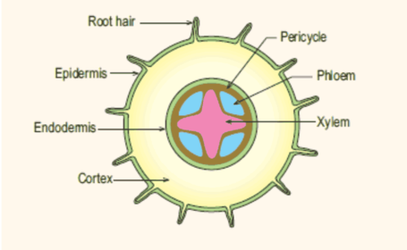
- muscles respire --> more CO2 --> oxyhaemoglobin dissociates --> releases oxygen - 102How are the phloem and xylem distributed in the roots?
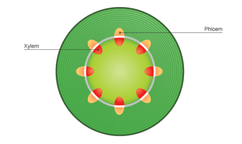
- 103How are the phloem and xylem distributed in the stem?
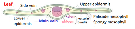
- 104How are the phloem and xylem distributed in a leaf?
- 105What is the structure of xylem?- hollow columns of dead xylem vessel elements
- lined end to end - continuous
- walls thickened with lignin - strength
- pits - water moves sideways
- narrow - capillary action - 106What is the structure of phloem?- sieve tube elements - very little cytoplasm, no nucleus, living cells form tube, end-to-end, sieve plates, thin walls
- companion cells - mitochondria for sucrose loading, plasmodesmata - 107How does water move from the root to the xylem?- minerals moved by AT by endodermis cells
- decreases water potential in xylem to water moves cortex --> xylem by osmosis
- symplast pathway
- apoplast pathway - 108What are the 2 pathways water can take to the xylem?1. Symplast pathway - through cell cytoplasm via plasmodesmata
2. Apoplast pathway - through cell walls until Casparian strip - 109What is the Casparian strip?- waxy, waterproof suberin layer
- endodermis cell layer of cortex
- water must travel through cytoplasm
- cell membranes can control - 110How does water move up the stem of a plant?1. Root pressure - moving minerals forces water in, pushes water up stem
2. Transpiration pull - cohesion, tension caused by low hydrostatic pressure
3. Capillary action - adhesion, xylem walls narrow so pull water up - 111Define transpirationThe loss of water vapour from aerial parts of the plant by diffusion into the atmosphere
- 112What factors affect transpiration?1. Light
2. Temperature
3. Humidity
4. Air movement - 113What exactly do potometers measure?Rate of water uptake (roughly equal to water lost by transpiration)
- 114How are xerophytes adapted?- smaller leaves/spines
- thicker waxy cuticle
- hairs
- pits containing stomata
- rolling leaves - 115How does sucrose loading happen?- companion cells use ATP to pump H+ ions out by active transport
- diffusion gradient - H+ diffuse back through contransporter proteins with sucrose molecules
- sucrose concentration in companion increases so diffuse into sieve tube - 116What is evidence for the translocation mechanism?- removing ring of bark
- radioactively labelled carbon dioxide
- movement much faster than diffusion
- pH of companion cells higher
- aphids - pressure gradient - 117What evidence is there against the translocation mechanism?- sucrose moves to all parts at same rate, not more quickly to lowest concentration
- sieve plates would be barrier
Informationen
- Beschreibung
- Flashcards according to OCR Biology specification
- Erstellt von
- C R
- Aufrufe
- 2.318
- Tags
- Bewertung