
Mindmap
Microscopes
Inhalt der Mindmap
Microscopes
TYPES
- Light
- Max Mag= x1500
- Max Res: 200nm
- light has wavelength of range 400- 750nm)
- can only distinguish between 2 points of light beam can pass thru them
- if 2 objs closer together than 1/2 wavelength of light (250nm)- cannot be distinguished
- will appear as 1 object
- if 2 objs closer together than 1/2 wavelength of light (250nm)- cannot be distinguished
- Lenses
- Four objective lenses
- x4, x10, x40, x100 (oil immersion lens)
- 1 eyepiece lens
- x10
- USING LIGHT MICROSCOPE
- Light passes thru multiple lenses to produce image at eyepieces
- light
- condenser lens
- thru specimen
- obj lens
- projector lens
- eyepiece lens
- eye
- eyepiece lens
- projector lens
- obj lens
- thru specimen
- condenser lens
- light
- Light passes thru multiple lenses to produce image at eyepieces
- Four objective lenses
- Electron
- TEM
- Electron beam PASSES through sample and give contrast
- E's pass thru denser bits less easily- contrast
- Final image: 2D
- Max Mag: x500 000
- Final image: 2D
- E's pass thru denser bits less easily- contrast
- Electron beam PASSES through sample and give contrast
- SEM
- Electron beam directed at sample
- Electrons bounce off sample
- Final image: 3D view of surface
- Max Mag: x100 000
- Final image: 3D view of surface
- Electrons bounce off sample
- Electron beam directed at sample
- Max Res: 0.1nm
- TEM
- Light
MAGNIFICATION AND RESOLUTION
- MAG= no. of times image is larger than object
- RES= ability to distinguish between 2 objects v.close together
- MAG= image size/actual size
Advantages of light microscope
- No skills/training required
- Can view many (living) specimens
- Easy to use
- PREPARATION
- STAINING
- Coloured stains- chemicals that bind to chemicals in/on specimen
- allows specimen to be seen
- some stains bind to specific cell structures
- Acetic orcein- DNA dark red
- Gentian violet- bacterial cell walls
- because a lot of biological material is not coloured- no detail
- Coloured stains- chemicals that bind to chemicals in/on specimen
- SECTIONING
- Specimens embedded in wax
- Thin sections cut w/out distorting structure
- Useful for making sections of soft tissue (brain)
- because material distorts when you cut it thinly
- Specimens embedded in wax
- STAINING
- PREPARATION
- Cheap
Disadvantages of light microscope
- Lower res
- Cannot view cell's ultrastructure
- Lower mag
- Lower res
ADVANTAGES OF ELECTRON MICROSCOPE
- Higher res
- Can view cell's ultrastructre
- more detailed image
- Higher res
DISADVANTAGES OF E MICROSCOPE
- EXPENSIVE
- HARD TO USE- SKILLS/TRAINING
- PREPARATION
- 1) Fix Specimen in glutaraldehyde to make tissue firm
- 2) dehydrate it- replace water w ethanol
- 3) fix this into solid resin
- 4) cut v.thinly with diamond knife
- 5) stain using lead salts- scatter e's differently= contrast
- 6) mount it on copper grid
- place specimen in vacuum
- 6) mount it on copper grid
- 5) stain using lead salts- scatter e's differently= contrast
- 4) cut v.thinly with diamond knife
- 3) fix this into solid resin
- 2) dehydrate it- replace water w ethanol
- "False colour" electron micrograph
- sometimes colour added because electron micrograph produced is B&W
- 1) Fix Specimen in glutaraldehyde to make tissue firm
- PREPARATION
- REQUIRES A VACUUM
- SPECIMEN MUST ALWAYS BE DEAD
- NOT ACCURATE REPRESENTATION OF LIVING SPECIMEN
- SPECIMEN MUST ALWAYS BE DEAD
- tissue observed undergoes several stages of prep
- each stage may alter real appearence
- PREP may introduce artefacts
- are structures not originally present/alter specimen from original condition
UNITS
- 1mm= 1000 micrometers
- 1 micrometer= 1000 nanometres (nm)
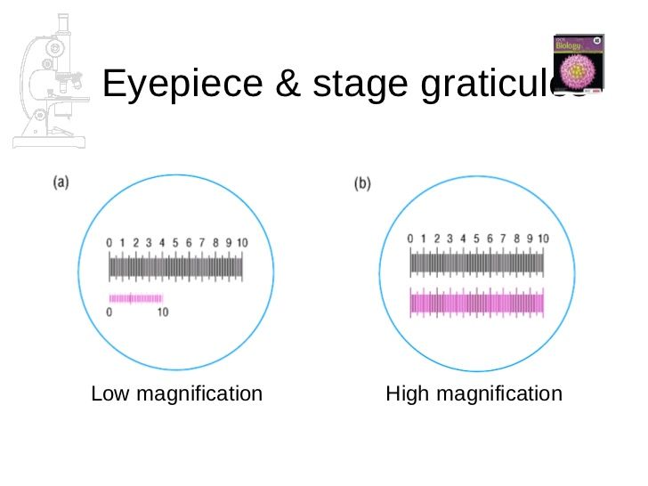
- Measuring cells/organelles w light microscope
- microscope eyepiece fitted with eyepiece graticule
- transparent w ruler etched on
- scale is arbitrary- represents diff lengths at diff mags
- must be CALIBRATED for each diff obj lens
- using a STAGE MICROMETER
- a microscopic rule on a slide placed on stage
- ruler is 1mm long and divided in 100 division
- each division is 0.01nm/10 um
- with x4 obj lens and x10 eyepiece- mag= x40
- 40 epu= 1000um --> 1epu=25 um
- ruler is 1mm long and divided in 100 division
- shows 'true' lengths
- a microscopic rule on a slide placed on stage
- using a STAGE MICROMETER
- must be CALIBRATED for each diff obj lens
- graticule remains constant at any mag
- microscope eyepiece fitted with eyepiece graticule
Bilder und Dateien der Mindmap
Informationen
- Beschreibung
- A-level Biology Mindmap am Microscopes, erstellt von jaja-princess am 29/03/2015.
- Erstellt von
- jaja-princess
- Aufrufe
- 0
- Bewertung