
5. Innate Immunity

Folien in diesem Satz (63)
-
- Understand the difference between pathogens and commensals
- Explain what is an antigen
- Appreciated the differences between the innate and adaptive immune system
- Summarise the properties of basic cell types of the immune system: Neutrophils, Macrophages, Dendritic Cells, B Lymphocytes, T Lymphocytes
- Become familiar with structure and function of tissues of the immune system: Lymphatic system, Secondary Lymphoid Tissues (Lymph, Nodes, Spleen), Primary Lymphoid Tissues (Thymus, Bone Marrow)
- distinctive features and functions of primary and secondary immune organs
- the topography of embryonic and post-natal haematopoiesis
- the structure of the thymus
- the structure and function of the spleen
- the structure of the lymphatic system
- the structure of lymph nodes
- the relationship between structure and function of immune organs
- characteristic features of immune cells in pathology specimens
-
- the principles underpinning innate immunity
- the principles of inflammation
- the mechanism of phagocytosis
- the basic biology of neutrophils
- the basic biology of macrophages
- the basic biology of dendritic cells
- the basic biology of natural killer cells and other innate lymphoid cells
- The principles underpinning complement activation and function
- How innate immune cells sense pathogen
- The type I interferon cascade and function of IFNb
- The NFkB cascade and function of selected cytokines
- How innate immune cells present antigen
- The significance of negative regulators of innate immunity
-
- Pathogen
- An organism that causes disease: virus, bacteria, fungi, parasite
- Roles in fermentation, digestion, absorption of nutrients
- Host/Microbe symbiosis (how they interact)
- Commensal= one organism benefits, other unaffected
- Mutalism= both orgainsms benefit
- Parasitism= one organisms benefit at expense of the other
- Microbes can be intracellular/extracellular, pathogenic/not- all need to multiply and spread
- Antigen
- Molecule capable of inducing an immune response
- Not always derived from a microbe (cell antigens), not all microbes have antigens(commensal bacteria do't cause immune response)
-
Mechanisms in Preventing Disease
- Mucosal barriers (physical barrier)- skin, respiratory tract, intestines
- Innate immune system- specific but not highly
- Immediate- what you loose in specificity, gain in speed
- Recognises foreign pathogens via germ-line encoded receptors (inherited receptors)
- No memory
- Adpative immune system- very specific
- Takes days to develop
- Highly specific requires rearrangement of receptor genes (not inherited),
- Generates memory
-
Mechanisms in Preventing Disease
- Upon encountering a microbe, a cell tries to:
- Clear the microbe or inhibit its replication- phagocytosis, interferon system
- Process the microbe and present to adaptive immune cells
- Alert and attract other cells that will help clear the infection- upregulation of surface receptors, cytokines, chemokines
- Kill itself
- All cells have the potential to perform these activities (to a certain extent), some cells are specifically made and programmed(armed) to do the first 3= innate immune cells
-
Microbes and the immune system
- No/short pathology= illness, clearance
- Acute disease= infection that is failed to clear
- Chronic disease/prolonged= failure to clear pathogen and failure to stop by immune system
-

- Primary and Secondary lymphoid tissue
- Immune cells develop in primary lymphoid organs (Thymus and Bone Marrow)
- Lymphocytes differentiate to express antigen receptors: Thymus= T lymphocytes, BM = B lymphocytes
- Immune responses occur in secondary lymphoid tissue (Spleen, LN, Peyer's Patch, Mucosal Associated Lymphoid Tissue (MALT), Gut Associated Lymphoid Tissue (GALT), Nasal Associated Lymphoid Tissue (NALT))
-
- Rules of Lymphoid Organs:
- Lymphocytes originate and are initially selected to fit the purpose in the primary lymphoid organs
- Lymphocytes are ultimately armed in secondary lymphoid organs (where response happens)
- Such effective, specific armament of lymphocytes takes place when the adaptive immune responses are initiated, e.g. in response to infection
- The divergent weaponries of lymphocytes, just as those of innate cells, are unleashed in the non-lymphoid target tissues
- Functional adaptive immunity requires orchestrated interactions of different immune cells in specific microenvironments within primary and secondary lymphoid organs.
- Functional immunity requires tightly regulated movement of immune cells between immune organs and non-lymphoid target tissues
- Organized structures of lymphocytes in target tissues and organs are often referred to as tertiary lymphoid organs (TLOs)
-
- Haematopoeisis- occurs in bone marrow
- Process of generation of all blood system from HSC
- Haematopoetic cells develop from a common precursor either a common myeloid progenitor or common lymphoid progenitor

-
- Haematopoesis through life
- Fetal life: occurs in yolk sack- from week 4 of development, liver until shortly before birth, spleen until cartilagenous bone vascularised
- Adult/infact life: occurs in BM in children, in adults occurs in BM of pelvis, sternum, vertebrate, cranial bones (less in long bones due to fat deposition)
- Emergency haematopoeisis: in adult life due to chronic inflammation, also occur in spleen, liver or LN (extramedullary haematopoesis)

- Bildunterschrift: Potential of different bones to be a source of blood cells during life, some bones maintain ability to host HSCs and some lose it (longer bones)
-
Primary Lymphoid Tissue: Thymus
- Educating T cells: Consists of epithelial cells that house train lymphocytes so they don't recognise self-antigens
- Immature T cells migrate from BM to thymus where they multiple and mature (only 2% of immature T cells achieve proper education to become mature)
- Reaches its peak size by puberty and decreases after (thymus involution)
-
Primary Lymphoid Tissue: Thymus
- Thymus Structure:
- Capsule= surrounds the 2 lobes holding them together
- Trabeculae= extensions of capsule into cortex and medulla, establish incomplete thymic lobules where blood vessels and nerves pass
- Cortex= Outer layer containing immature T cells from BM
- Medulla= Inner layer mature T cells and Hassals corpuscles (epithelial structures)
- Thymic evolution: with aging= decrease of size and production of naive T cells, increase in adipose tissue, decrease immune function

-
Secondary Lymphoid Tissue: Spleen
- Highly organised structure:
- Areas rich in immune cells (white pulp)
- Contains specific lymphoid microenvironments (T and B cell zones)
- Contains (PALS) periarteriolar lymphoid sheath in T cell zone- Proliferating lymphocytes around central arterioles, which are branches of the splenic artery(connects spleen to rest of body)
- Areas rich in RBC (red pulp)
- Filters blood, removed old RBCs and other cells, contains macrophages, carry out extramedullary haematopoesis
- Directs immune responses to antigens in the blood
- Hyposplenism= under developed spleen, predisposes to infection
- Splenomegaly= enlarged spleen, common in chronic inflammatory diseases (malaria)
-
Secondary Lymphoid Tissue: Lymphatics
- Drainage system involved in fluid balance: returns to the blood excess interstitial fluid, plasma proteins, tumour cells use this pathway for spreading from its primary site metastasis (due to inc flow)
- Lymph capillaries: Originate as closed tubes in almost all tissues (except CNS, epidermis and cartilage)
- Capillary wall is constructed of overlapping endothelial cells that respond to fluid pressure
- Lymph vessels: Carry lymph and cells from peripheral tissues to draining LNs
- Lymph:
- Plasma containing proteins and solutes that filters out of venules+ capillaries due to hydrostatic/oncotic pressure
- Tissue derived pathogens may be in lymph
- Cells carried in lymph: lymphocytes, DCs, macrophages, phagocytes
-
Secondary Lymphoid Tissue: Lymphatics

- 2 types of lymphatics based on where they are:
- 1. Superficial Lymphatics: follow superficial veins, connect to primary LNs (axillary, inguinal, cervical), drain into deep lymphatics
- LNs strategically placed near areas that serve as entry points of infection
- Symmetrical-2 of each
- Trunk can be divided into 4 quadrants with different drainage by vertical and horizontal lines through umbilicus
-
Secondary Lymphoid Tissue: Lymphatics

- 2. Deep Lymphatics: follow main vessels, connect to LN deeper in organs (oara-aoartic nodes either side of the aorta drain the paired organs, nodes anterior the gut etc (pre-aortic)
- Thoracic duct- longest lymph vessel connecting upper LN network to lower network:
- Lymph from 3/4 of body drains into left brachiocephalic vein via thoracic duct
- Lymph from upper righ quadrant enters to righ brachiocephalic vein
-
Secondary Lymphoid Tissue: Lymphatics
- Lymph Nodes
- Pea sized immune structures strategically placed around the body
- Central component of host defence: represent meeting place for immune cells and their exposure to antigen
- Structure of LN:
- Medulla: contains lymphatic sinuses, plasma cells, B cells, macrophages
- T cell Paracortex
- B cell follicles:
- Primary follicle= mature, naive B cells
- Secondary follicle= Germinal Center reaction: GCs develop in response to Ag stimulation, site of B cell proliferation, selection, affinity maturation and differentiation (where antigenic responses develop)
-
Cell Movement to and out of LNs

- Flow of fluid is directional
- Naïve lymphocytes enter via high endothelial venules and exit via efferent(E=EXIT) lymphatics
- APCs enter via afferent lymphatics(A=arrival), scan for lymphocytes and exit via different lymphatics
-
Cell Movement to and out of LNs

- How B and T cells are separated:
- Cell movement caused by membrane receptors pulling cell towards areas rich in their ligand= chemotaxis
- Distinct structural (stroma) cells in LN produce distinct chemokines that attract B or Tcells
- B cell areas- CXCL13 (ligand of CXCR5 receptor on B cells)
- T cell areas- CCL21 and CCL19 (ligands of CCR7 receptor on naive T cells and DCs - DCs APCs attracted to same place as T cell
-
Cell Movement to and out of LNs
- Chemotaxis: Directed movement of cells or organisms in response to chemicals. The cells are attracted or repelled by substances (chemoattractants), commonly forming gradients
- Chemokines: chemotactic cytokines (secreted soluble proteins)
-
Similar Structure of Spleen and LN

- Similarities:
- Entry and exit points
- B cell follicles, T cell zones
- Same ligands
- Organised in a similar way since this structure is very efficient to optimise immune response
-
Mucosa Associated Lymphoid Tissue

- Similar organisation to spleen and LNs
- Large organised aggregates of lymphoid tissue in digestive, respiratory and genitourinary tracts
- Peyer's Patch:
- Organised aggregates of lymphoid cells in ileum
- Essential for monitoring bacteria populations, preventing growth of pathogenic bacteria in intestine
- Contain large population of Ab-producing plasma cells (exceeding amount in the spleen, lymph nodes and bone marrow combined)
-
Pathology- how we study tissues
-
- Antigen non-specific:
- Phagocytic cells (macrophages) detect foreign material and seek to remove
- Aided by low number of receptors that recognise broad molecular patterns (not present in euk)
- Macrophages (first line of defence):
- Induce inflammation through secreting soluble factors (cytokines+chemokines)
- Phagocytic
- Found in all peripheral tissue
- Neutrophils:
- Most common WBC in blood
- Migrate rapidly to site of inflammation
- Phagocytic- ingest and kill bacteria
- Release toxic granules
-
-
Adaptive Immune System Overview
- Antigen Specific: B and T cells require help from T helper cells to respond
- B and T cells express antigen-specific receptors, both are highly variable, specficity confined to a single clone
- Ab on B cells recognise 3D structures and linear peptides
- T cell receptor (TCR) only recognise linear peptides (ensure dont self attack)
- Clonal Expansion:
- Activation of antigen receptor, B and T cells expand, mature and differentiate into highly specialised cell populations
-
Adaptive Immune System Overview
- DCs: Interphase of Innate and adaptive immunity
- Phagocytic
- Characterised by presence of long dendrite process
- Main function: activation of adaptive response
- Actively survey peripheral tissues, once activated migrate to LN to activate antigen specific naive Tcells
- Lymphatics vessel network:
- Fluid drainage from peripheral tissues, LNs found at drainage points
- If destroyed= Surgically induced lymphodema (associated with breast cancer surgery), elephantiasis(induced by parasitic worms that block draining lymphatic)
- LN= highly organised, B cell follicle- rich in B cells Ab response generated, T cell zone- rich in T cells T cell-DC interactions, T helper response helps make antibodies
-
- Steps of adaptive immune response:
- Activation of tissue DCs
- Migration to LN- attracted to same place as T cells due to chemokines
- Activation of antigen specific T helper cells
- Activate B cell- antibody producing cell
- B cell proliferates and produces plasma cells, plasma cell produces Ab
-
- Occurs as result of infection/ damage(wound)- aim to clear infection and repair(tissue) damage
- Characteristics: Redness, swelling, pain
- A prerequisite for a successful immune response
- Also a potentially harmful process: Components of inflammation that are capable of destroying microbes can also injury bystander normal tissue (if the response is too strong or too long)
-
- Roles of inflammation in combating infection:
- Deliver effector cells and molecules to site of infection/damage- enhance microbe clearance
- Blood clotting- form physical barrier and prevent spreading of infection to bloodstream
- Enhancement of tissue repair process (resolution of inflammation part of process)
- Three changes to blood vessels during inflammation:
- Increased diameter- dilate, enhance number of cells and molecules reaching site
- Increased adhesion molecules that bind leukocytes- sticky around site of damage
- Increased vascular permeability- allows exit of fluid and proteins into tissue= edema (swelling)
- Extravasation= Attachment of leukocytes to the endothelium followed by their migration through blood vessels to infected tissue
-

- Myeloid lineage innate immune cells
- Neutrophils, macrophages, myeloid DCs
- Derived from common myeloid progenitor (CMP)
- Others cells from the lymphoid lineage are derived from common lymphoid progenitor (CLP)
-
- Neutrophils: first at the scene
- Form polymorphonuclear family together with basophil and eosinophils
- Most abundant WBCs = 40-75%
- Recruited to site of infection of injury within minutes
- Predominant cells in pus
- Short lived, quick acting, motile
- Kill pathogens in 3 ways: Phagocytosis, Degranulation, Neutrophil extracellular traps (NET)
-
- Targets (microbes/infected cells) coated in opsonins- protein tag (eg Ab FcR) that enhances phagocytosis
- Requires membrane rearrangement
- Steps:
- Attachment/adhesion and engulfment
- Phagosome formation- invaginations of membrane
- Recruitment of lysosomes
- Phagosome-lysosome fusion
- Microbe killing due to low pH in phagosome and high conc of: ROS, Defensins (antimicrobial peptides), Hydrolytic enz

-
- Degranulation
- Once neutrophil is activated-> secrete toxic granules
- Release azurophilic (primary) or specific (secondary) granules- contain proteases, defensins, ROS
- Efficient way of clearing surrounding space in non-highly specific manner
- Neutrophil Extracellular Traps (NETs)
- Usually associated with bacterial infections
- Networks of extracellular fibres primarily composed of DNA and antimicrobial proteins (histones, neutrophil elastase)
- Creates a sticky environment and traps microbes/pathogens- makes them less motile
- Kill them through localised high conc of antimicrobial proteins
-
- Macrophages: mature form of monocytes
- Reside in tissue (monocytes are in circulation): eg Microglia = brain, Kuppfer cells= liver, Osteoclasts= bone
- Upon infection: tissue-resident macrophages phagocytose microbes, monocytes are recruited to the infected tissue and mature into macrophages
- Activated through: direct interaction with pathogens, factors secreted by T-helper lymphocytes (eg IFNϒ, IL4= cytokines)
- Function:
- Highly pathogenic: engulf and destroy pathogens
- Secrete cytokines that orchestrate inflammatory response
- Present antigens to T lmyphocytes
- Contribute to tissue repair and formation of new blood vessels
-
- Myeloid Dendritic Cells: Professional APCs- directly link innate and adaptive
- Long finger- like projections
- Immature DCs migrate from bloodstream to tissues or are tissue-resident
- Phagocytic but not as much as macrophages
- Scan for presence of potential microbe and become activated (mature) upon encounter
- During maturation:
- Become less phagocytic and endocytic
- Proliferate
- Process microbial proteins to small peptides and present to T lymphocytes
- Up-regulate the expression of surface proteins and cytokines for optimal antigen presentation
- Secrete type I interferons: inhibit pathogen replication, induce apoptosis of nearby infected cells, enhance presentation
- Migrate to nearest LN through lymphatics system
-
- Plasmacytoid DCs (pDCs): Subset of DCs derived from CLP that look like plasma cells
- Rare- less than 0.5% of PBMCs
- Antigen sensing/recognition similar to myeloid DCs
- Main function: NOT antigen presentation but production of type I interferons(IFNα and IFNβ), important in antiviral responses
-
- Innate Lymphoid Cells (ILCs):
- Newly discovered family of lymphoid cells that translate cytokine signals into effector cytokines w/out expressing antigen specific receptor (produce cytokines to kill a cell)
- ILCs mirror the phenotypes and functions of T cells:
- Make similar things upon activation but no antigen receptor (do not directly respond to antigen) and no clonal selection upon stimulation
- ILCs respond to signals from infected/injured tissues and produce cytokines
- NK cells -> cytotoxic CD8+ T cells = eg Natural killer cells mirror what CD8+ T cells do
- ILC1 -> CD4+ T helper 1 (TH1) cells
- ILC2 -> CD4+ T helper 2 (TH2) cells
- ILC3 -> CD4+ T helper 17 (TH17) cells
- Next slide- both innate and adaptive cell mirrors
-

- CD8 vs NK:
- CD8 cells characterised by ability to make 3 things: Granzymes, Perforin, IFNϒ
- NK cells once activated make the same thing but much earlier and less specific
- ILCs vs T cells
- ILCs sense signal cytokines eg IL12, IL15, IL33 etc
- Translate that signal into an effect
- T cells do this but in a specific and sustained manner
-
- Natural Killer Cells: Type of ILC
- Terminally differentiated cell of lymphoid lineage
- Larger than B and T lymphocytes
- Characterised by cytoplasmic granules containing cytotoxic proteins (granzyme and perforin) that are secreted to kill infected cells
- Also secrete type II IFNϒ cytokine and kill infected cells before cytotoxic T lymphocytes

-
- NK cells become activated in response to:
- Type I interferons (IFNα and IFNβ) secreted by other innate (myeloid) cells (macrophages and DCs)
- Other cytokines produced by an infected tissue (eg IL15)
- Their cytotoxic activity is controlled through inhibitory and activating receptors on their cell membrane
- 2 main functions of NK (mechanisms of clearing infected cells):
- Clearance of 'missing self 'cells
- Antibody-dependent cellular cytotoxicity
-
- Missing self :
- Almost every cell in body expresses MHC-I on surface (immune system recognises 'self')
- Many pathogens down-regulate MHC-I expression in infected cells- to escape recognition, avoid clearance by cytotoxic T lymphocytes, also common in cancer cells
- NK cells kill any cells with no/low MHC-I expression (missing-self)

-
- NK cells Antibody-Dependent Cellular Cytotoxicity (ADCC):
- Another way to kill labelled cells
- NK cells carry receptors for the Fc region of Ab
- Microbes/ cells coated with these antibodies trigger NKs to release cytotoxic granules,+ increase phagocytosis

-
- A system of soluble pattern-recognition receptors and effector molecules that can detect and destroy microbes
- 3 main receptors that activate the cascade:
- C1q(classical pathway)- complement receptor interacts with Ab coated pathogen surface
- Mannose-binding Lectin (MBL) and ficolins (lectin pathway)- Recognises carbohydrates on pathogen surfaces
- C3 (alternative pathway)- Undergoes spontaneous hydrolysis leading to deposition of C3 convertase on pathogen surface, spontaneous activation of cascade through accumulation of C3 protein on pathogen surface
-
- The complement activation cascade:
- Circulating proteins react with eachother to fight infection- many are inactive precursor proteases(zymogens)
- At site of infection:
- Initiation of complement system through 1 of 3 ways
- Activates complement enzyme (through proteolytic cleavage of its zymogen
- Activated enzyme cleaves it substrate (another complement zymogen), activating it
- Amplification= activation of small number of complement proteins at start of pathway is amplified-> rapid generation of large complement response
- Has to be strongly regulated to prevent uncontrolled complement activation
-
-
- Complement system helps Ab and phagocytes clear microbes form an organism in 3 ways:
- Opsonisation- enhancing phagocytosis, complement molecule bound to Ab makes phagocytosis easier
- Chemotaxis- some complement proteins act as chemoattractants to recruit more phagocytes to site of complement activation
- Pathogen lysis- complement compounds directly damage bacteria by creating pores in bacterial membrane
- There is a limited number of complement molecules that can perform all 3 functions
-
Complement: C3 Convergence Pathway

- Activation of C3 is key event in complement activation
- Mediates effector functions, initated by all 3 pathways
-
Complement: Immune Complex Clearance
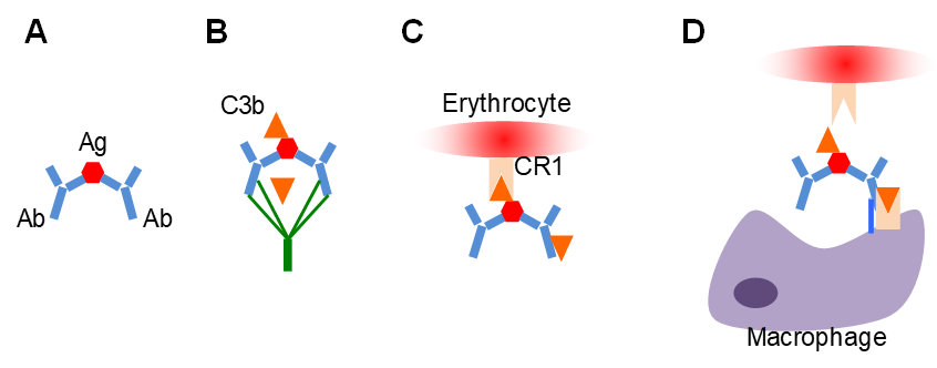
- A. Antigen-antibody complex formed in the circulation
- B. Complement activation-> deposition of C3b on complex
- C. Complement receptor CR1 on erythrocytes binds immune complex through C3b
- D. Erythrocytes go to spleen and liver, phagocytosis physically removes immune complex from erythrocyte surface
-
Pathogen Recognition by Innate Cells
- Broad Pathogen Associated Molecular Patterns (PAMPs) are recognised by a small number of Pattern Recognition Receptors (PRRs)
- PRRs are inherited (germline encoded), do not change / adaptive immunity receptors are specific and change
- PRRs are less specific than adaptive receptors- specific enough to distinguish gram -ve from gram +ve but not gram +ve from gram +ve
-
-
- Found on nearly all surfaces of a pathogen class
- Eg LPS on surface of Gram -ve bacteria
- Eg dsRNA in dsRNA viruses
- PAMPs are structures essential for the microbes survival
-
- A few proteins can be used to sense millions of pathogens
- All cells express SOME PRRs, DCs and macrophages express ALL PRRs
- 4 families of PRRs:
- Toll-like Receptors (TLRs)
- 10 in humans, cover any type of potential pathogen
- Localisation on endosome: TLR3, TLR7, mainly dealing with viruses (have to enter cell because obligate parasites)
- Or localisation on cell membrane: Cell membrane= TLR2 (recognises Gram +ve bacteria- peptidoglycan), TLR4 (recognises Gram -ve bacteria- LPS)
- Nucleotide-binding oligomerisation domain (NOD)- like receptors (NLRs)
- C-type lectin receptors (CLRs)
- RIG-1 like receptors (RLRs)
-
-
PRR Activation: Kinase Cascade

- Exposure of cell to PAMP
- Kinase signalling cascade (with adaptor proteins)
- Activate TF to translocate to nucleus
- Targets:
- Amplifiers
- Antipathogen molecules/mechanisms
- Messengers to activate adaptive response
- Negative regulators to prevent overwhelming activation, eg. ubiquitin ligases ubiquitinate proteins leading to their degradation
-
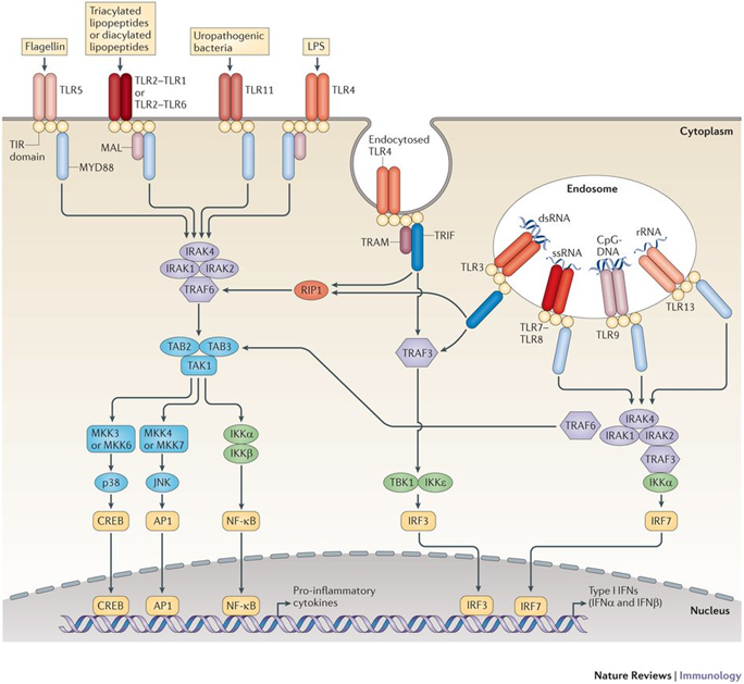
Example: TLR4 Activation at Cell Mem

- TLR4 binds LPS
- Kinase cascase
- Activation of 3 transcription families (IRF3, NF-KB, AP-1)
- Downstream of IR3= transcription of IFN-B (type I interferon)
- NF-kB and AP-1 pathways lead to production of inflammatory regulators
- Positive feedback- IFNB activates more cells that express IFNB
- IFNB also activates second wave of TF (STAT family) that contribute to antipathogen gene expression
-
TLR4 Activation Depends on Localisation
- TLR4 only TLR localised on both endosome and membrane
- MyD88 and TRIF are important adaptor proteins
- MyD88= early event in activation, essential for any TLR response (except TLR3)
- TRIF= shared between TLR3 and TLR4 (only TLR4 when in endosome)
-
NF-kB Activation Downstream PRRs

- Induces transcription of:
- Chemokines- attract other adaptive and innate immune cells
- Cytokines- promote inflammation eg IL1B, TNFa, IL6, IL8 (which are induced rapidly/ early)
-
IRF3 + IRF7 Activation Downstream PRRs
- Induce transcription of IFNβ and IFNα and ISGs
- Secreted type I IFNs bind IFNAR (interferon A receptor)= Positive feedback (production of early IFNs)
- Induces second wave of interferon stimulated genes (ISGs) through the action of other TFs of IRF and STAT

-
Function of Type I interferons
- Functions in immune response:
- Inhibition of pathogen (virus, parasite) replication through inhibition of protein translation in the infected cell
- Induction of cell-death (apoptosis) of infected and neighbouring cells
- Activation of other innate immune cells (eg natural killer cells)
- Activation of antigen presentation machinery (make DCs and macrophages present antigens to T cells)= initiation of adaptive immunity
-
Summary: 2 Pathways in Innate Activation
- The type I interferon pathway
- Driven by activation of the IRF family TFs
- Leads to production of IFNβ and other interferon stimulated genes (ISGs)
- The NF-κB pathway
- Driven by the activation of the NF-κB TF
- Leads to the production of cytokines (e.g. IL1b, TNFa, IL6)
- Transcription of several inflammatory cytokines + surface receptors can be activated downstream by either pathway
-
Antigen Presentation Machinery
- Distinct pathways for intracellular and extracellular antigens:
- Components of both systems (including MHC molecules) are upregulated in response to type I interferons (produced in response to PAMP)
- MHC-I proteins have cargo (peptide): self peptide or microbial (antigenic) peptide

-
- Innate immune gene expression programme induces its own negative regulators in later stage:
- Anti-inflammatory cytokines eg IL10
- Phosphatases- stop signalling cascade eg dephosphorylation of p38 by DUSPs
- Intracellular factors: inhibit inflammatory TFs eg lκB (ubiquitin ligase) prevents NFκB from going to the nucleus
- MicroRNAs: non-coding, suppress translation of inflammatory mRNAs eg suppresion of TRAF6 and IRAK1 by miR-146a
- Prolonged response can lead to tissue damage and disease caused by immune system:
- Eg non-cytopathic viruses (hepatitis virus) that establish chronic infection
- Lead to chronic inflammation, creates long, non-resolving response that will damage liver


