7. Specialised Cells: CD4 T Cells
Folien in diesem Satz (50)
-
- What distinguishes the main subsets of CD4 T cell?
- What is “signal 3”?
- And what is signal 3 for Th1 + Th2 induction?
- What intracellular signalling pathways cause Th1 + Th2 induction?
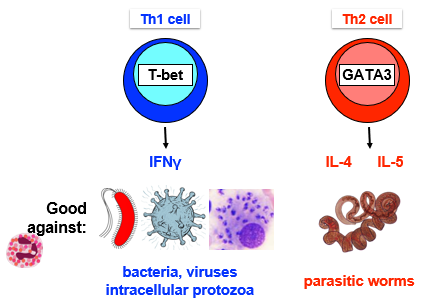
- What pathogens are Th1 + Th2 cells effective against?
- How do Th1 + Th2 “help” B cell class switching?
- How do Th1 cells “help” macrophage activation?
- How do CD4 T cells “help” CD8 T cell responses?
- Which pathogens are killed by Th17 cells?
- What is “Signal 3” for Th17 differentiation and what are the downstream signalling pathways?
- What are Th17 effector mechanisms?
- Why do we need regulatory T cells?
- What are natural and induced Tregs?
- What is the relationship between Tregs and Th17 cells?
- How do Tregs suppress immune responses?
-
-
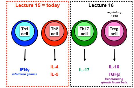
- Bildunterschrift: CD4 T cell subsets make signature cytokines
-
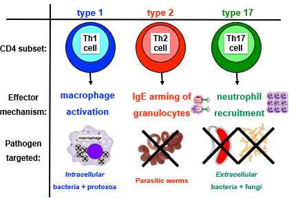
- Distinct subsets of CD4 T cells coordinate distinct immune responses to target different pathogens, subsets are distinguished by signature cytokines
- Cytokines= proteins secreted by immune and non-immune cells that influence immune function: Interleukins, interferons, tumour necrosis factors, transforming growth factor
- CD4 T cell differentiation events:
- Production of polarising cytokines - signal 3
- Cytokine signalling events in CD4 T cells
- Effector functions of CD4 T cell subsets
-
1. Produce Polarising Cytokines: Th1
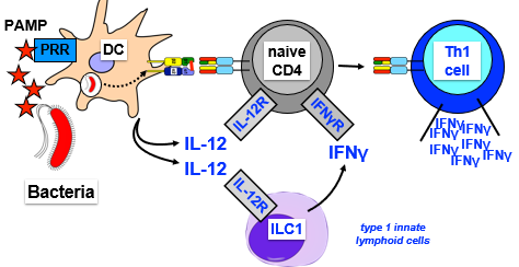
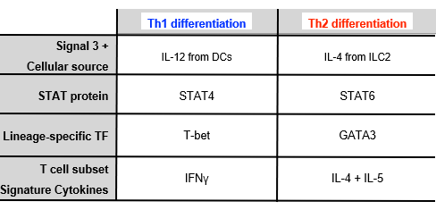
- Signal 3-Th1 differentiation, supported by ILC1-derived IFNϒ
- Summary: PAMPs stimulate DCs to produce IL12-> main driver of Th1 differentiation (signal 3)
- DC recognises bacterial PAMP- presents peptide on MHC-II
- PAMP stimulates DC to produce cytokine IL-12
- IL-12 binds IL-12R on naive T cells and elicits signal in naive CD4 T cell
- ILC1 aLso express IL12R and will bind IL12 and produce IFNϒ
- IFNϒ binds to IFNϒR on naive CD4 T cell
- Sum of signals (1 +2 ) + 3= cytokines (IL12 and IFNϒ) = cause naive T cell to be activated to Th1 cell that produces signature cytokine IFNϒ
-
1. Produce Polarising Cytokines: Th1
-
1. Produce Polarising Cytokines: Th2
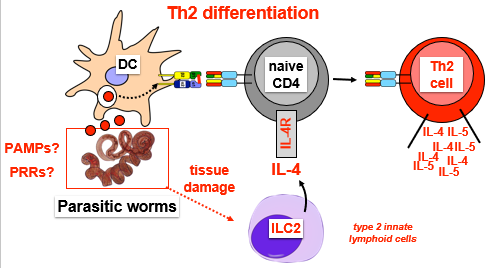
- Signal 3-Th2 differentiation
- Th2 is response induced by parasitic worms
- DC present peptide on MHC-11 (dont know PAMP or PRR)
- Parasitic worms cause tissue damage which activates ILC2 to produce IL-4 (signal 3)
- IL-4 binds IL-4R on naive CD4 T cell
- Sum of signals (1 +2 ) + 3= cytokines (IL4 = cause naive T cell to be activated to Th2 cell that produces signature cytokines IL4 and IL5
-
1. Produce Polarising Cytokines: Th2
-
2. Cytokine Signalling in CD4 T Cells
- How does Il-12 induce Th1 differentiation and how does IL-4 induce Th2 differentiation
- T cell signalling: JAK-STAT pathway GENERAL
- JAK is associated with cytokine receptor
- When cytokine binds receptor, JAKs activated, phosphorylate cytokine receptor
- Phosphorylation acts as docking site for STAT proteins and recruit them to receptor
- STATs close to JAKs and get phosphorylated, then translocate to nucleus
- STATs bind to promoter of and transcribe lineage specific genes: cytokines, cytokine receptors, TFs
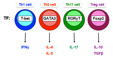
- Lineage specific TFs promote the production of signature cytokines by different CD4 T cell subsets
-
2. Cytokine Signalling in CD4 T Cells
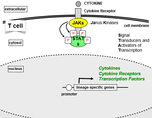
- Bildunterschrift: JAK-STAT pathway
- Bildunterschrift: Lineage specific TFs induce signature cytokines
-
2. Cytokine Signalling in CD4 T Cells: Th1
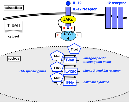
- IL-12 signal cytokine binds IL-12R, phosphorylation through JAKs of intracellular portion of IL12R
- Recruit STAT4, phosphorylated and translocates to nucleus
- Activated STAT4 binds to TH1-specific genes:
- T-bet= lineage specific TF- promotes more response to IL12
- IL12R- signal 3 cytokine receptor- promotes cell to be more responsive to IL12
- IFNϒ= hallmark cytokine of Th1 cells
- T-bet binds to all 3 gene loci, activates, reinforces and stabilises Th1 differentiation, promoting expression of all 3
-
2. Cytokine Signalling in CD4 T Cells: Th1
-
2. Cytokine Signalling in CD4 T Cells: Th2
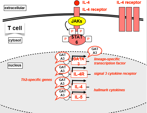
- IL-4 signal cytokine binds IL-4R, phosphorylation through JAKs of intracellular portion of IL12R
- Recruit STAT6, phosphorylated and translocates to nucleus
- Activated STAT6 binds to TH2-specific genes:
- GATA3= lineage specific TF- promotes more response to IL-4
- IL4R- signal 3 cytokine receptor- promotes cell to be more responsive to IL-4
- IL4 +IL5= hallmark cytokine of Th2 cells
- GATA binds to all 3 gene loci, activates, reinforces and stabilises Th1 differentiation, promoting expression of all 3
-
2. Cytokine Signalling in CD4 T Cells: Th2
-
Summary of Differentiation so far
-
Overview of Th1 + Th2 Effector Function
-
3. Effector Functions of CD4 T cells
-
3. Effector Functions of CD4 T cells
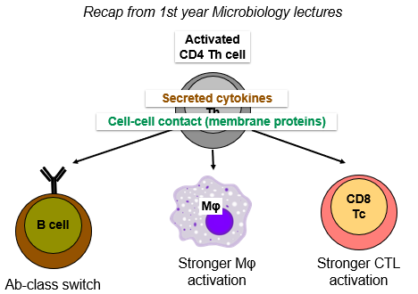
- 1. Activated CD4 T cells help Ab class switch
- Ab's have different isotypes-> starts as IgM and IgD but class switch into IgG, IgE, IgA
- Isotypes have different heavy chain constant regions:
- Bind different Fc receptors
- Differing ability to activate complement
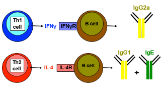
- CD4 T cell signature cytokines direct Ab class switching:
- Th1 cell produced IFNϒ and B cells express the IFNϒR- Th1 cell IFNϒ induces B cells to class switch to IgG2a
- Th2 cell IL-4 induces B cell to class switch to IgG1 + IgE
-
Activated CD4 T cells help Ab class switch
-
Activated CD4 T cells help Ab class switch
- Eg of Importance of class switching:
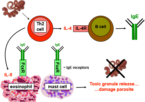
- Parasitic worms induce Th2 cell to produce IL4, induces class switch to IgE
- Innate immune cells (granulocytes) express receptor for IgE (FcεR)
- Immune cells bind IgE, armed with IgE (receptor)
- May see pathogen molecules that IgE recognises and bind-> activate immune cells
- Th2 also produces IL5 cytokine= survival factor and differentiation factor for eosinophils
- Produces lots of eosinophils in blood all armed with IgE
- Mast cells will degranulate- release toxic granules onto parasite to attack
-
Activated CD4 T cells help Ab class switch
-
3. Effector Functions of CD4 T cells
- 2. Activated CD4 T cells help macrophages
- CD4 T cell activated by DC in LN, macrophage far away in peripheral tissue fighting infection
- Activated T cell needs to migrate from LN to peripheral tissue
-
Activated CD4 T cells help macrophages
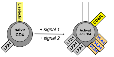
- Activation-induced changes in CD4 T cell surface proteins
- Naive CD4 T cells have lots of L-selectin (to enter HEV) and integrin LFA1 that gets them into LN
- Following activation:
- Loss of L-selectin
- Increase integrins (more LFA1 and new VLA-4)
- Induction of CD40L ligand
-
Activated CD4 T cells help macrophages
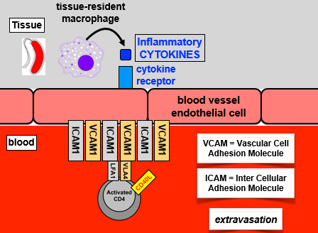
- T cells migrate from blood into inflamed tissues:
- Tissue resident macrophage produces inflammatory cytokines recognised by receptor on endothelial cell
- Endothelium activated, expressed high levels of adhesion molecules: ICAM1 (binds LFA1), VCAM1 (binds VLA4)
- Binding of adhesion molecules to integrins arrests activated T cell on endothelial cell and migration into tissue controlled by chemokine gradient
-
Activated CD4 T cells help macrophages
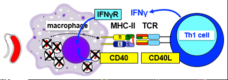
- Macrophages present antigens, re-stimulate activated CD4 T cell
- Present antigens on MHC-II
- T cell with correct antigen specificity
- TCR signals delivered by MHC-II:
- Th1 produce IFNγ + express high levels of CD40L
- CD40L binds CD40 on mac
- Activate macrophage to kill intracellular pathogens
-
3. Effector Functions of CD4 T cells
- 3. CD4 T cells help CD8 CTL activation:
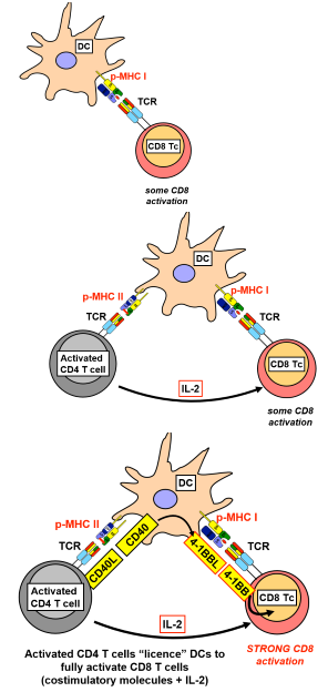
- Activated CD4 T cells licence DCs to fully activate CD8 T cells:
- DC presents peptide on MHC-I and binds TCR on CD8 T cell = weak activation
- Simultaneous presentation of MHC-II:peptide to CD4 T cell (different peptide derived from same pathogen), activated CD4 T cell produces IL2= further activation CD8
- CD40L (induced by cross-talk between CD4 + DC) binds CD40 on DC and causes upregulation of other co-stimulatory molecules on DC for CD8 = strong activation of CD8
-
-
CD4 T cells help CD8 CTL activation
-
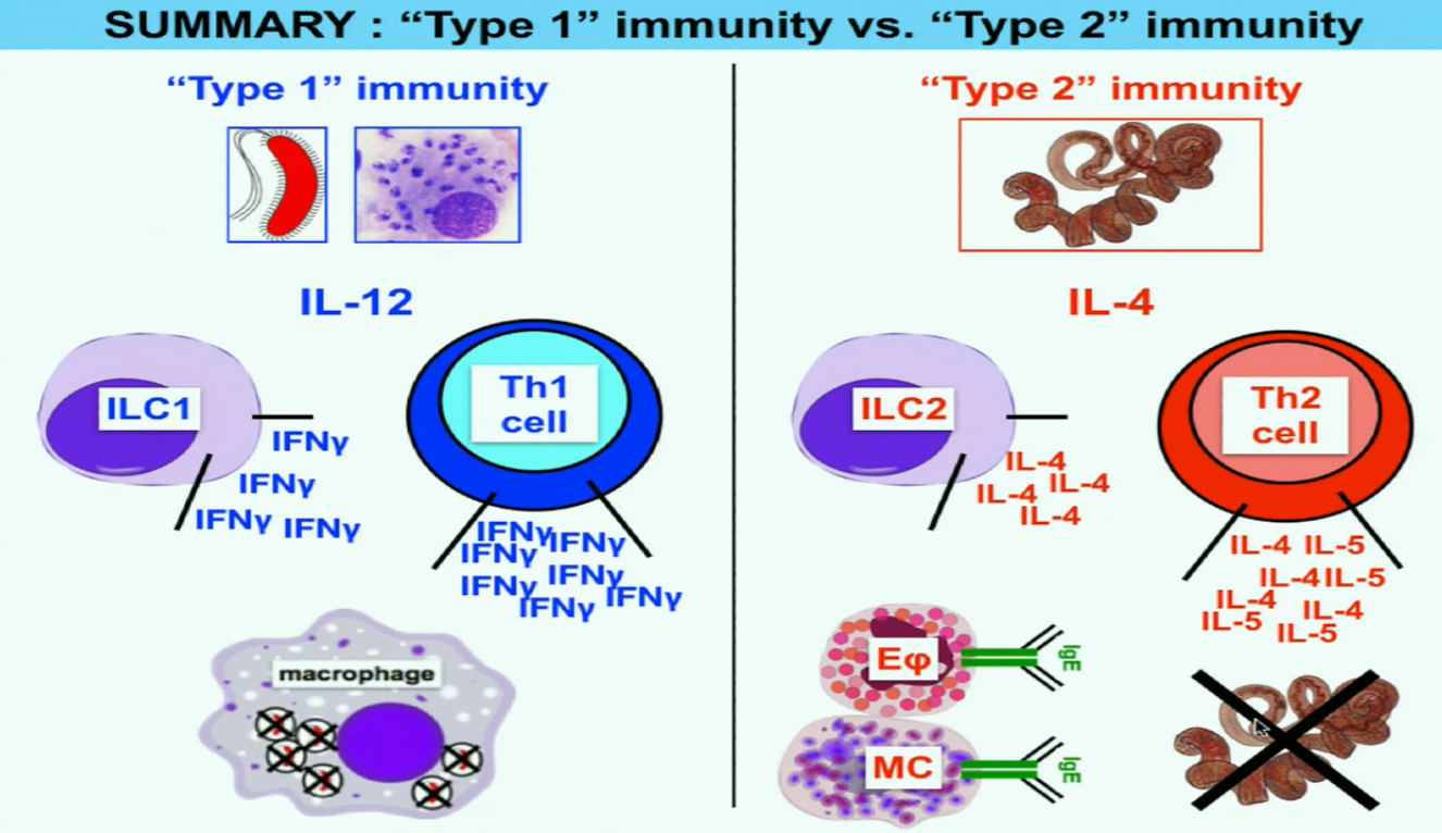
- Th1 and Th2 cells are coordinators- directors of broader immune responses (Type 1 and Type 2 immunity)
- Type 1 immunity:
- Bacteria activates DC and macrophages to produce IL12
- ILC1 produce IFNϒ, supports differentiation into Th1
- Lots of production of IFNϒ- Th1 and ILC1
- Activation of macrophages to kill intracellular pathogens
- Type 2 immunity:
- Worm activates IL4 production
- ILC2 produces IL4 which supports differentiation of Th2 cells
- Th2 produce IL4 and IL5 cytokines- lead to activation of B cells to produce IgE class switch Ab
- When eosinophils and mast cells recognise parasite they degranulate and kill pathogen
-
-
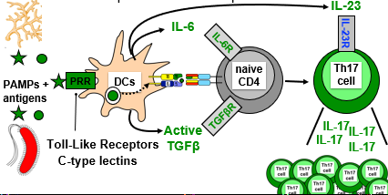
- Make large amounts of cytokine IL-17
- Initiate effector immune responses against pathogens not dealt with by Th1 and Th2 cells: Extracellular bacteria and fungi
-
- PAMPs bind DC PRR, uptake antigens present on MHC-II
- Trigger DCs to produce signal 3= active TGFβ and IL-6 (inflammatory cytokine)
- Initial Th17 differentiation
- DCs present peptides to CD4 T cell, which binds as well as cytokines to receptors on CD4 T cell triggering initial differentiation into Th17
- Th17 characterised by production of IL-17
- Need additional signal to keep alive- DCs produce IL-23 cytokine as part of signal 3
-
-
Cytokine-induced Signalling in Th17 Cells
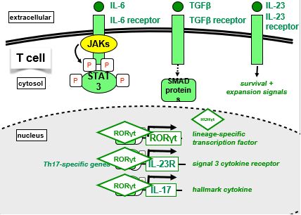
- IL-6 binds IL-6R on T cell, activates JAKs to phsophorylate intracellular portion of IL-6R
- Phosphotyrosine acts as docking site for STAT3, phosphorylated by JAKs, translocates to nucleus and bind Th17 specfic genes
- STAT3 alone not sufficient to induce gene expression
- TGFβ binds to receptor on T cell and activates SMAD proteins
- SMADs and STAT3 bind and induce expression of Th17 specific genes
- IL23 (not there upon initial activation) binds to its receptor on T cell and signals for survival and expansion
- RORγT TF protein is expressed and will take over to stabilise expression of all 3 genes (RORγT TF , IL-23R, IL-17)
-
Cytokine-induced Signalling in Th17 Cells
-
- Recruited to initial sites of inflammation:
- Infection of extracellular pathogen causes DC to mature and present, DC migrate down afferent lymphatic to draining LN
- Interacts with T cell with correct specificity, differentiation into Th17
- Some Th17 cells leave via efferent lymphatic, end up in blood and recruited back to inflamed tissue
- Tissue resident macrophage induced by pathogen to secrete inflammatory cytokines, act on endothelial wall of blood vessel, activate expression of adhesion molecules, chemokine gradient pulls Th17 cells into tissue
-
Th17 Effector Functions: Neutrophils
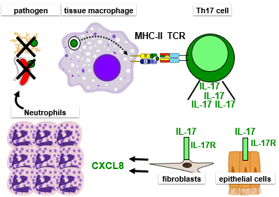
- Once at site of inflammation, recruit neutrophils:
- Interaction between mac and specific Th17 cell via MHC-II:peptide- reactivates Th-17 (first in LN), produces IL-17
- IL-17 binds to receptor (broadly expressed by non-immune cells (stromal: fibroblast, eptihelial cells)
- Triggers non-immune cells to produce lots of chemokine CXCL8
- CXCL8 recruits neutrophils from blood to site to kill pathogen
-
Th17 Effector Functions: Neutrophils
-
- DC produce active TGFβ, IL-6, IL-23 in response to fungi or extracellular bacteria = Signal 3 for Th17 differentiation
- TGFβ + IL-6 induce initial Th17 differentiation
- IL-23 promotes expansion + survival of Th17 cells
- Lineage-specific TF for Th17 cells is RORγT
- Th17 cells produce IL-17
- This induces non-immune cells at sites of infection to produce CXCL8 = neutrophil chemoattractant
- Recruited neutrophils kill pathogen
-
Summary of T Cell Subset so far
-
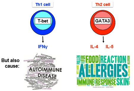
- Immune responses are inflammatory and potentially pathogenic... autoimmunity and immunopathology (damages tissue even after clearance)
- Central tolerance: deletion of self-reactive T cells in thymus
- Some self-reactive T cells escape this negative selection, so need peripheral tolerance
- Peripheral tolerance: stop activation of mature self-reactive T cells
- Via co-stimulation- in absence of infection APCs have low level of CD80/CD86
- Not always efficient, need specialised cells to turn off immune responses.. Regulatory T cells
-
2 Types of Regulatory T Cell (Treg)
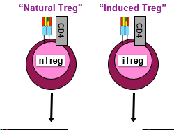
- Still have TCR and express CD4 like normal T cells
- Difference=repressive, turn off effector T cells, APCs etc
-
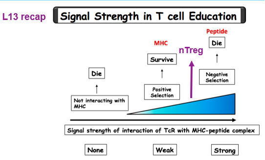
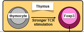
- nTregs differentiate in thymus in response to stronger than normal TCR stimulation
- TCR binds to self-peptide:MHC stronger than normal but not strong enough to be deleted
- Induces expression of Treg lineage-specific TF: Foxp3
- T cells exit thymus and circulate through LN and spleen
- Function: they recognise self-peptides and have stronger than normal self-reactivity, recognise T cells that escaped negative selection and recirculating-> directly inhibit autoreactive T cell
-
-
- iTregs differentiate in the periphery
- Signal 3 for iTreg is TGFB (in absence of inflammatory signal)
- DC take up antigen, produce active TGFB which binds receptor on naive CD4 and upregulates foxp3 positive iTreg
- iTreg potentially specific for any antigen: pathogen, self-antigen, commensal
-
Differentiation of iTreg vs Th17 Cells
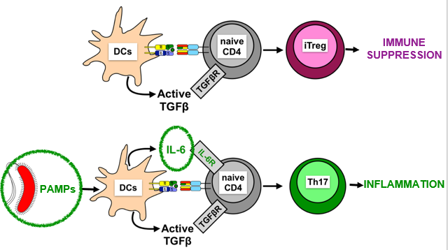
- Pathogens induce production of inflammatory cytokines (eg IL-6) that switch immune suppression to Th17 inflammatory response
-
iTregs Supress Immune Response
- Via cell surface molecules:
- Treg express high levels of CTLA-4 (co-inhibitory molecule)
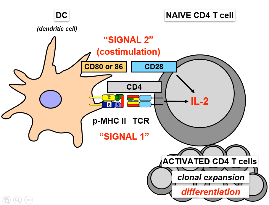
- CTLA-4 binds and sequesters DC CD80/86 from naive cells - no signal 2
- CTLA-4 can physically rip CD80/86 from APCs= no ability co-stimulate
- Tregs express high levels of IL-2R
- Act as sink for IL-2, take it away from activated cells so they cant proliferate
- Outcompete activated T cells for IL-2
-
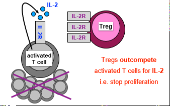
- Bildunterschrift: Express IL-2R: Outcompete activated T cells for IL-2
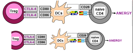
- Bildunterschrift: Express CTLA-4: no co-stimulation (no signal 2)
-
iTregs Supress Immune Response
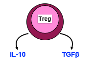
- Via secretion of immunosuppressive cytokines:
- Inhibit Il-12 production by DCs (no Th1 response)
- Reduce MHC-II + CD80/86 levels on DCs
- Directly inhibit T cell proliferation
- Generate more Tregs (TGFβ)
-
- Mutations to Foxp3 gene of X chromosome = IPEX syndrome
- Cant make functional Foxp3 protein, lack Tregs, die by autoimmune diseases
- Fatal early (childhood) onset autoimmune disease in males:
- Diabetes, anemia, splenomegaly, skin inflammation, cytokine storm
-
- Tregs are essential for peripheral tolerance
- nTregs differentiate in thymus + turn-off self-reactive T cells
- In absence of inflammatory cytokines, TGFβ induces iTregs
- iTregs suppress immune responses through surface + secreted molecules