
Karteikarten
Microscopy
Karten in diesem Stapel (31)
- 1Why is microscopy important?-Allows examination of cellular structures
-Microscopy first revealed the existence of microbial life and subcellular structures
-Magnification and staining determines what can be observed
-Staining distinguishes cell from non cell or cellular structure from other parts of a cell - 2What allows you to see from 10 micro metres to 1 Angstrom?TEM
- 3What allows you to see from 1 mm to 10 nm?SEM
- 4What allow you to see from 1 mm to 1 micro metre?Bright field
- 5What does a light (compound) microscope see?
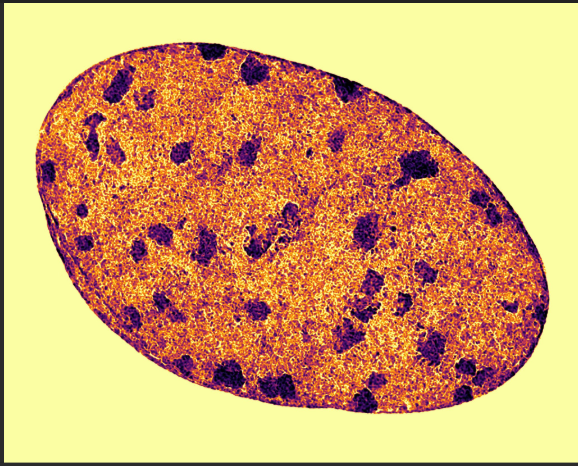
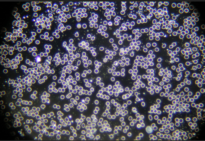
- 6What does a Dark field microscope see?-Light reaches from the side, then scattered by the specimen; specimen is a light image on a dark background
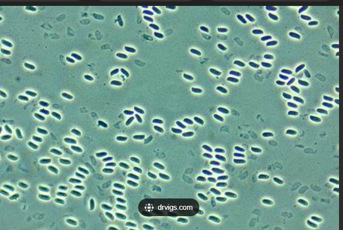
- 7What does a Phase contrast microscope see?-Light passing through a cell differs from light passing in the surrounding liquid ; cells differ in refractive index from surrounding, the factor by which light is slowed as it passes through. Dark image on a light background
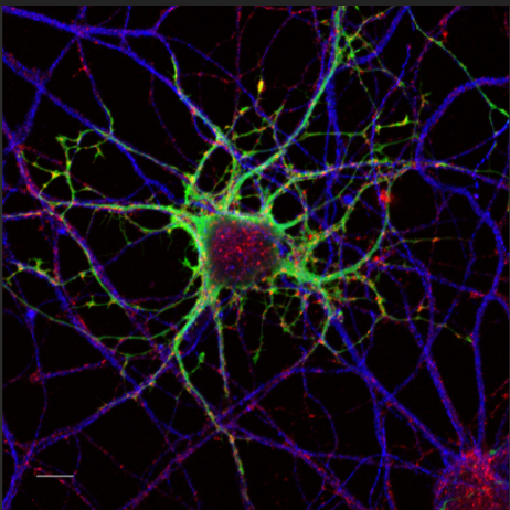
- 8What does a fluorescent microscope see?-sample-labelled with fluorescent dye
- 9What does an electron microscope see?Electron beam; investigate ultra structure
SEM = scanning electron microscope
TEM = transmission electron microscope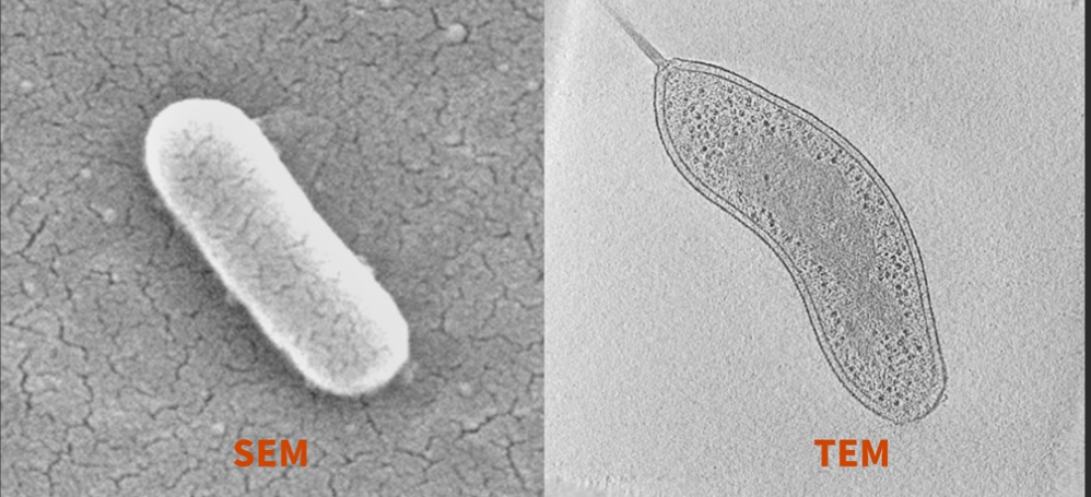
- 10How does the light compound microscope work?Combination of magnification of ocular (10x) and objective lens is the total magnification
-Objective lenses ; low power 10x, High power 40x, Oil immersion 100X
(HIGHEST MAGNIFICATION = 1000x)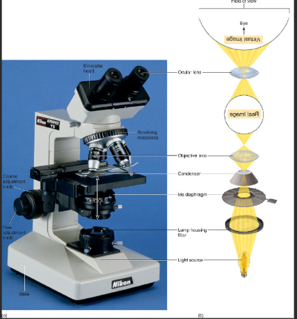
- 11How does oil immersion lens work?-oil immersion to stop light that disperses (refracts) when it passes from glass slide to air
-The oil used has the same density (refractive index) as glass - result- greater clarity of image
(OIL must be removed from 100x lens immediately after use using lens tissue)
(Avoid oil touching other lens)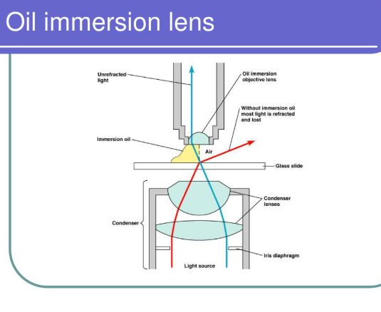
- 12Why do we stain microbial cells?-To visualise the specimen clearly under the microscope
-To contrast the microbial cell against its background
-Without staining, the specimen under the microscope will look transparent or will not be visible clearly
-Allows us to see the shape and size of microbial cells - 13What stains are used?Stains are chromophores
-Most stains acidic/basic and a few neutral
-Basic dye is positively charged at neutral pH and can bind to negatively charged bacteria staining the cell
-Acid dye stays out of cell and stains medium in which cell exists (the liquid surrounding bacterial cell) - 14What is an example of a simple stain?Methylene blue- visualise microscopic cells
- 15What do gram stain do?Differentiate between bacterial cell types
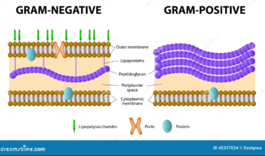
- 16What stains gram positive (thick cell wall) bacteria?Purple (crystal violet stain remains)
- 17What stains gram negative (thin cell wall) bacteria?Pink (counterstained with safranin)
- 18What is acid fast stain?High mycolic acid content of cell walls resists normal staining -used to identify Mycobacteria (TB)
- 19How is a slide smeared1. Spread culture in thin film over slide, dry in air
2. Heat fixing and staining (pass slide through flame to heat fix)
3. Flood slide with stain; rinse and dry
4. Check under microscope - 20What does gram positive and gram negative bacteria look like?
- 21How does the gram stain work?1. Flood the heat-fixed smear with crystal violet for 1 min (Both stained purple)
2. Add iodine solution for 1 min. Mordant makes dye less soluble so it adheres to cell walls
3. Decolorize with alcohol briefly - about 20 sec. Decolorization washes strain from gram negative cell walls
4. Counterstain with safranin. Allows dye adherence to gram negative cell walls
THEREFORE, gram negative = pink and gram positive = purple - 22Which bacterium is an exception to gram stain?Mycobacteria
-the mycobacteria has a waxy surface which is impervious to many stains
-ACID FAST STAIN - high mycolic acid content of cell walls resists normal staining
-Carbol fuchsin -primary dye (basic, lipid soluble, red)
-Acid wash =decolourizer
-Methylene blue - counter stain - 23What is agar?it is a seaweed extract used to solidify nutrient medium used to grow microorganisms in
- 24How is microbial colony morphology used to identify microorganisms?Depending on
-SIZE
-SHAPE
-ELEVATION
-CONSISTENCY
-SURFACE
-EDGE
-OPACITY
-COLOUR - 25What are the different morphologies visible on solid medium?
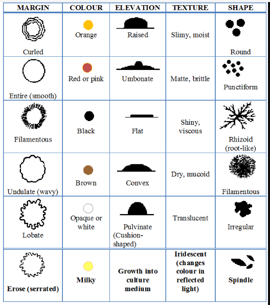
- 26What appearance does Pseudomonas aeruginosa have?Smooth, translucent, irregular, spreading edge
- 27What appearance does serratia marcescens on MacConkey agar have?red = lactose positive. Bile salts precipitate - hazy outer edge
- 28What appearance does bacillus subtilis have?dry, flat and irregular, with lobate margins
- 29What appearance does micrococcus luteus have on blood agar?Circular, pin head, convex, smooth
- 30What does microbial morphology look like in liquid culture?-Cloudy or even distribution (Turbidity)
-Growth on top surface (pellicle)
-Growth on side of vessel (biofilm)
-Flocs (floating masses) or sedimentation
-Slime production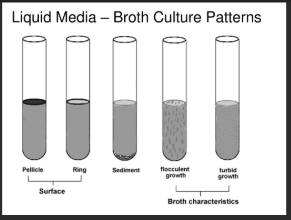
- 31Why is growth of microorganisms on solid and liquid media important?Differentiate between cells and be a first step to identification
->Hospital labs
->Process and environmental quality control in industry
Informationen
- Beschreibung
- University Lab Skills 2 Karteikarten am Microscopy, erstellt von Adrianna Bialecka am 26/01/2025.
- Erstellt von
- Adrianna Bialecka
- Aufrufe
- 0
- Bewertung