
Flashcards
micro
Cards in this deck (72)
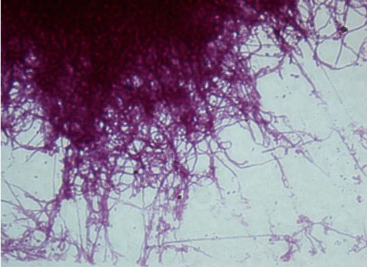
- 1—Actinomyces, Grams staining, pure culture
Gram stain technique
1. The heat fixed bacterial smear is flooded with crystal violet and left on for 2 minutes.
2. Then it is flooded with Lugol’s iodine (mordant) which is left on for 1 minute.
3. The smear is then decolourized with ethanol (96%) for 30 seconds up to one minute.
4. Afterwards it is flooded with safranin which is left on for 2-3 minutes.
5. Finally it is washed with water, dried and examined under oil immersion. - 2—Actinomyces, Grams staining, pure culture
Gram stain technique
1. The heat fixed bacterial smear is flooded with crystal violet and left on for 2 minutes.
2. Then it is flooded with Lugol’s iodine (mordant) which is left on for 1 minute.
3. The smear is then decolourized with ethanol (96%) for 30 seconds up to one minute.
4. Afterwards it is flooded with safranin which is left on for 2-3 minutes.
5. Finally it is washed with water, dried and examined under oil immersion.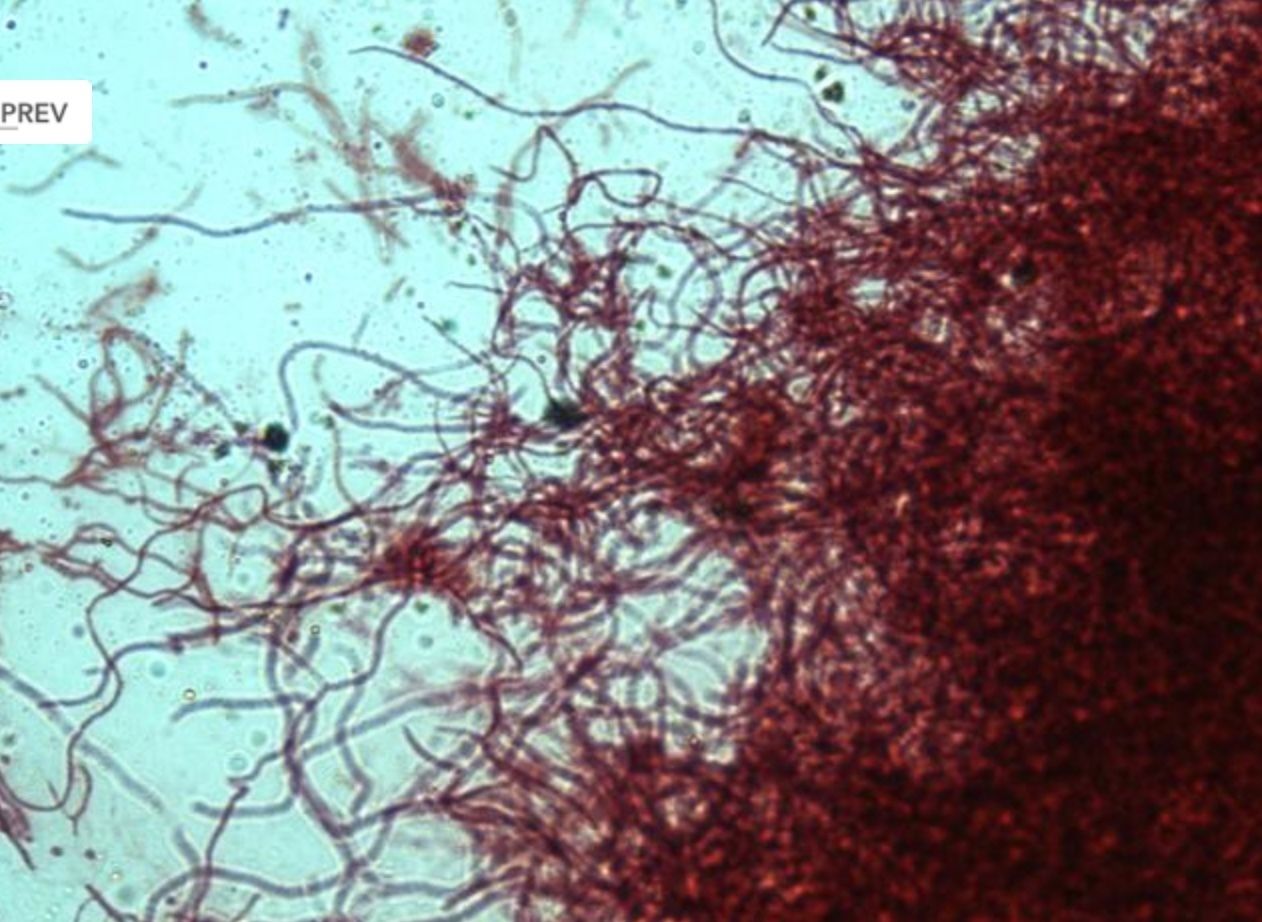
- 3—Actinomyces, Grams staining, pure culture
Gram stain technique
1. The heat fixed bacterial smear is flooded with crystal violet and left on for 2 minutes.
2. Then it is flooded with Lugol’s iodine (mordant) which is left on for 1 minute.
3. The smear is then decolourized with ethanol (96%) for 30 seconds up to one minute.
4. Afterwards it is flooded with safranin which is left on for 2-3 minutes.
5. Finally it is washed with water, dried and examined under oil immersion.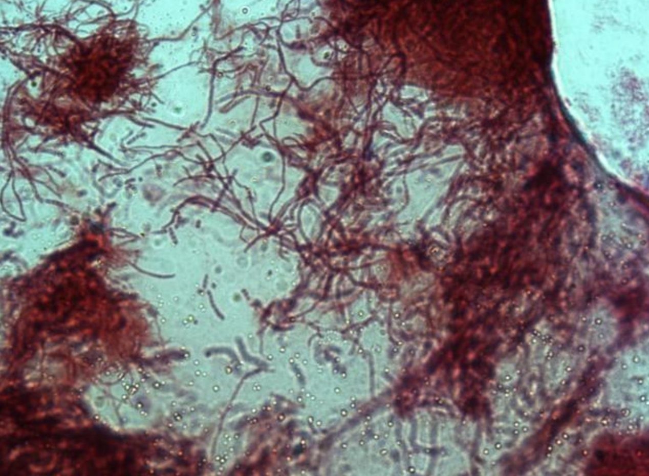
- 4—Actinomyces, Grams staining, pure culture
Gram stain technique
1. The heat fixed bacterial smear is flooded with crystal violet and left on for 2 minutes.
2. Then it is flooded with Lugol’s iodine (mordant) which is left on for 1 minute.
3. The smear is then decolourized with ethanol (96%) for 30 seconds up to one minute.
4. Afterwards it is flooded with safranin which is left on for 2-3 minutes.
5. Finally it is washed with water, dried and examined under oil immersion.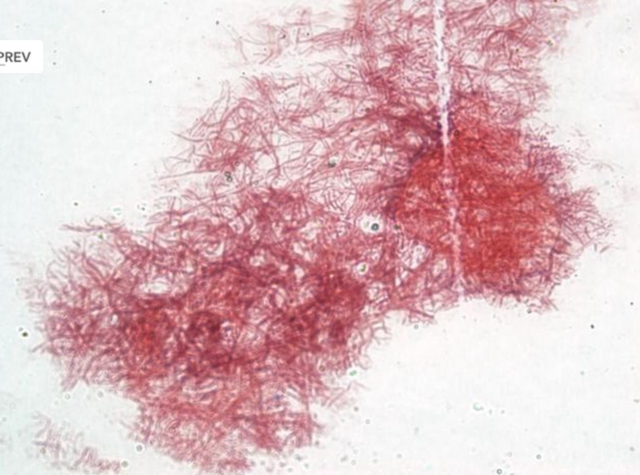
- 5—Bacillus subtilis (endospores), methylene blue, pure culture
Simple staining
Fixed smears are stained using neutral and basic aniline dyes.
Only one dye is used, for example, methylene blue, safranin, etc.
All bacteria will stain the same colour, however it will be possible to evaluate the shape, size and arrangement of bacteria.
The slide is placed on a stand over the container and flooded with methylene blue. The dye should be left on for 5 minutes. When using safranin, the dye should be left on for 4 minutes.
Afterwards the slide is washed with water and dried. Only dry slides are examined under oil immersion.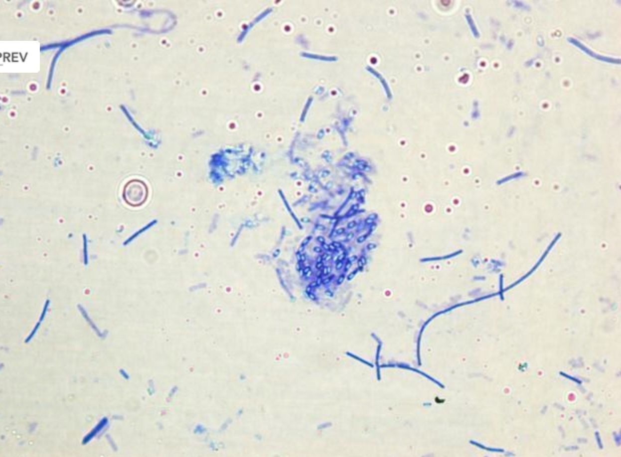
- 6—Bacillus subtilis (endospores), methylene blue, pure culture
Simple staining
Fixed smears are stained using neutral and basic aniline dyes.
Only one dye is used, for example, methylene blue, safranin, etc.
All bacteria will stain the same colour, however it will be possible to evaluate the shape, size and arrangement of bacteria.
The slide is placed on a stand over the container and flooded with methylene blue. The dye should be left on for 5 minutes. When using safranin, the dye should be left on for 4 minutes.
Afterwards the slide is washed with water and dried. Only dry slides are examined under oil immersion.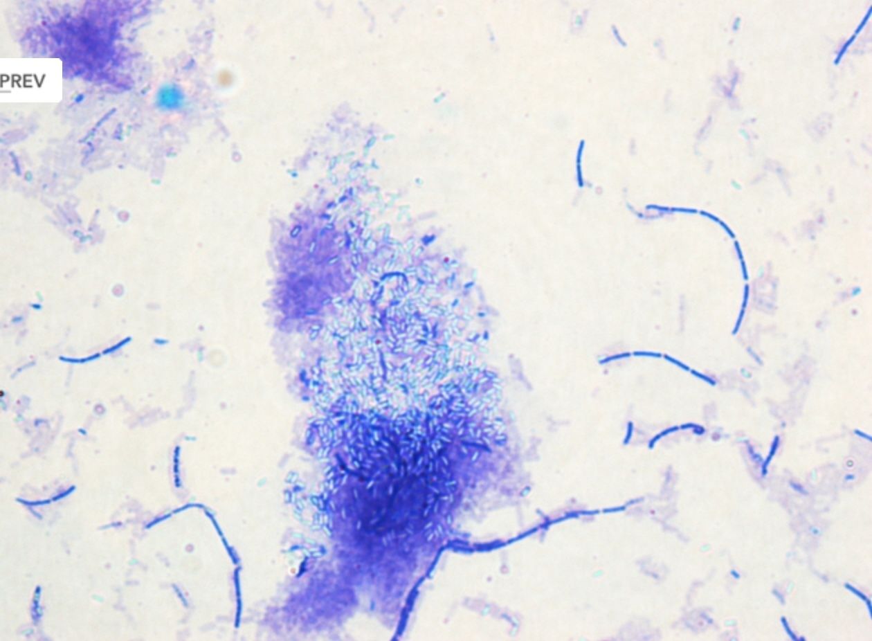
- 7—Corynebacterium diphtheriae, Neissers staining, pure culture
Neisser staining is used infrequently, however it is possible to discern inclusion bodies, granules of volutin, in Corynebacterium diphtheriae, the causative
agent of diphtheria.
A solution: methylene blue, glacial acetic acid, ethanol, distilled water.
B Solution: crystal violet, Ethanol, distilled water
C solution: chrysoidin Y solution and distilled water.
1) Prepare fixed smear.
2) Flood with 2 parts of solution A and 1 part of solution B for 15 sec.
3) Allow due to flow off.
4) add solution C for 46 sec. Rinse with water
4) Let it dry and examine under the microscope with oil immersion.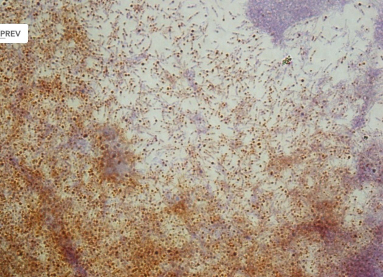
- 8—Corynebacterium diphtheriae, Neissers staining, pure culture
Neisser staining is used infrequently, however it is possible to discern inclusion bodies, granules of volutin, in Corynebacterium diphtheriae, the causative
agent of diphtheria.
A solution: methylene blue, glacial acetic acid, ethanol, distilled water.
B Solution: crystal violet, Ethanol, distilled water
C solution: chrysoidin Y solution and distilled water.
1) Prepare fixed smear.
2) Flood with 2 parts of solution A and 1 part of solution B for 15 sec.
3) Allow due to flow off.
4) add solution C for 46 sec. Rinse with water
4) Let it dry and examine under the microscope with oil immersion.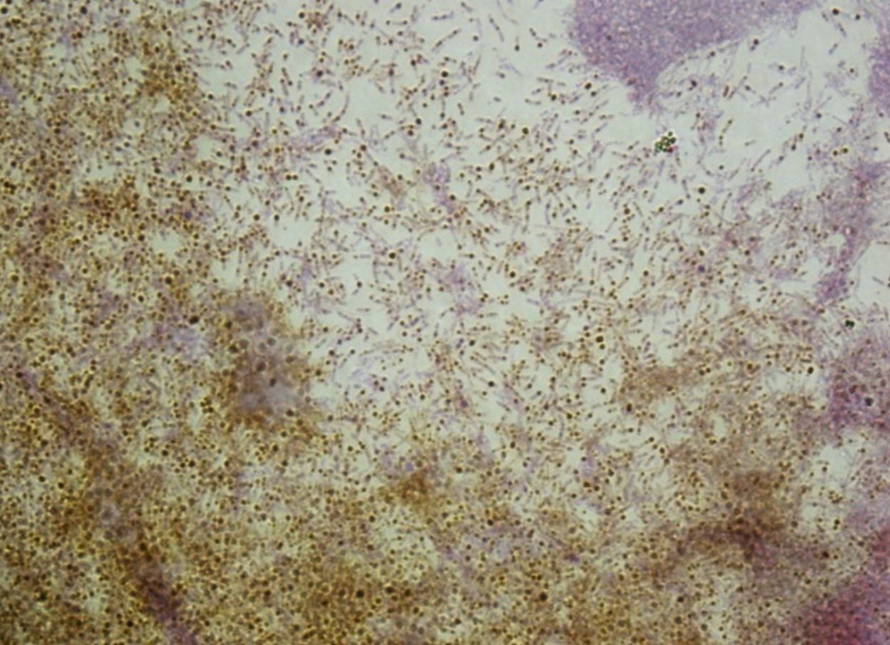
- 9—Corynebacterium diphtheriae, Neissers staining, pure culture
Neisser staining is used infrequently, however it is possible to discern inclusion bodies, granules of volutin, in Corynebacterium diphtheriae, the causative
agent of diphtheria.
A solution: methylene blue, glacial acetic acid, ethanol, distilled water.
B Solution: crystal violet, Ethanol, distilled water
C solution: chrysoidin Y solution and distilled water.
1) Prepare fixed smear.
2) Flood with 2 parts of solution A and 1 part of solution B for 15 sec.
3) Allow due to flow off.
4) add solution C for 46 sec. Rinse with water
4) Let it dry and examine under the microscope with oil immersion.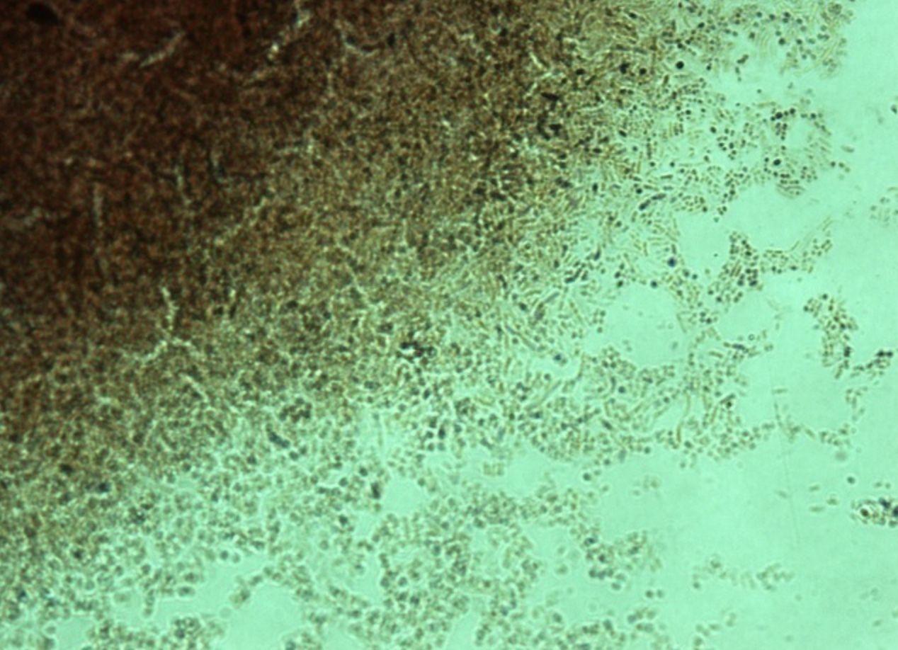
- 10—Corynebacterium diphtheriae, Neissers staining, pure culture
Neisser staining is used infrequently, however it is possible to discern inclusion bodies, granules of volutin, in Corynebacterium diphtheriae, the causative
agent of diphtheria.
A solution: methylene blue, glacial acetic acid, ethanol, distilled water.
B Solution: crystal violet, Ethanol, distilled water
C solution: chrysoidin Y solution and distilled water.
1) Prepare fixed smear.
2) Flood with 2 parts of solution A and 1 part of solution B for 15 sec.
3) Allow due to flow off.
4) add solution C for 46 sec. Rinse with water
4) Let it dry and examine under the microscope with oil immersion.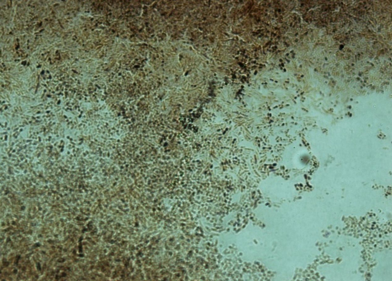
- 11—Corynebacterium diphtheriae, Neissers staining, pure culture
Neisser staining is used infrequently, however it is possible to discern inclusion bodies, granules of volutin, in Corynebacterium diphtheriae, the causative
agent of diphtheria.
A solution: methylene blue, glacial acetic acid, ethanol, distilled water.
B Solution: crystal violet, Ethanol, distilled water
C solution: chrysoidin Y solution and distilled water.
1) Prepare fixed smear.
2) Flood with 2 parts of solution A and 1 part of solution B for 15 sec.
3) Allow due to flow off.
4) add solution C for 46 sec. Rinse with water
4) Let it dry and examine under the microscope with oil immersion.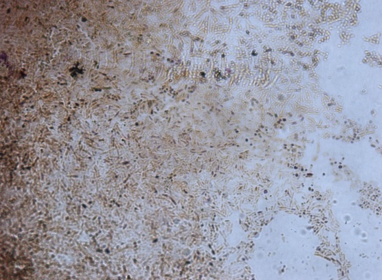
- 12—Corynebacterium diphtheriae, Neissers staining, pure culture
Neisser staining is used infrequently, however it is possible to discern inclusion bodies, granules of volutin, in Corynebacterium diphtheriae, the causative
agent of diphtheria.
A solution: methylene blue, glacial acetic acid, ethanol, distilled water.
B Solution: crystal violet, Ethanol, distilled water
C solution: chrysoidin Y solution and distilled water.
1) Prepare fixed smear.
2) Flood with 2 parts of solution A and 1 part of solution B for 15 sec.
3) Allow due to flow off.
4) add solution C for 46 sec. Rinse with water
4) Let it dry and examine under the microscope with oil immersion.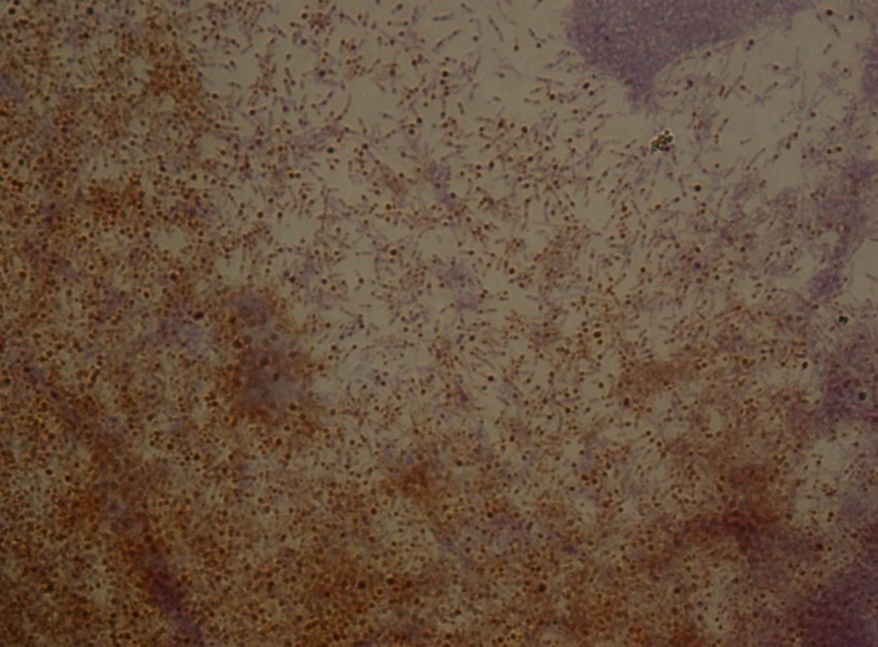
- 13—Candida albicans, methylene blue, pure culture
Simple staining
Fixed smears are stained using neutral and basic aniline dyes.
Only one dye is used, for example, methylene blue, safranin, etc.
All bacteria will stain the same colour, however it will be possible to evaluate the shape, size and arrangement of bacteria.
The slide is placed on a stand over the container and flooded with methylene blue. The dye should be left on for 5 minutes. When using safranin, the dye should be left on for 4 minutes.
Afterwards the slide is washed with water and dried. Only dry slides are examined under oil immersion.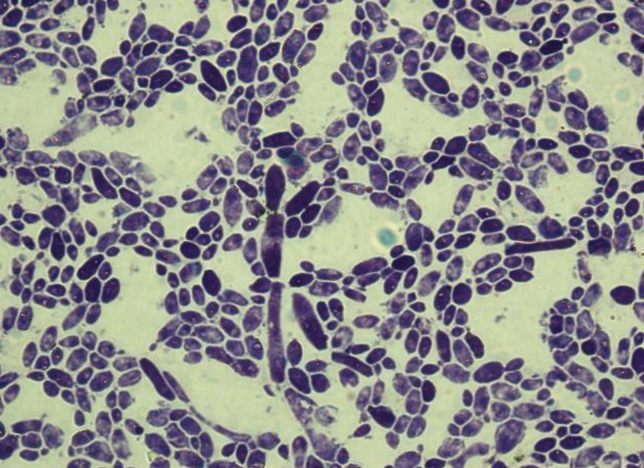
- 14—C.albicans, methylene blue, pure culture
Simple staining
Fixed smears are stained using neutral and basic aniline dyes.
Only one dye is used, for example, methylene blue, safranin, etc.
All bacteria will stain the same colour, however it will be possible to evaluate the shape, size and arrangement of bacteria.
The slide is placed on a stand over the container and flooded with methylene blue. The dye should be left on for 5 minutes. When using safranin, the dye should be left on for 4 minutes.
Afterwards the slide is washed with water and dried. Only dry slides are examined under oil immersion.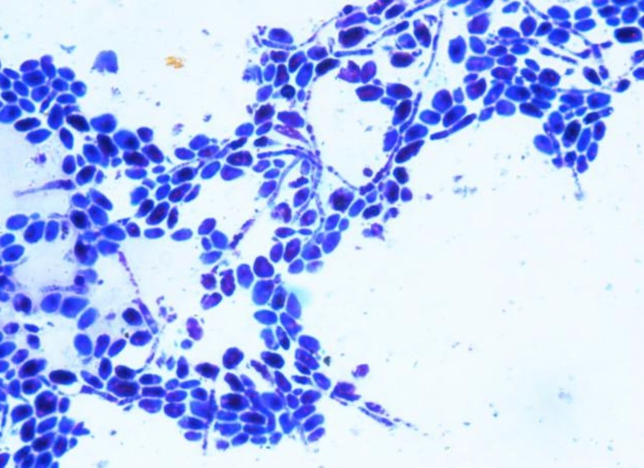
- 15—C.albicans, methylene blue, pure culture
Simple staining
Fixed smears are stained using neutral and basic aniline dyes.
Only one dye is used, for example, methylene blue, safranin, etc.
All bacteria will stain the same colour, however it will be possible to evaluate the shape, size and arrangement of bacteria.
The slide is placed on a stand over the container and flooded with methylene blue. The dye should be left on for 5 minutes. When using safranin, the dye should be left on for 4 minutes.
Afterwards the slide is washed with water and dried. Only dry slides are examined under oil immersion.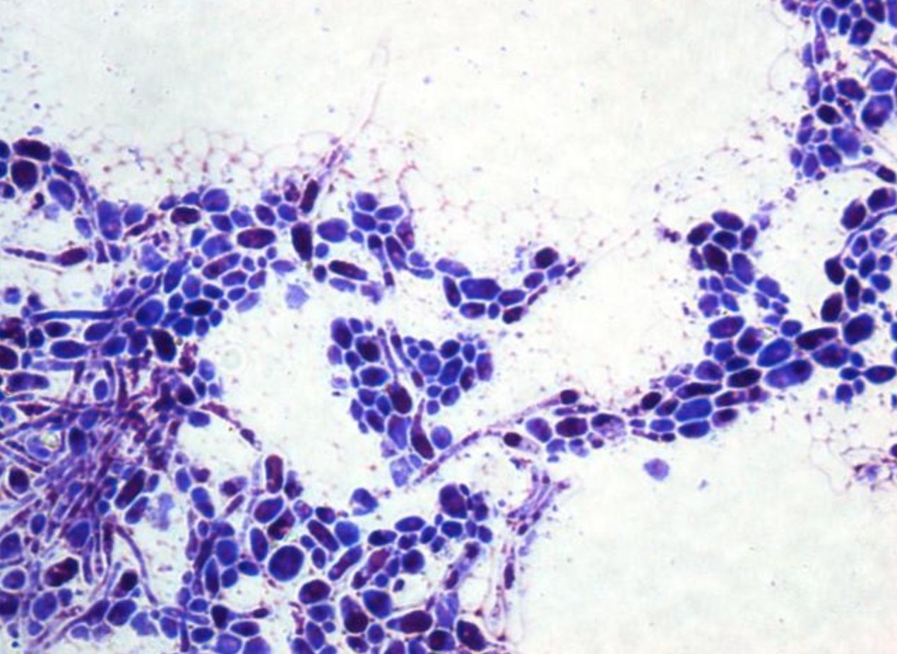
- 16—C.albicans, methylene blue, pure culture
Simple staining
Fixed smears are stained using neutral and basic aniline dyes.
Only one dye is used, for example, methylene blue, safranin, etc.
All bacteria will stain the same colour, however it will be possible to evaluate the shape, size and arrangement of bacteria.
The slide is placed on a stand over the container and flooded with methylene blue. The dye should be left on for 5 minutes. When using safranin, the dye should be left on for 4 minutes.
Afterwards the slide is washed with water and dried. Only dry slides are examined under oil immersion.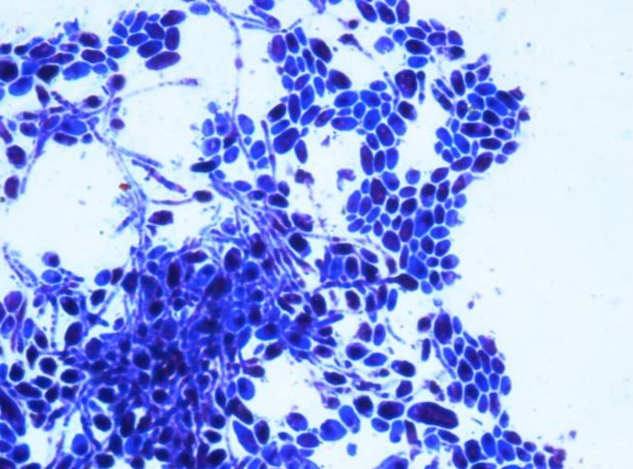
- 17—Corynebacterium diphtheriae, Neissers staining, pure culture
Neisser staining is used infrequently, however it is possible to discern inclusion bodies, granules of volutin, in Corynebacterium diphtheriae, the causative
agent of diphtheria.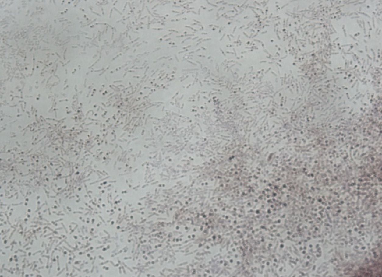
- 18—Corynebacterium diphtheriae, Neissers staining, pure culture
Neisser staining is used infrequently, however it is possible to discern inclusion bodies, granules of volutin, in Corynebacterium diphtheriae, the causative
agent of diphtheria. - 19—E.coli, Grams staining, pure culture
Gram stain technique
1. The heat fixed bacterial smear is flooded with crystal violet and left on for 2 minutes.
2. Then it is flooded with Lugol’s iodine (mordant) which is left on for 1 minute.
3. The smear is then decolourized with ethanol (96%) for 30 seconds up to one minute.
4. Afterwards it is flooded with safranin which is left on for 2-3 minutes.
5. Finally it is washed with water, dried and examined under oil immersion.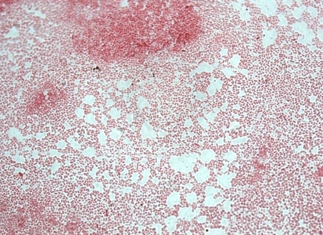
- 20—E.coli, Grams staining, pure culture
Gram stain technique
1. The heat fixed bacterial smear is flooded with crystal violet and left on for 2 minutes.
2. Then it is flooded with Lugol’s iodine (mordant) which is left on for 1 minute.
3. The smear is then decolourized with ethanol (96%) for 30 seconds up to one minute.
4. Afterwards it is flooded with safranin which is left on for 2-3 minutes.
5. Finally it is washed with water, dried and examined under oil immersion.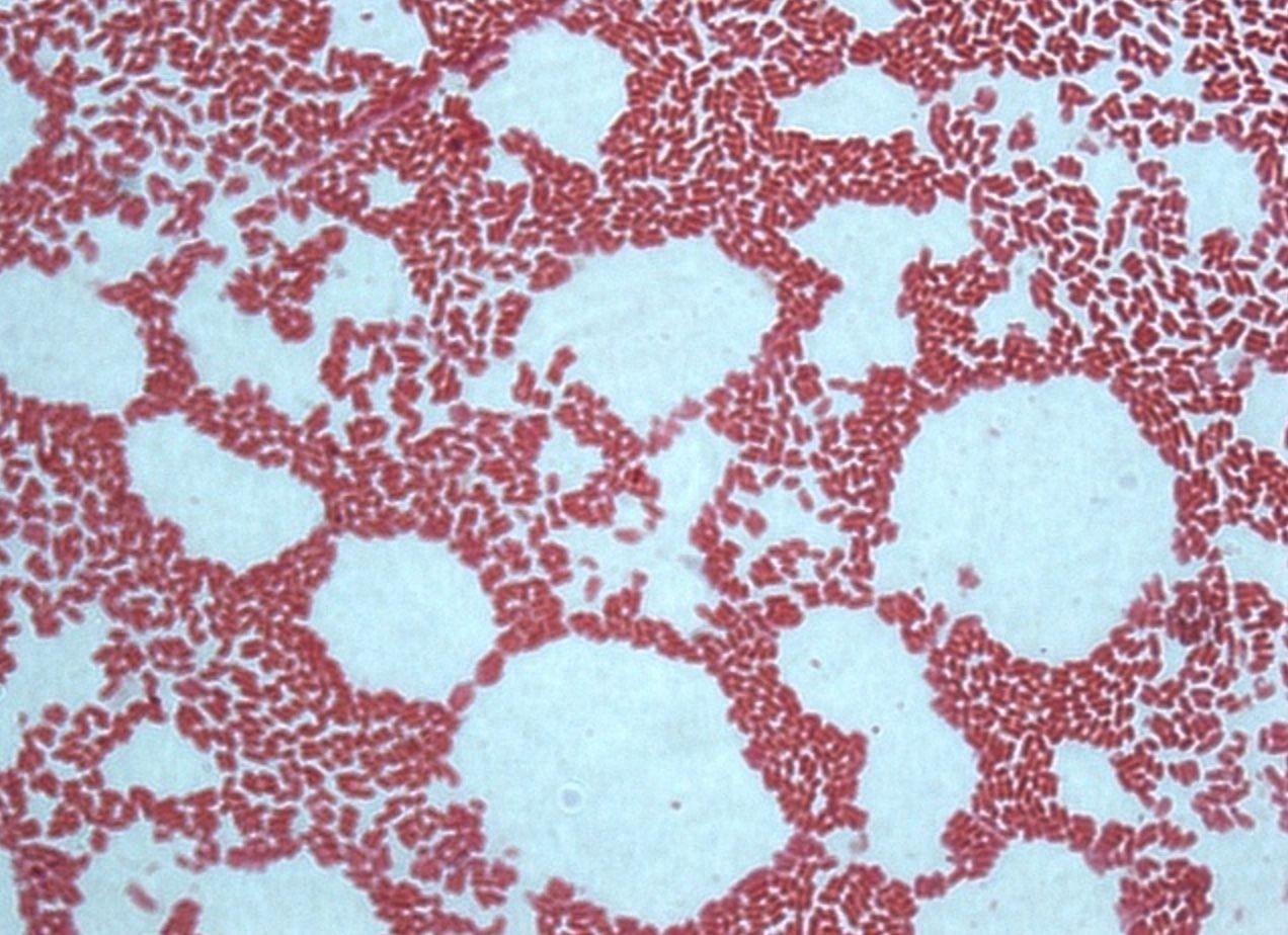
- 21—E.coli, Grams staining, pure culture
Gram stain technique
1. The heat fixed bacterial smear is flooded with crystal violet and left on for 2 minutes.
2. Then it is flooded with Lugol’s iodine (mordant) which is left on for 1 minute.
3. The smear is then decolourized with ethanol (96%) for 30 seconds up to one minute.
4. Afterwards it is flooded with safranin which is left on for 2-3 minutes.
5. Finally it is washed with water, dried and examined under oil immersion.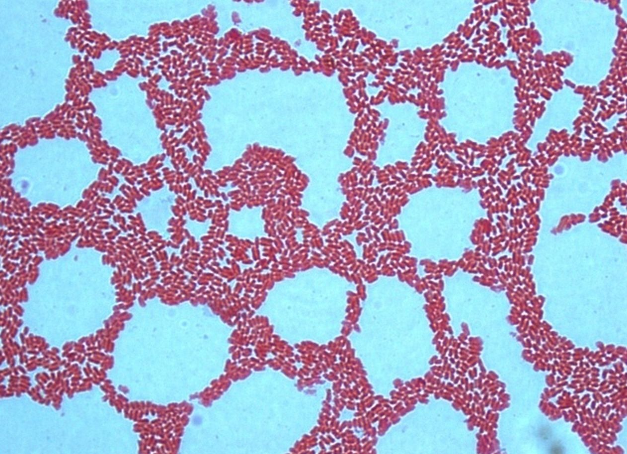
- 22—E.coli, Grams staining, pure culture
Gram stain technique
1. The heat fixed bacterial smear is flooded with crystal violet and left on for 2 minutes.
2. Then it is flooded with Lugol’s iodine (mordant) which is left on for 1 minute.
3. The smear is then decolourized with ethanol (96%) for 30 seconds up to one minute.
4. Afterwards it is flooded with safranin which is left on for 2-3 minutes.
5. Finally it is washed with water, dried and examined under oil immersion.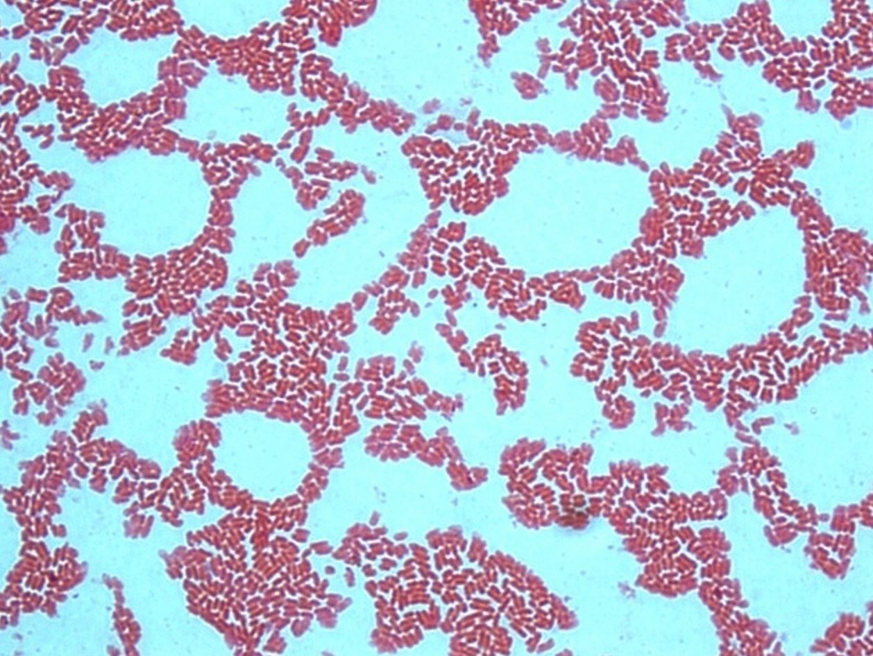
- 23—E.coli, Grams staining, pure culture
Gram stain technique
1. The heat fixed bacterial smear is flooded with crystal violet and left on for 2 minutes.
2. Then it is flooded with Lugol’s iodine (mordant) which is left on for 1 minute.
3. The smear is then decolourized with ethanol (96%) for 30 seconds up to one minute.
4. Afterwards it is flooded with safranin which is left on for 2-3 minutes.
5. Finally it is washed with water, dried and examined under oil immersion.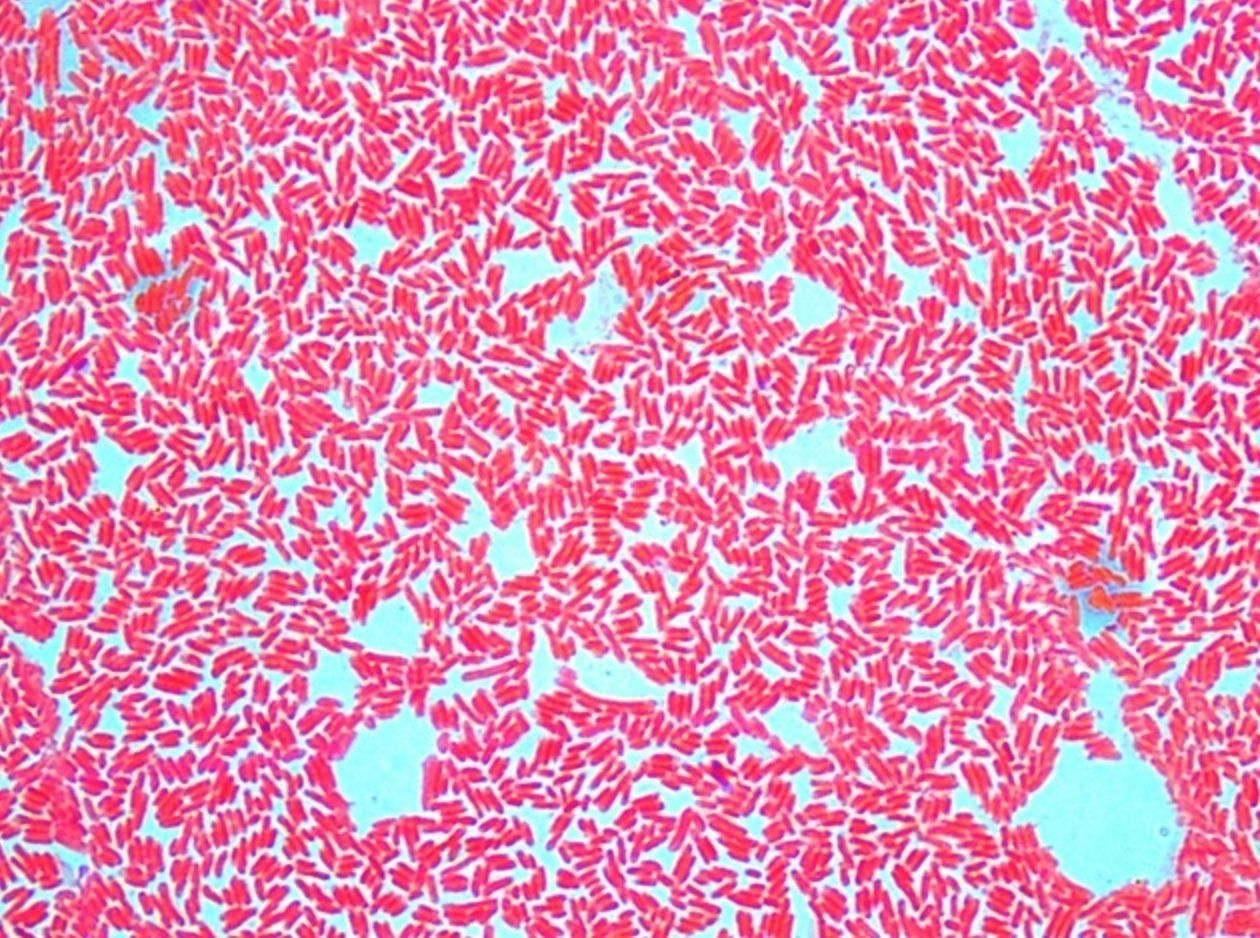
- 24—E.coli, Grams staining, pure culture
Gram stain technique
1. The heat fixed bacterial smear is flooded with crystal violet and left on for 2 minutes.
2. Then it is flooded with Lugol’s iodine (mordant) which is left on for 1 minute.
3. The smear is then decolourized with ethanol (96%) for 30 seconds up to one minute.
4. Afterwards it is flooded with safranin which is left on for 2-3 minutes.
5. Finally it is washed with water, dried and examined under oil immersion.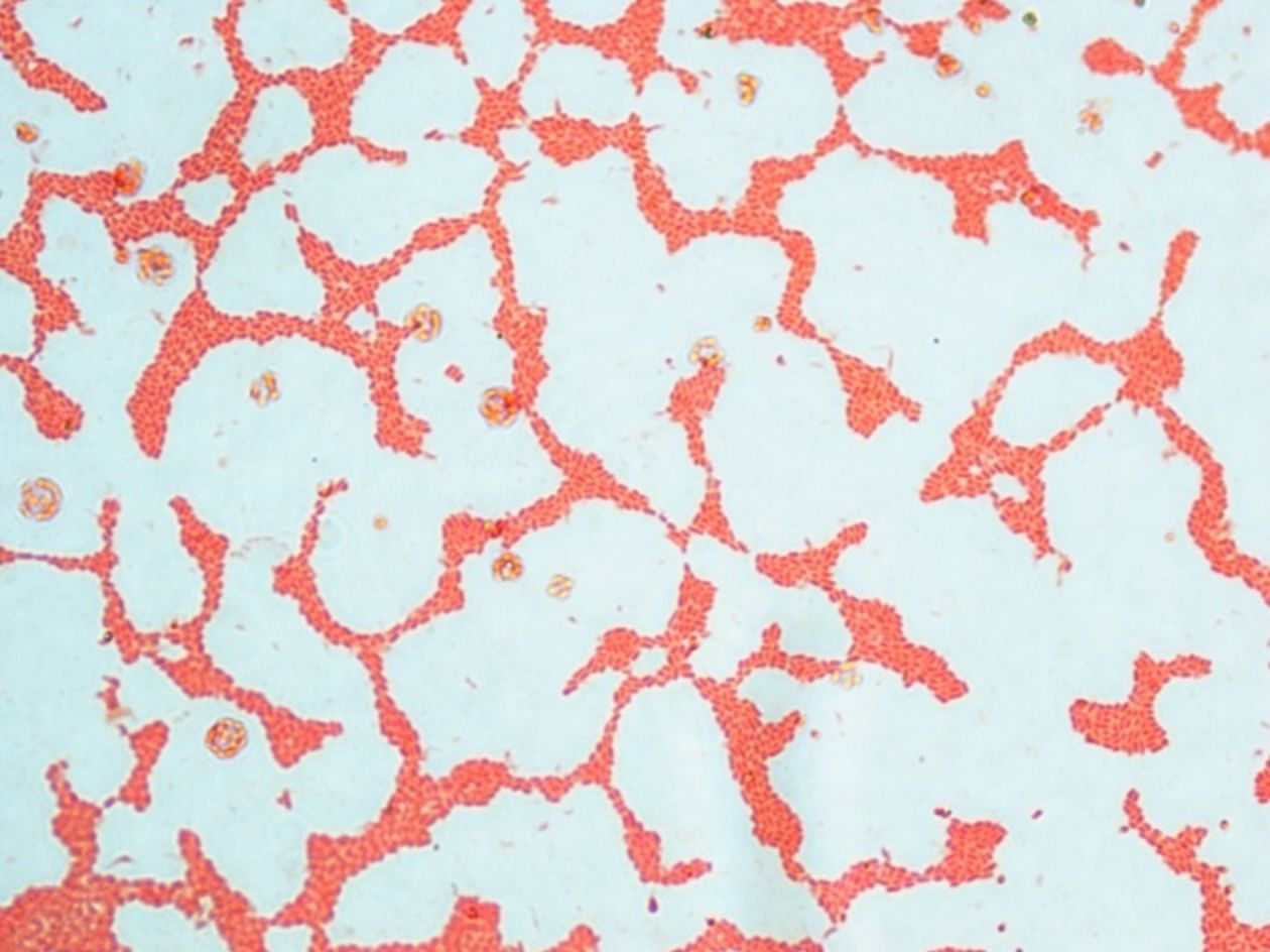
- 25—E.coli, Grams staining, pure culture
Gram stain technique
1. The heat fixed bacterial smear is flooded with crystal violet and left on for 2 minutes.
2. Then it is flooded with Lugol’s iodine (mordant) which is left on for 1 minute.
3. The smear is then decolourized with ethanol (96%) for 30 seconds up to one minute.
4. Afterwards it is flooded with safranin which is left on for 2-3 minutes.
5. Finally it is washed with water, dried and examined under oil immersion.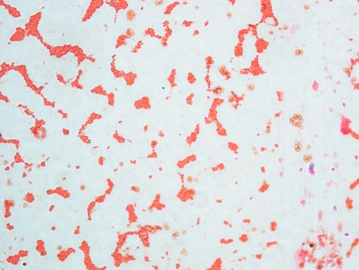
- 26—E.coli, Grams staining, pure culture
Gram stain technique
1. The heat fixed bacterial smear is flooded with crystal violet and left on for 2 minutes.
2. Then it is flooded with Lugol’s iodine (mordant) which is left on for 1 minute.
3. The smear is then decolourized with ethanol (96%) for 30 seconds up to one minute.
4. Afterwards it is flooded with safranin which is left on for 2-3 minutes.
5. Finally it is washed with water, dried and examined under oil immersion.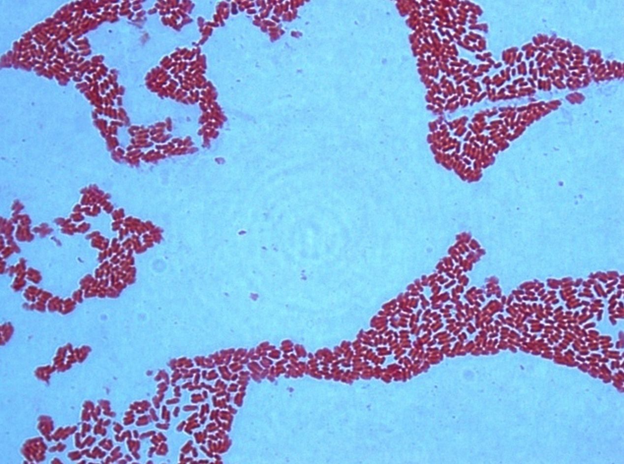
- 27—E.coli, Grams staining, pure culture
Gram stain technique
1. The heat fixed bacterial smear is flooded with crystal violet and left on for 2 minutes.
2. Then it is flooded with Lugol’s iodine (mordant) which is left on for 1 minute.
3. The smear is then decolourized with ethanol (96%) for 30 seconds up to one minute.
4. Afterwards it is flooded with safranin which is left on for 2-3 minutes.
5. Finally it is washed with water, dried and examined under oil immersion.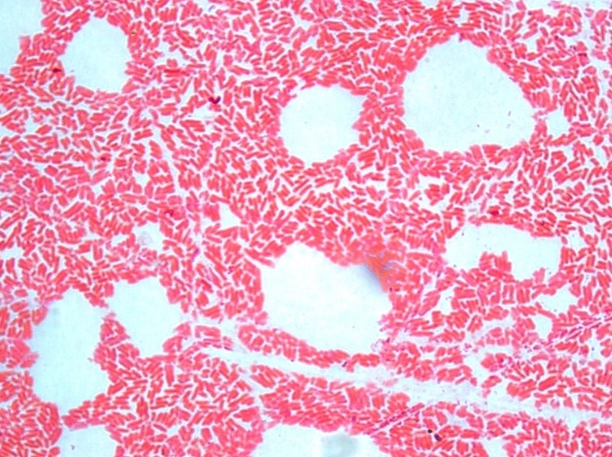
- 28—E.coli et Staphylococcus, Grams staining, mixed culture
Gram stain technique
1. The heat fixed bacterial smear is flooded with crystal violet and left on for 2 minutes.
2. Then it is flooded with Lugol’s iodine (mordant) which is left on for 1 minute.
3. The smear is then decolourized with ethanol (96%) for 30 seconds up to one minute.
4. Afterwards it is flooded with safranin which is left on for 2-3 minutes.
5. Finally it is washed with water, dried and examined under oil immersion.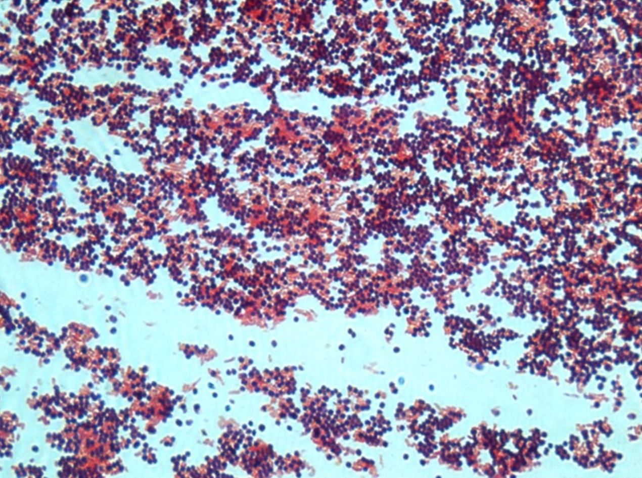
- 29—E.coli et Staphylococcus, Grams staining, mixed culture
Gram stain technique
1. The heat fixed bacterial smear is flooded with crystal violet and left on for 2 minutes.
2. Then it is flooded with Lugol’s iodine (mordant) which is left on for 1 minute.
3. The smear is then decolourized with ethanol (96%) for 30 seconds up to one minute.
4. Afterwards it is flooded with safranin which is left on for 2-3 minutes.
5. Finally it is washed with water, dried and examined under oil immersion.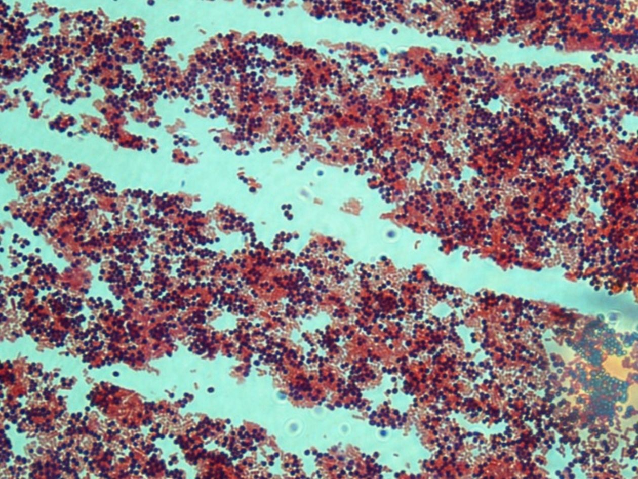
- 30—E.coli, Grams staining, pure culture
Gram stain technique
1. The heat fixed bacterial smear is flooded with crystal violet and left on for 2 minutes.
2. Then it is flooded with Lugol’s iodine (mordant) which is left on for 1 minute.
3. The smear is then decolourized with ethanol (96%) for 30 seconds up to one minute.
4. Afterwards it is flooded with safranin which is left on for 2-3 minutes.
5. Finally it is washed with water, dried and examined under oil immersion.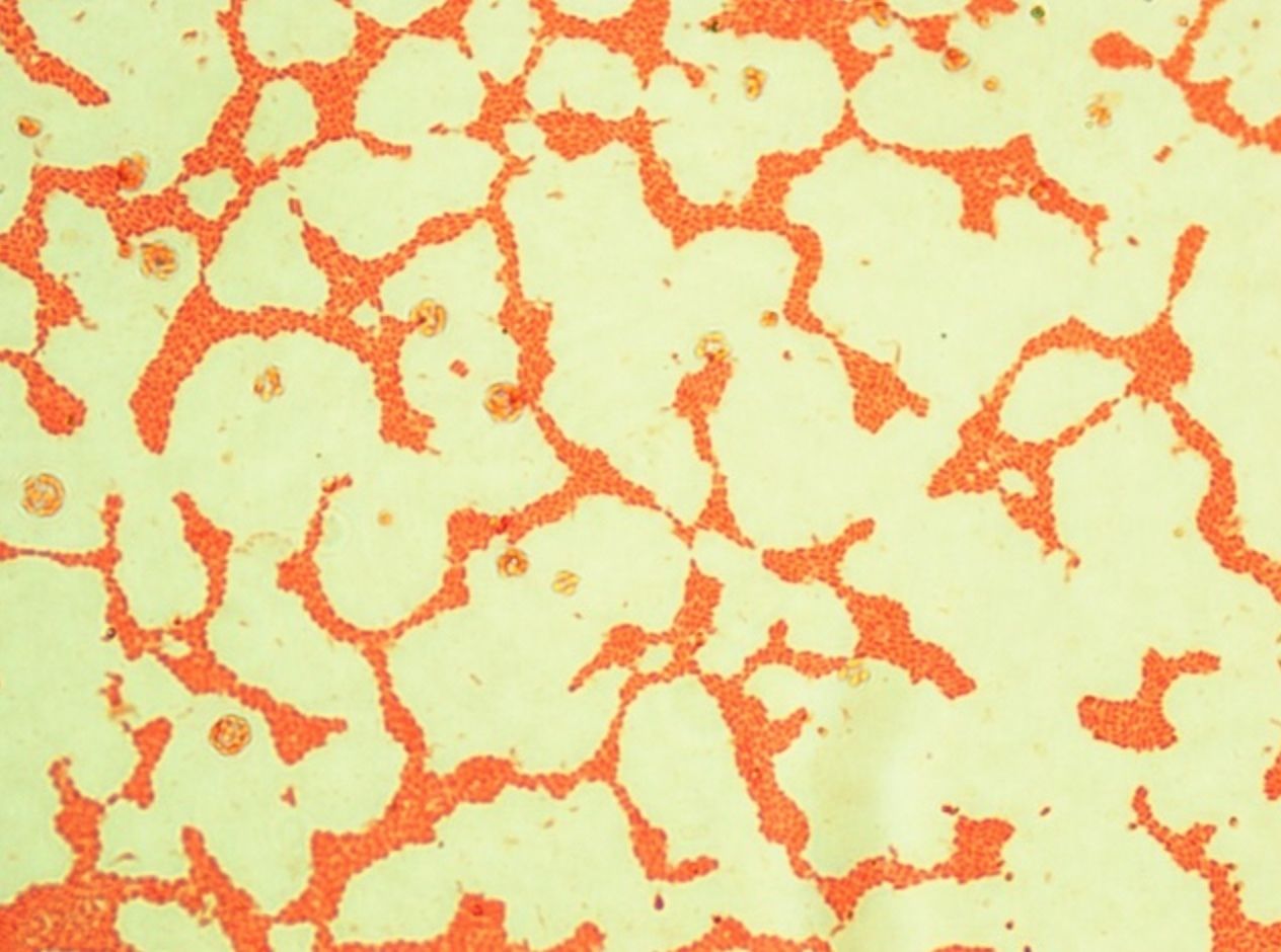
- 31—E.coli, Grams staining, pure culture
Gram stain technique
1. The heat fixed bacterial smear is flooded with crystal violet and left on for 2 minutes.
2. Then it is flooded with Lugol’s iodine (mordant) which is left on for 1 minute.
3. The smear is then decolourized with ethanol (96%) for 30 seconds up to one minute.
4. Afterwards it is flooded with safranin which is left on for 2-3 minutes.
5. Finally it is washed with water, dried and examined under oil immersion.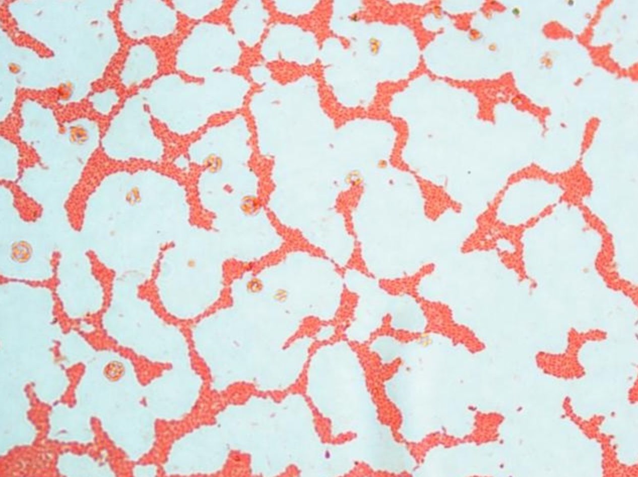
- 32—Bacillus subtilis (endospores), methylene blue, pure culture
Simple staining
Fixed smears are stained using neutral and basic aniline dyes.
Only one dye is used, for example, methylene blue, safranin, etc.
All bacteria will stain the same colour, however it will be possible to evaluate the shape, size and arrangement of bacteria.
The slide is placed on a stand over the container and flooded with methylene blue. The dye should be left on for 5 minutes. When using safranin, the dye should be left on for 4 minutes.
Afterwards the slide is washed with water and dried. Only dry slides are examined under oil immersion.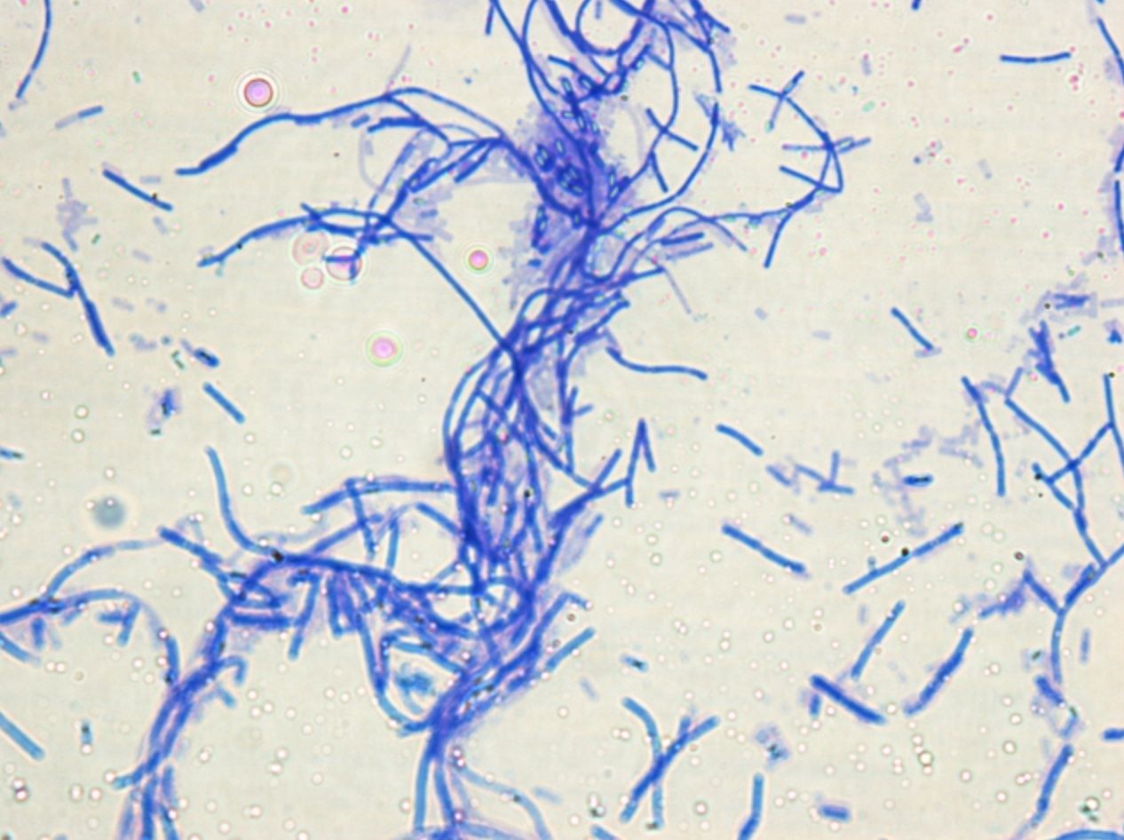
- 33—Bacillus subtilis (endospores), methylene blue, pure culture
Simple staining
Fixed smears are stained using neutral and basic aniline dyes.
Only one dye is used, for example, methylene blue, safranin, etc.
All bacteria will stain the same colour, however it will be possible to evaluate the shape, size and arrangement of bacteria.
The slide is placed on a stand over the container and flooded with methylene blue. The dye should be left on for 5 minutes. When using safranin, the dye should be left on for 4 minutes.
Afterwards the slide is washed with water and dried. Only dry slides are examined under oil immersion.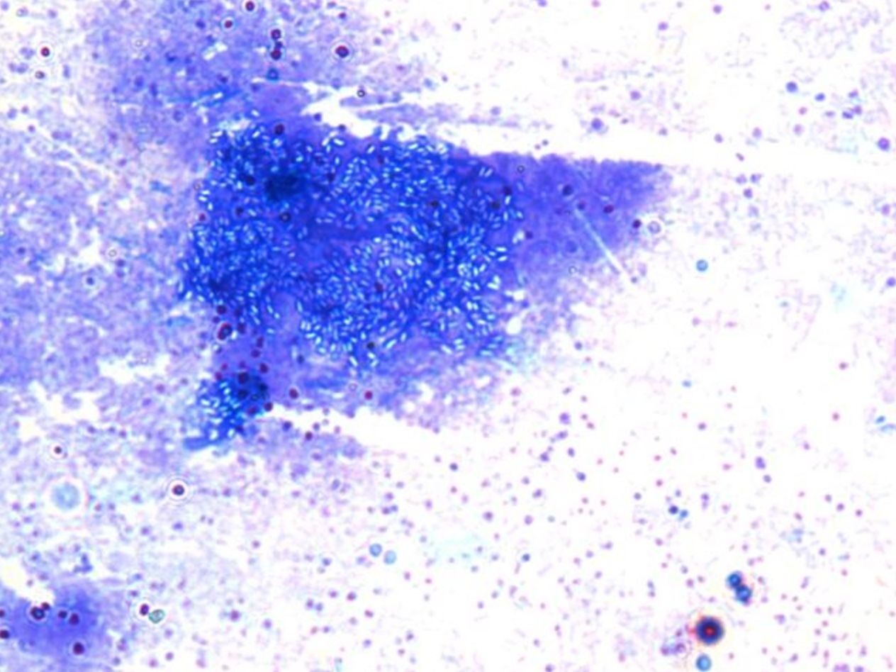
- 34—Bacillus subtilis (endospores), methylene blue, pure culture
Simple staining
Fixed smears are stained using neutral and basic aniline dyes.
Only one dye is used, for example, methylene blue, safranin, etc.
All bacteria will stain the same colour, however it will be possible to evaluate the shape, size and arrangement of bacteria.
The slide is placed on a stand over the container and flooded with methylene blue. The dye should be left on for 5 minutes. When using safranin, the dye should be left on for 4 minutes.
Afterwards the slide is washed with water and dried. Only dry slides are examined under oil immersion.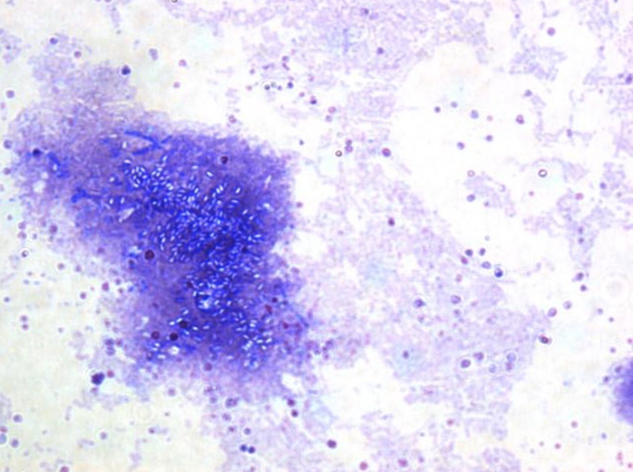
- 35—Klebsiella pneumoniae, Giemsa (Gin) staining, pure culture
The Gin Stain is used to visualize bacterial capsules. As a result the background stains dark brown, bacteria stain red/pink but the capsule does not stain and is visible as a light area around the red/pink bacterium.
1) Thin film of the specimen is inoculated on a microscope slide and fixed with methanol for 30 seconds, by immersing it or by putting a few drops of methanol on the slide.
2) Slide is immersed in 5% Giemsa stain solution for 20–30 minutes.
3) Rinse with water and dry.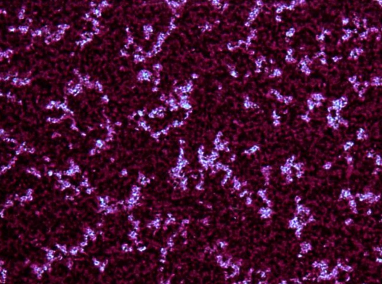
- 36—Klebsiella pneumoniae, Giemsa (Gin) staining, pure culture
The Gin Stain is used to visualize bacterial capsules. As a result the background stains dark brown, bacteria stain red/pink but the capsule does not stain and is visible as a light area around the red/pink bacterium.
1) Thin film of the specimen is inoculated on a microscope slide and fixed with methanol for 30 seconds, by immersing it or by putting a few drops of methanol on the slide.
2) Slide is immersed in 5% Giemsa stain solution for 20–30 minutes.
3) Rinse with water and dry.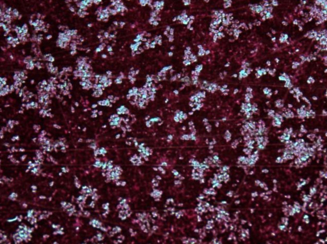
- 37—Klebsiella pneumoniae, Giemsa (Gin) staining, pure culture
The Gin Stain is used to visualize bacterial capsules. As a result the background stains dark brown, bacteria stain red/pink but the capsule does not stain and is visible as a light area around the red/pink bacterium.
1) Thin film of the specimen is inoculated on a microscope slide and fixed with methanol for 30 seconds, by immersing it or by putting a few drops of methanol on the slide.
2) Slide is immersed in 5% Giemsa stain solution for 20–30 minutes.
3) Rinse with water and dry.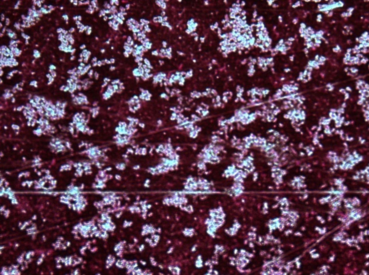
- 38—Myobacterium tuberculosis, acid-fast
staining (Ziehl-Neelsen) patient's sample
The cell walls of acid-fast bacteria contain high concentration of lipids that after being stained with concentrated carbol fuchsin do not decolourize with an acid or an alcohol-acid solution (they stain red). Other bacteria stain blue.
1. A filter paper is placed on a heat fixed bacterial smear which is then flooded with a carbol fuchsin stain. The smear is heated over the flame of an alcohol lamp until vapour appears. The heating process is repeated two more times.
2. The filter paper is taken off, the slide is washed with water and decolourized with a 5% sulphuric acid solution or an ethanol and hydrochloric acid mix (100 ml of ethanol (96%), 3 ml of hydrochloric acid (25%)) until the smear decolourizes.
3. The smear is washed with water.
4. Then it is flooded with methylene blue that is left on for 2 minutes.
5. Finally the smear is washed with water, dried and examined under oil immersion.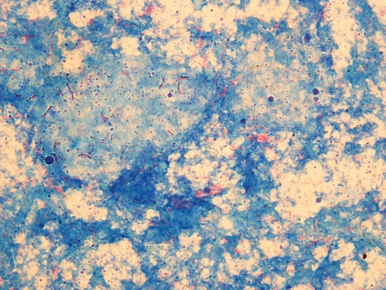
- 39—Myobacterium tuberculosis, acid-fast
staining, patient's sample
The cell walls of acid-fast bacteria contain high concentration of lipids that after being stained with concentrated carbol fuchsin do not decolourize with an acid or an alcohol-acid solution (they stain red). Other bacteria stain blue.
1. A filter paper is placed on a heat fixed bacterial smear which is then flooded with a carbol fuchsin stain. The smear is heated over the flame of an alcohol lamp until vapour appears. The heating process is repeated two more times.
2. The filter paper is taken off, the slide is washed with water and decolourized with a 5% sulphuric acid solution or an ethanol and hydrochloric acid mix (100 ml of ethanol (96%), 3 ml of hydrochloric acid (25%)) until the smear decolourizes.
3. The smear is washed with water.
4. Then it is flooded with methylene blue that is left on for 2 minutes.
5. Finally the smear is washed with water, dried and examined under oil immersion.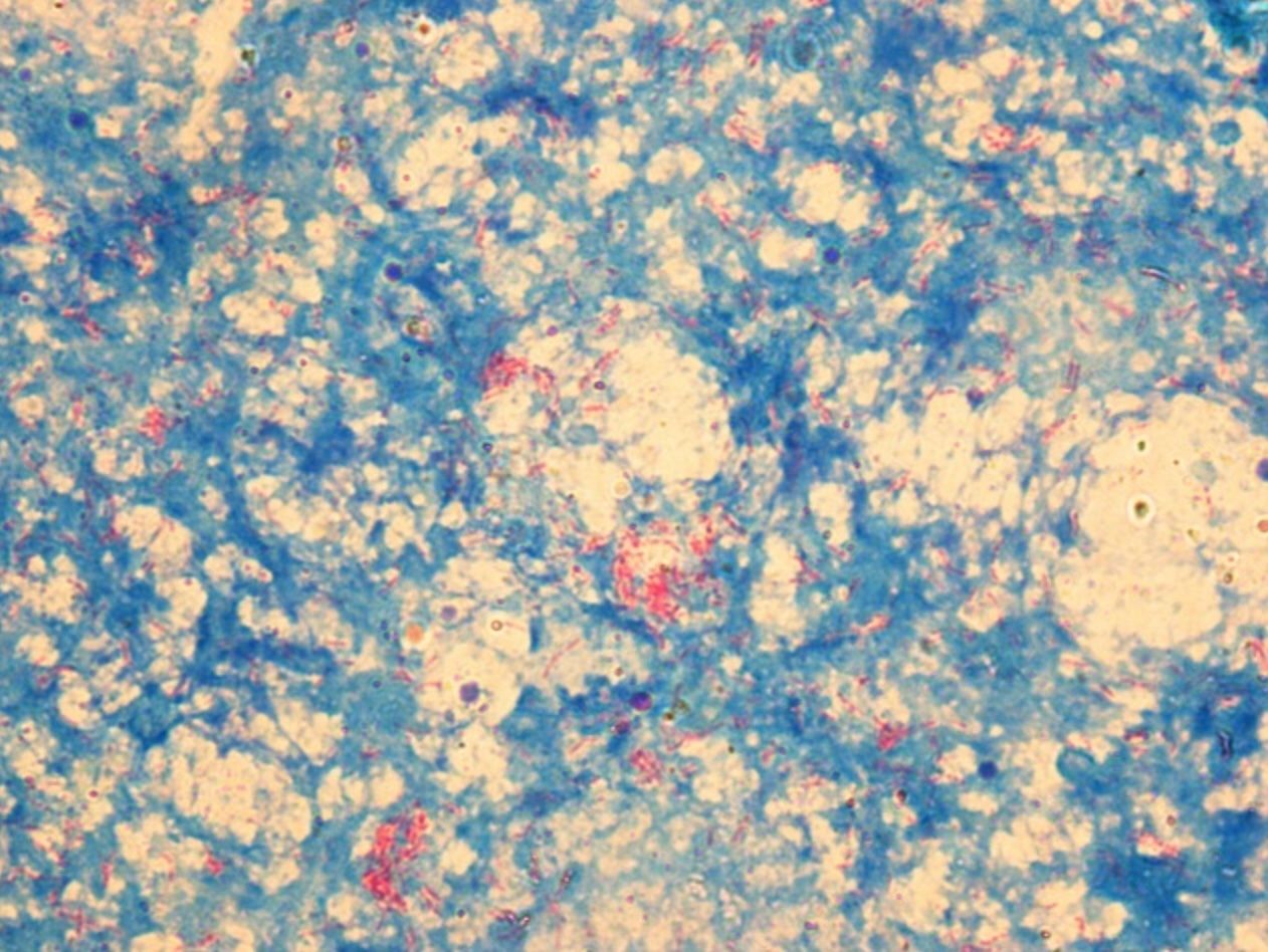
- 40—Myobacterium tuberculosis, acid-fast
staining, patient's sample
The cell walls of acid-fast bacteria contain high concentration of lipids that after being stained with concentrated carbol fuchsin do not decolourize with an acid or an alcohol-acid solution (they stain red). Other bacteria stain blue.
1. A filter paper is placed on a heat fixed bacterial smear which is then flooded with a carbol fuchsin stain. The smear is heated over the flame of an alcohol lamp until vapour appears. The heating process is repeated two more times.
2. The filter paper is taken off, the slide is washed with water and decolourized with a 5% sulphuric acid solution or an ethanol and hydrochloric acid mix (100 ml of ethanol (96%), 3 ml of hydrochloric acid (25%)) until the smear decolourizes.
3. The smear is washed with water.
4. Then it is flooded with methylene blue that is left on for 2 minutes.
5. Finally the smear is washed with water, dried and examined under oil immersion.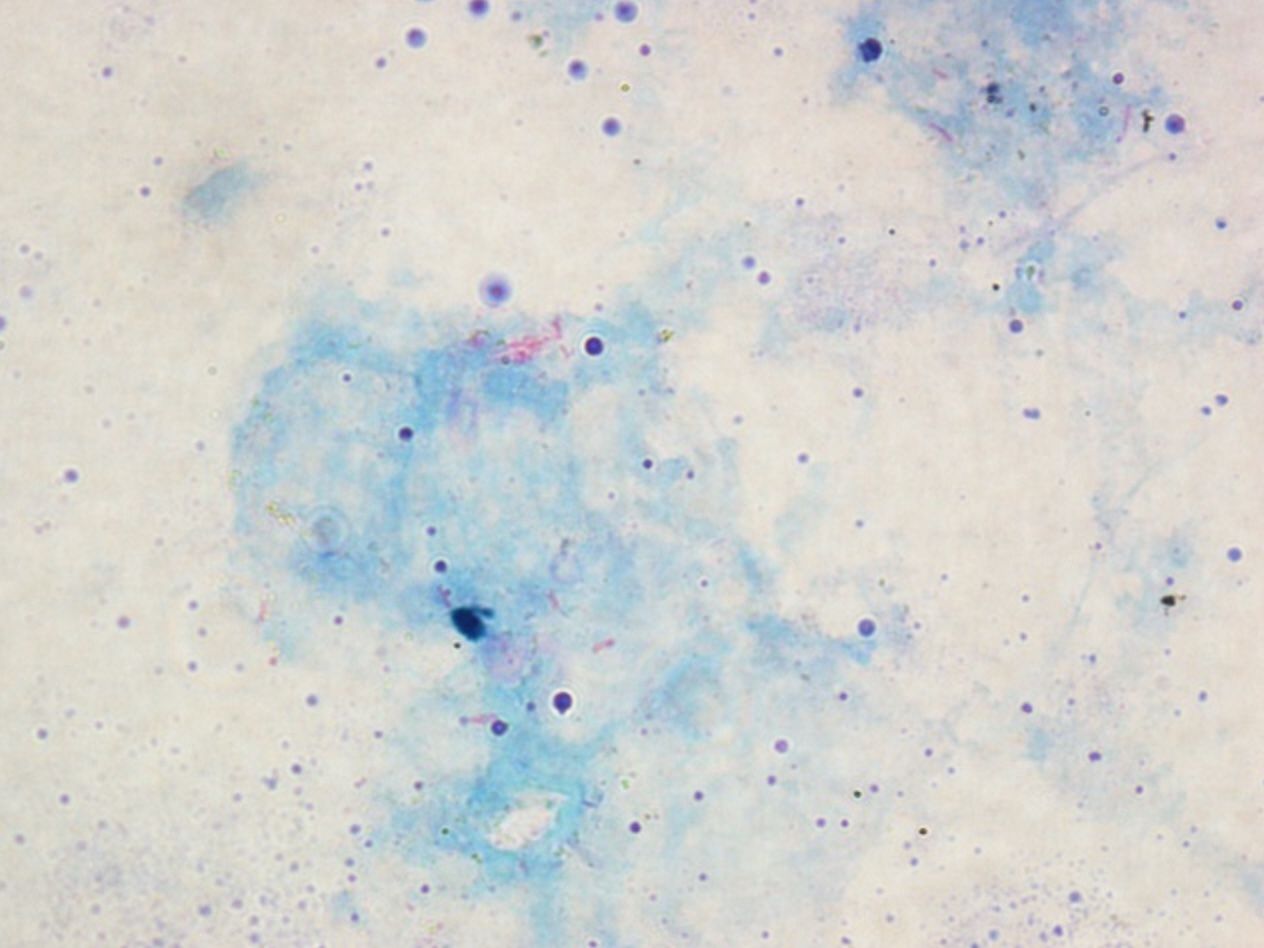
- 41—Myobacterium tuberculosis, acid-fast
staining, patient's sample
The cell walls of acid-fast bacteria contain high concentration of lipids that after being stained with concentrated carbol fuchsin do not decolourize with an acid or an alcohol-acid solution (they stain red). Other bacteria stain blue.
1. A filter paper is placed on a heat fixed bacterial smear which is then flooded with a carbol fuchsin stain. The smear is heated over the flame of an alcohol lamp until vapour appears. The heating process is repeated two more times.
2. The filter paper is taken off, the slide is washed with water and decolourized with a 5% sulphuric acid solution or an ethanol and hydrochloric acid mix (100 ml of ethanol (96%), 3 ml of hydrochloric acid (25%)) until the smear decolourizes.
3. The smear is washed with water.
4. Then it is flooded with methylene blue that is left on for 2 minutes.
5. Finally the smear is washed with water, dried and examined under oil immersion.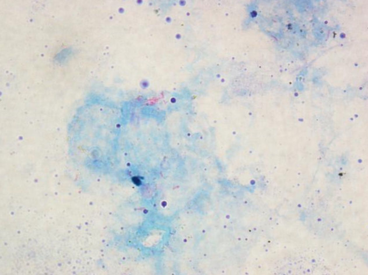
- 42—Myobacterium tuberculosis, acid-fast
staining, patient's sample
The cell walls of acid-fast bacteria contain high concentration of lipids that after being stained with concentrated carbol fuchsin do not decolourize with an acid or an alcohol-acid solution (they stain red). Other bacteria stain blue.
1. A filter paper is placed on a heat fixed bacterial smear which is then flooded with a carbol fuchsin stain. The smear is heated over the flame of an alcohol lamp until vapour appears. The heating process is repeated two more times.
2. The filter paper is taken off, the slide is washed with water and decolourized with a 5% sulphuric acid solution or an ethanol and hydrochloric acid mix (100 ml of ethanol (96%), 3 ml of hydrochloric acid (25%)) until the smear decolourizes.
3. The smear is washed with water.
4. Then it is flooded with methylene blue that is left on for 2 minutes.
5. Finally the smear is washed with water, dried and examined under oil immersion.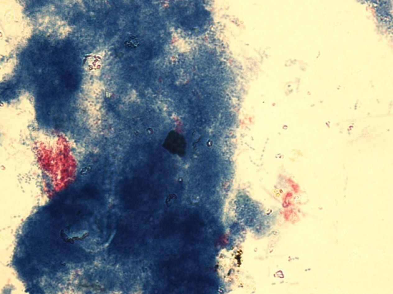
- 43—Myobacterium tuberculosis, acid-fast
staining, patient's sample
The cell walls of acid-fast bacteria contain high concentration of lipids that after being stained with concentrated carbol fuchsin do not decolourize with an acid or an alcohol-acid solution (they stain red). Other bacteria stain blue.
1. A filter paper is placed on a heat fixed bacterial smear which is then flooded with a carbol fuchsin stain. The smear is heated over the flame of an alcohol lamp until vapour appears. The heating process is repeated two more times.
2. The filter paper is taken off, the slide is washed with water and decolourized with a 5% sulphuric acid solution or an ethanol and hydrochloric acid mix (100 ml of ethanol (96%), 3 ml of hydrochloric acid (25%)) until the smear decolourizes.
3. The smear is washed with water.
4. Then it is flooded with methylene blue that is left on for 2 minutes.
5. Finally the smear is washed with water, dried and examined under oil immersion.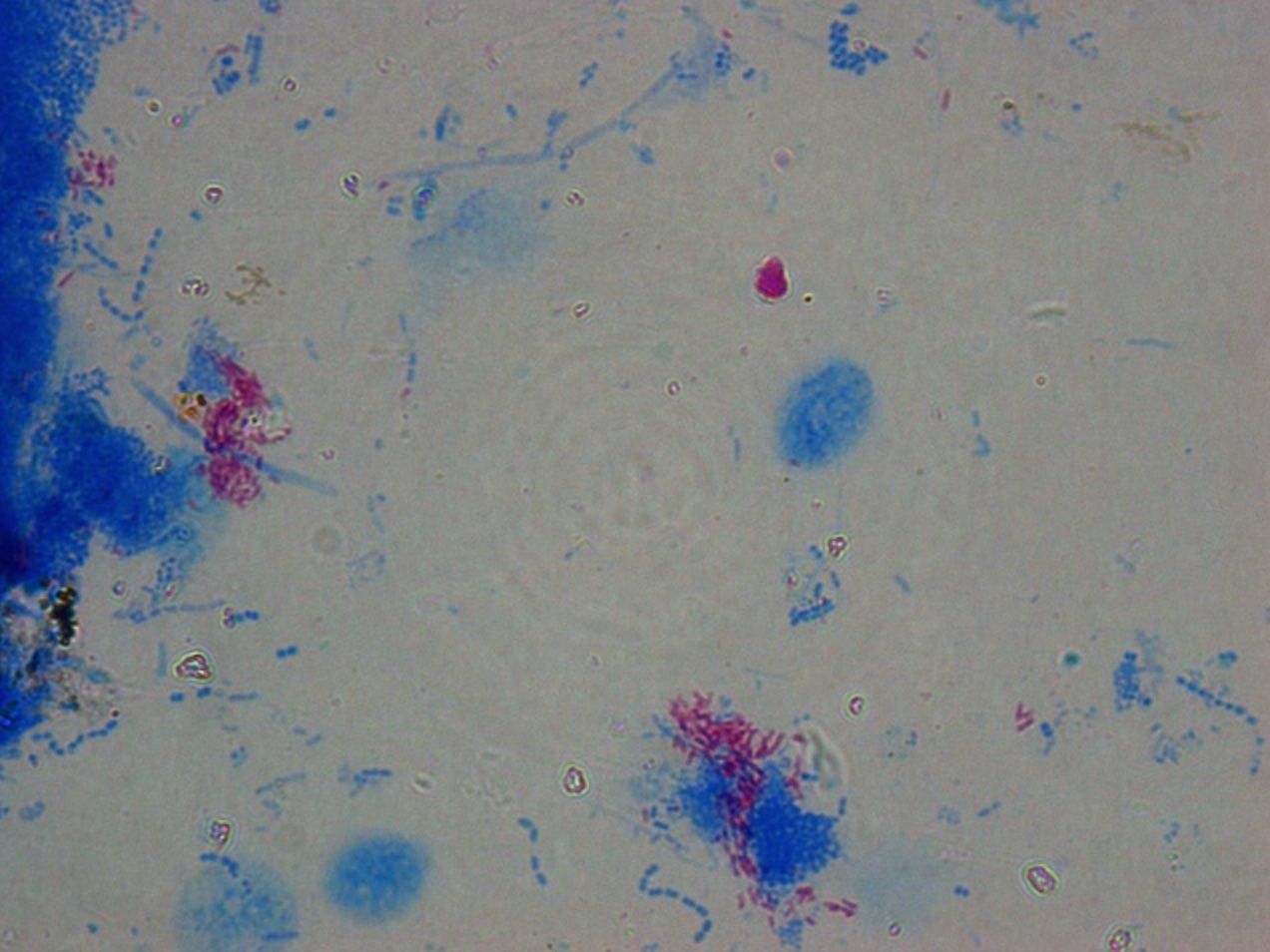
- 44—Myobacterium tuberculosis, acid-fast
staining, patient's sample
The cell walls of acid-fast bacteria contain high concentration of lipids that after being stained with concentrated carbol fuchsin do not decolourize with an acid or an alcohol-acid solution (they stain red). Other bacteria stain blue.
1. A filter paper is placed on a heat fixed bacterial smear which is then flooded with a carbol fuchsin stain. The smear is heated over the flame of an alcohol lamp until vapour appears. The heating process is repeated two more times.
2. The filter paper is taken off, the slide is washed with water and decolourized with a 5% sulphuric acid solution or an ethanol and hydrochloric acid mix (100 ml of ethanol (96%), 3 ml of hydrochloric acid (25%)) until the smear decolourizes.
3. The smear is washed with water.
4. Then it is flooded with methylene blue that is left on for 2 minutes.
5. Finally the smear is washed with water, dried and examined under oil immersion.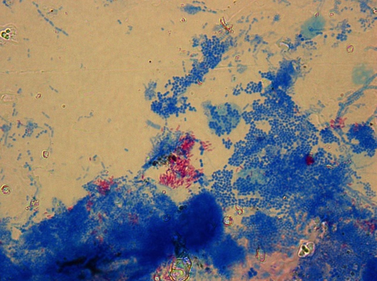
- 45—Myobacterium tuberculosis, acid-fast
staining, patient's sample
The cell walls of acid-fast bacteria contain high concentration of lipids that after being stained with concentrated carbol fuchsin do not decolourize with an acid or an alcohol-acid solution (they stain red). Other bacteria stain blue.
1. A filter paper is placed on a heat fixed bacterial smear which is then flooded with a carbol fuchsin stain. The smear is heated over the flame of an alcohol lamp until vapour appears. The heating process is repeated two more times.
2. The filter paper is taken off, the slide is washed with water and decolourized with a 5% sulphuric acid solution or an ethanol and hydrochloric acid mix (100 ml of ethanol (96%), 3 ml of hydrochloric acid (25%)) until the smear decolourizes.
3. The smear is washed with water.
4. Then it is flooded with methylene blue that is left on for 2 minutes.
5. Finally the smear is washed with water, dried and examined under oil immersion.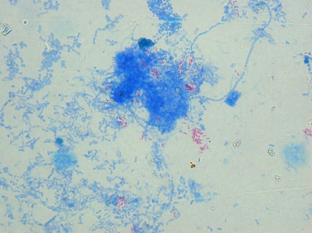
- 46—Saccharomyces, methylene blue, pure culture
Simple staining
Fixed smears are stained using neutral and basic aniline dyes.
Only one dye is used, for example, methylene blue, safranin, etc.
All bacteria will stain the same colour, however it will be possible to evaluate the shape, size and arrangement of bacteria.
The slide is placed on a stand over the container and flooded with methylene blue. The dye should be left on for 5 minutes. When using safranin, the dye should be left on for 4 minutes.
Afterwards the slide is washed with water and dried. Only dry slides are examined under oil immersion.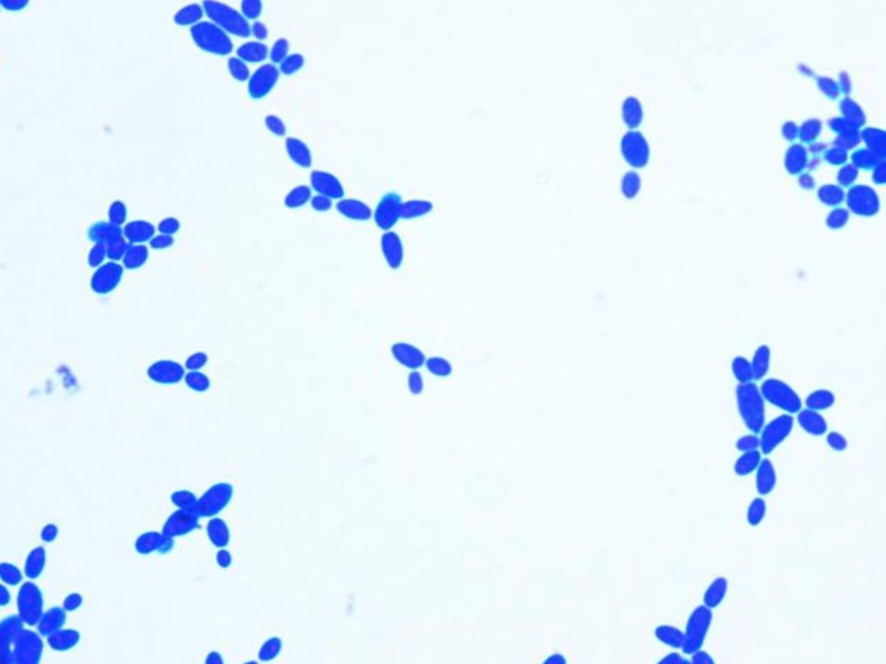
- 47—Saccharomyces, methylene blue, pure culture
Simple staining
Fixed smears are stained using neutral and basic aniline dyes.
Only one dye is used, for example, methylene blue, safranin, etc.
All bacteria will stain the same colour, however it will be possible to evaluate the shape, size and arrangement of bacteria.
The slide is placed on a stand over the container and flooded with methylene blue. The dye should be left on for 5 minutes. When using safranin, the dye should be left on for 4 minutes.
Afterwards the slide is washed with water and dried. Only dry slides are examined under oil immersion.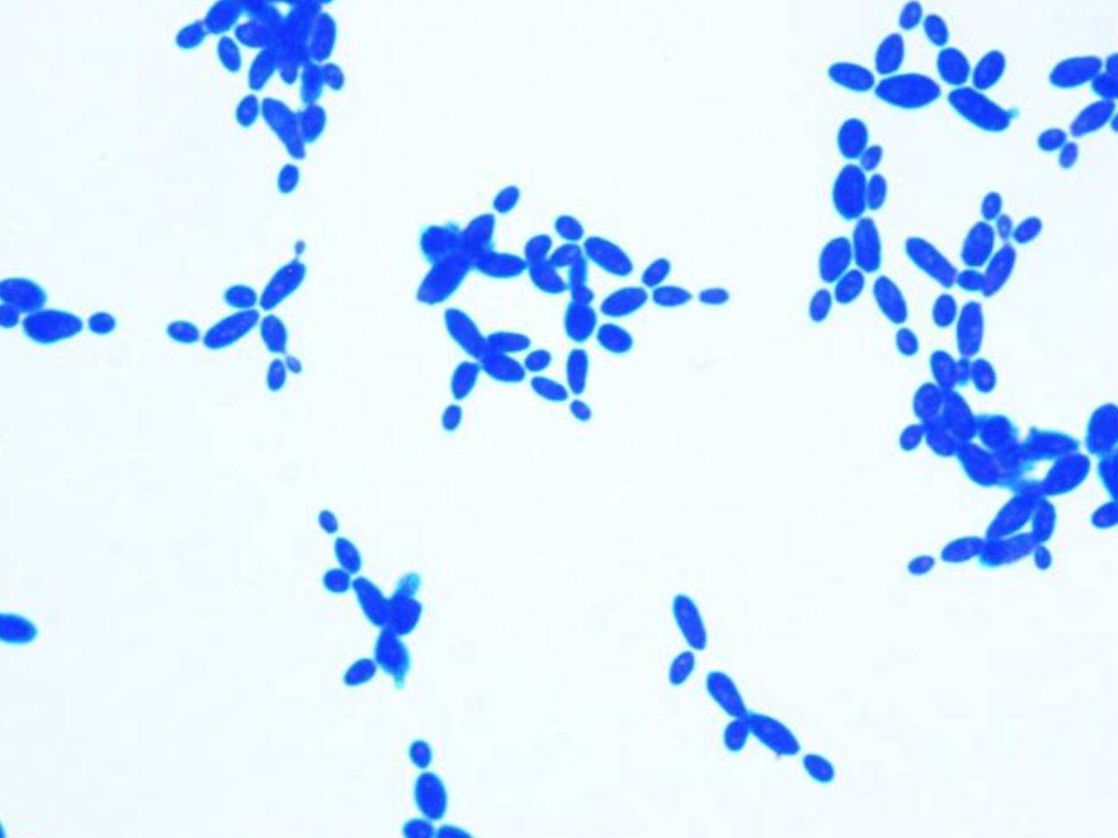
- 48—Saccharomyces, methylene blue, pure culture
Simple staining
Fixed smears are stained using neutral and basic aniline dyes.
Only one dye is used, for example, methylene blue, safranin, etc.
All bacteria will stain the same colour, however it will be possible to evaluate the shape, size and arrangement of bacteria.
The slide is placed on a stand over the container and flooded with methylene blue. The dye should be left on for 5 minutes. When using safranin, the dye should be left on for 4 minutes.
Afterwards the slide is washed with water and dried. Only dry slides are examined under oil immersion.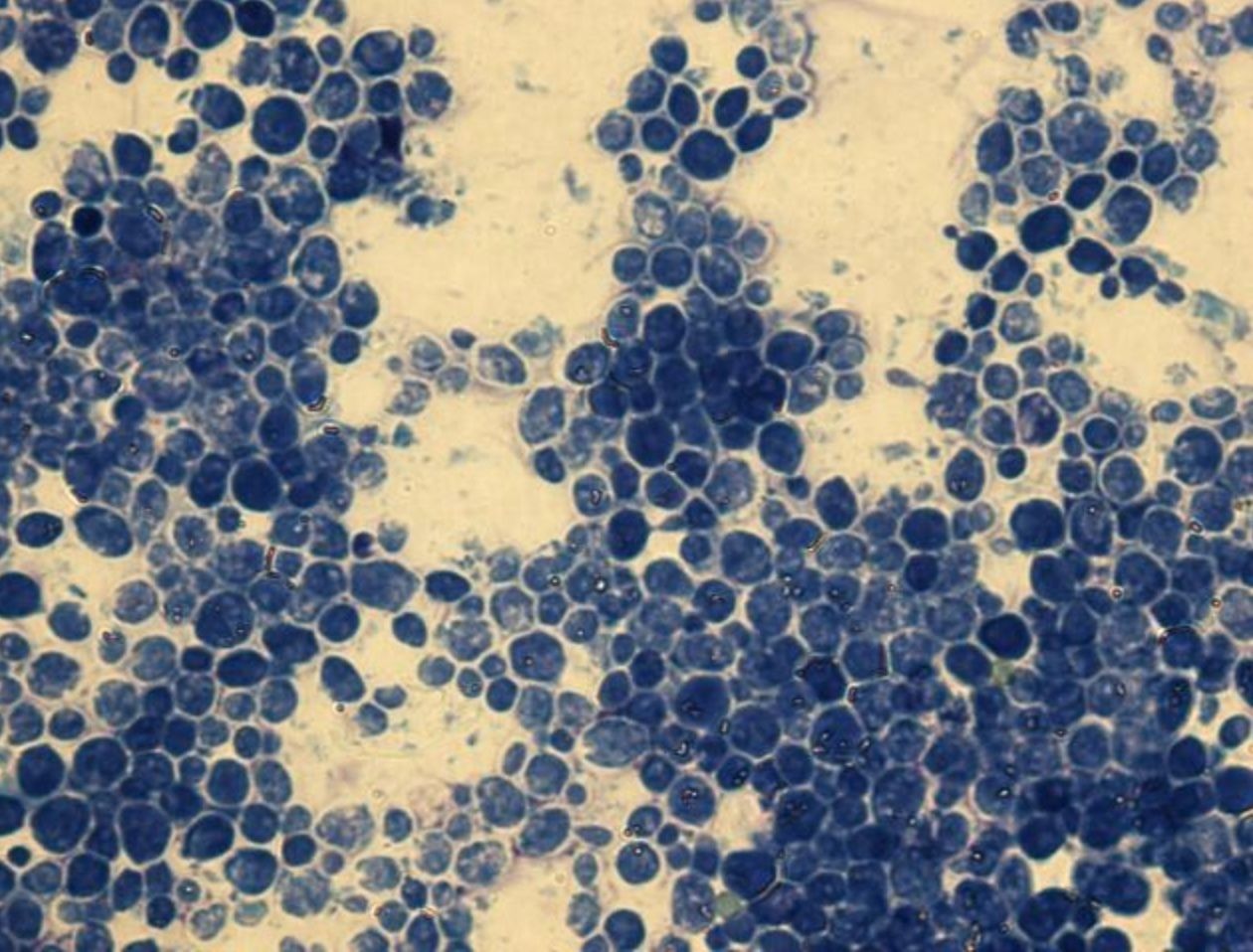
- 49—Sarcina, methylene blue, pure culture
Simple staining
Fixed smears are stained using neutral and basic aniline dyes.
Only one dye is used, for example, methylene blue, safranin, etc.
All bacteria will stain the same colour, however it will be possible to evaluate the shape, size and arrangement of bacteria.
The slide is placed on a stand over the container and flooded with methylene blue. The dye should be left on for 5 minutes. When using safranin, the dye should be left on for 4 minutes.
Afterwards the slide is washed with water and dried. Only dry slides are examined under oil immersion.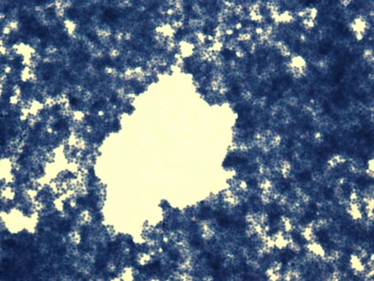
- 50—Sarcina, methylene blue, pure culture
Simple staining
Fixed smears are stained using neutral and basic aniline dyes.
Only one dye is used, for example, methylene blue, safranin, etc.
All bacteria will stain the same colour, however it will be possible to evaluate the shape, size and arrangement of bacteria.
The slide is placed on a stand over the container and flooded with methylene blue. The dye should be left on for 5 minutes. When using safranin, the dye should be left on for 4 minutes.
Afterwards the slide is washed with water and dried. Only dry slides are examined under oil immersion.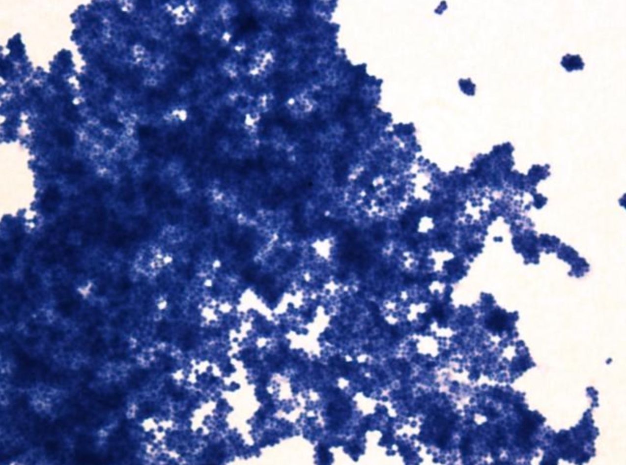
- 51—Sarcina, methylene blue, pure culture
Simple staining
Fixed smears are stained using neutral and basic aniline dyes.
Only one dye is used, for example, methylene blue, safranin, etc.
All bacteria will stain the same colour, however it will be possible to evaluate the shape, size and arrangement of bacteria.
The slide is placed on a stand over the container and flooded with methylene blue. The dye should be left on for 5 minutes. When using safranin, the dye should be left on for 4 minutes.
Afterwards the slide is washed with water and dried. Only dry slides are examined under oil immersion.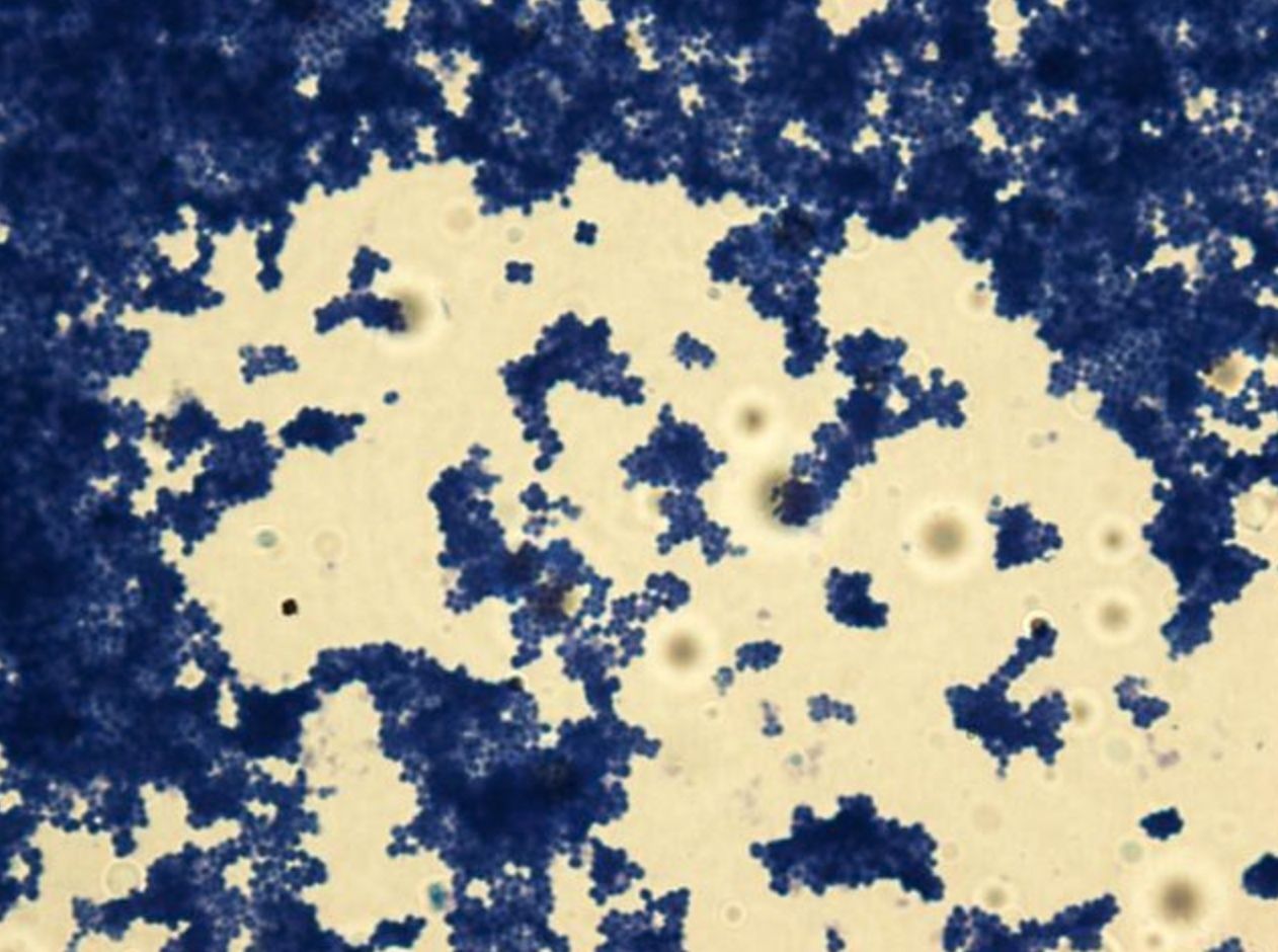
- 52—Sarcina, methylene blue, pure culture
Simple staining
Fixed smears are stained using neutral and basic aniline dyes.
Only one dye is used, for example, methylene blue, safranin, etc.
All bacteria will stain the same colour, however it will be possible to evaluate the shape, size and arrangement of bacteria.
The slide is placed on a stand over the container and flooded with methylene blue. The dye should be left on for 5 minutes. When using safranin, the dye should be left on for 4 minutes.
Afterwards the slide is washed with water and dried. Only dry slides are examined under oil immersion.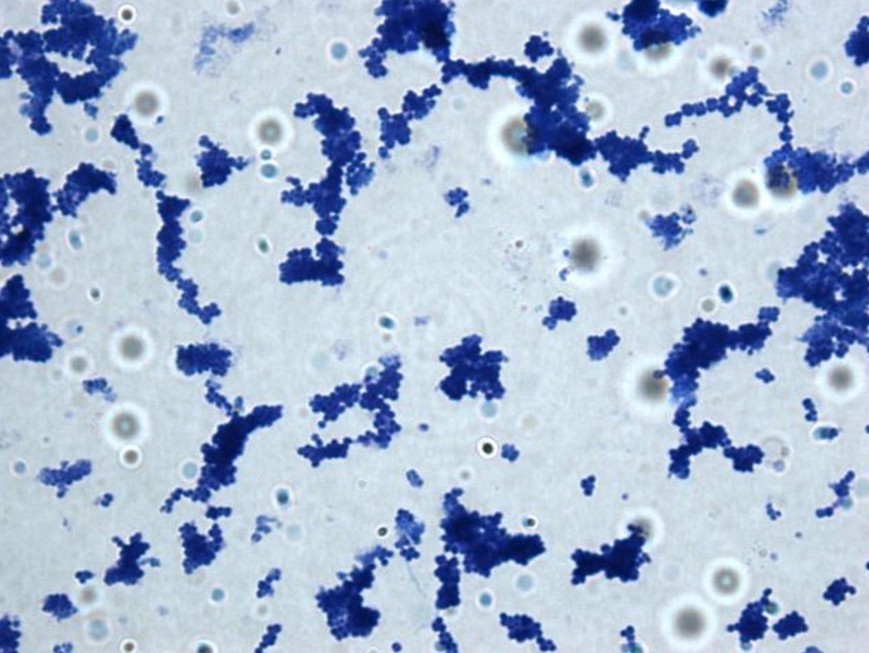
- 53—Sarcina, methylene blue, pure culture
Simple staining
Fixed smears are stained using neutral and basic aniline dyes.
Only one dye is used, for example, methylene blue, safranin, etc.
All bacteria will stain the same colour, however it will be possible to evaluate the shape, size and arrangement of bacteria.
The slide is placed on a stand over the container and flooded with methylene blue. The dye should be left on for 5 minutes. When using safranin, the dye should be left on for 4 minutes.
Afterwards the slide is washed with water and dried. Only dry slides are examined under oil immersion.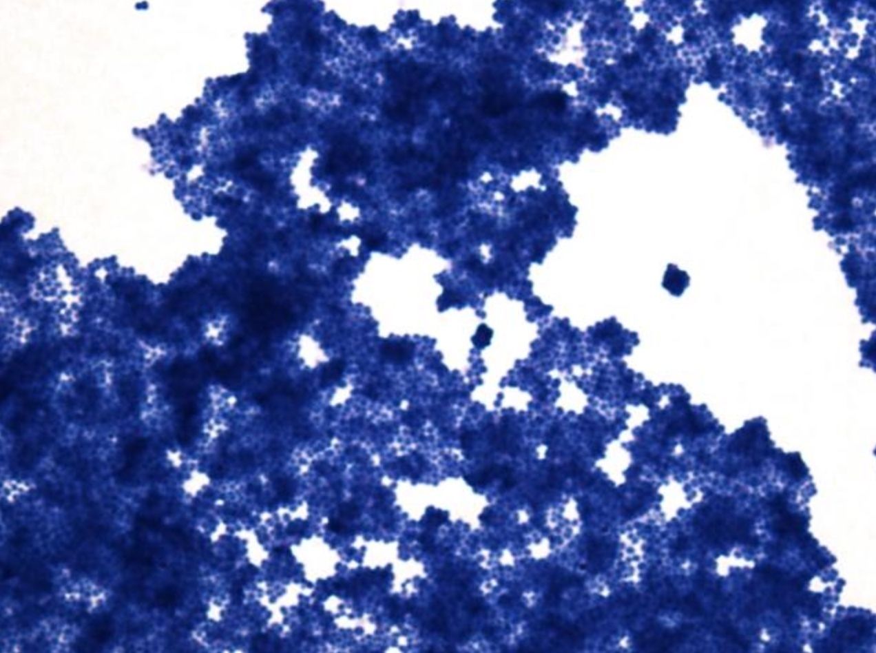
- 54—Sarcina, methylene blue, pure culture
Simple staining
Fixed smears are stained using neutral and basic aniline dyes.
Only one dye is used, for example, methylene blue, safranin, etc.
All bacteria will stain the same colour, however it will be possible to evaluate the shape, size and arrangement of bacteria.
The slide is placed on a stand over the container and flooded with methylene blue. The dye should be left on for 5 minutes. When using safranin, the dye should be left on for 4 minutes.
Afterwards the slide is washed with water and dried. Only dry slides are examined under oil immersion.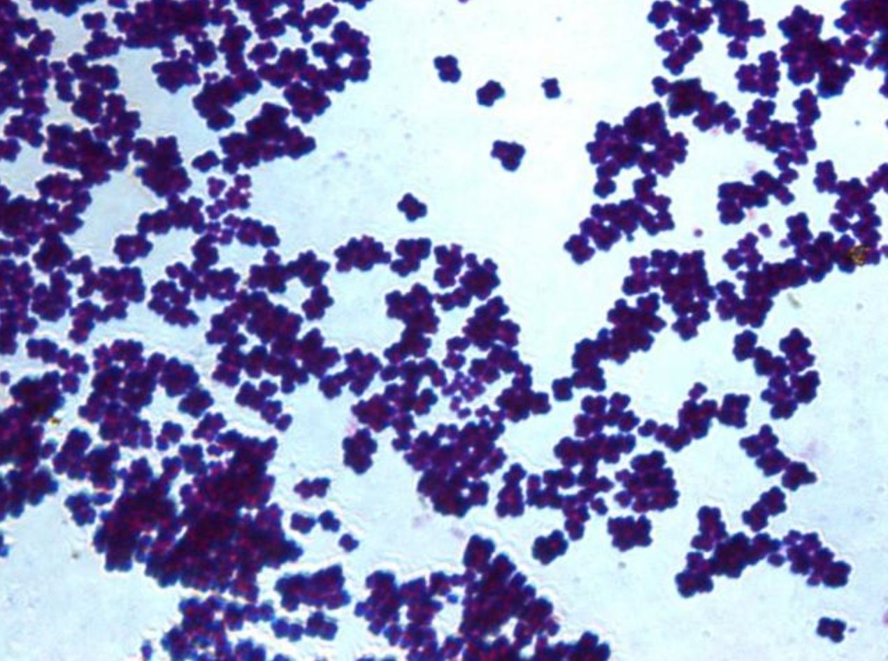
- 55—Staphylococcus, Grams staining, pure culture
Gram stain technique
1. The heat fixed bacterial smear is flooded with crystal violet and left on for 2 minutes.
2. Then it is flooded with Lugol’s iodine (mordant) which is left on for 1 minute.
3. The smear is then decolourized with ethanol (96%) for 30 seconds up to one minute.
4. Afterwards it is flooded with safranin which is left on for 2-3 minutes.
5. Finally it is washed with water, dried and examined under oil immersion.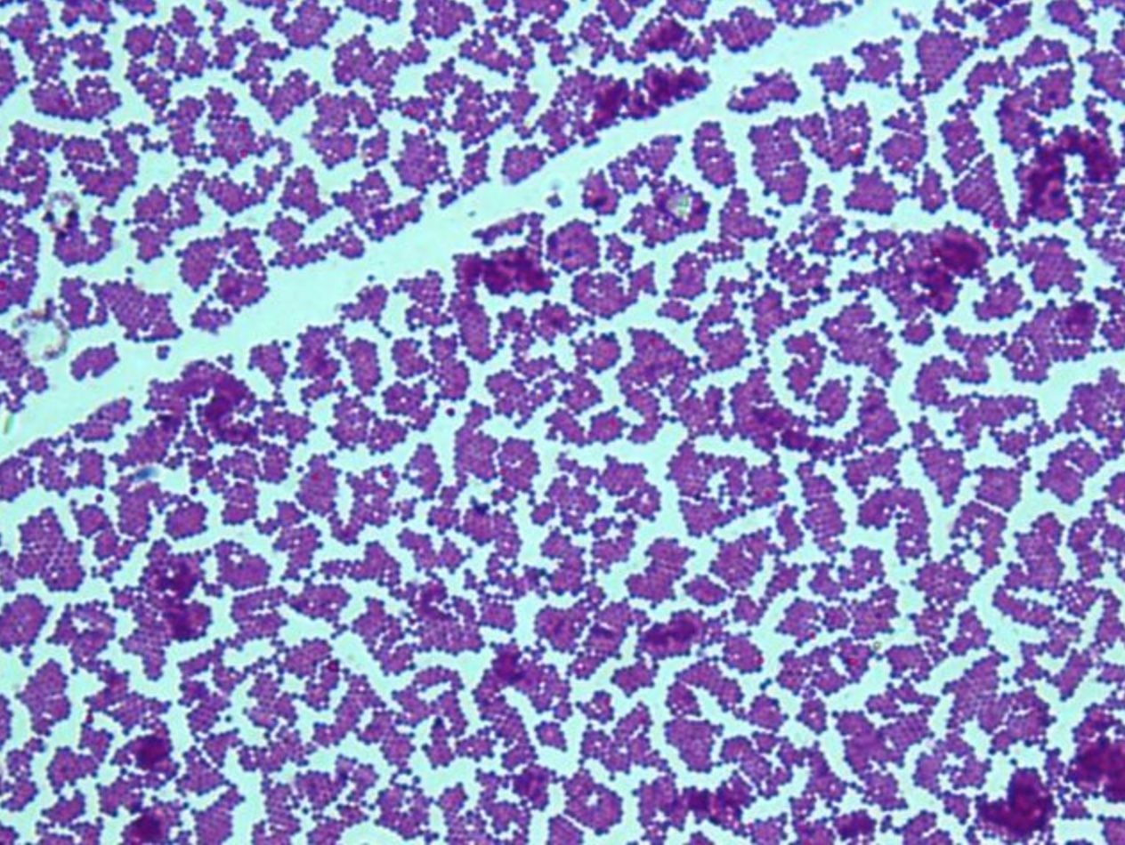
- 56—Staphylococcus, Grams staining, pure culture
Gram stain technique
1. The heat fixed bacterial smear is flooded with crystal violet and left on for 2 minutes.
2. Then it is flooded with Lugol’s iodine (mordant) which is left on for 1 minute.
3. The smear is then decolourized with ethanol (96%) for 30 seconds up to one minute.
4. Afterwards it is flooded with safranin which is left on for 2-3 minutes.
5. Finally it is washed with water, dried and examined under oil immersion.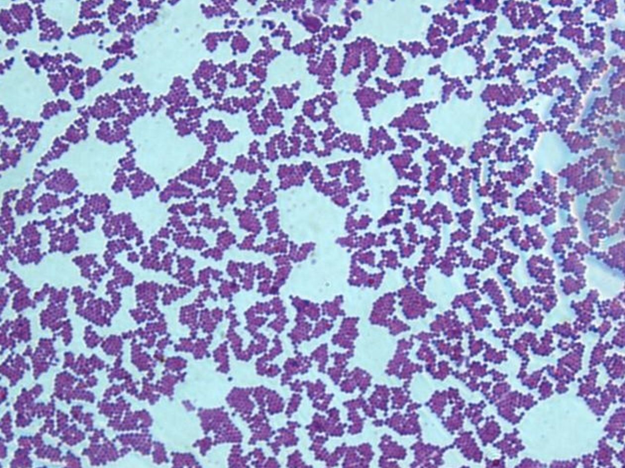
- 57—Staphylococcus, Grams staining, pure culture
Gram stain technique
1. The heat fixed bacterial smear is flooded with crystal violet and left on for 2 minutes.
2. Then it is flooded with Lugol’s iodine (mordant) which is left on for 1 minute.
3. The smear is then decolourized with ethanol (96%) for 30 seconds up to one minute.
4. Afterwards it is flooded with safranin which is left on for 2-3 minutes.
5. Finally it is washed with water, dried and examined under oil immersion.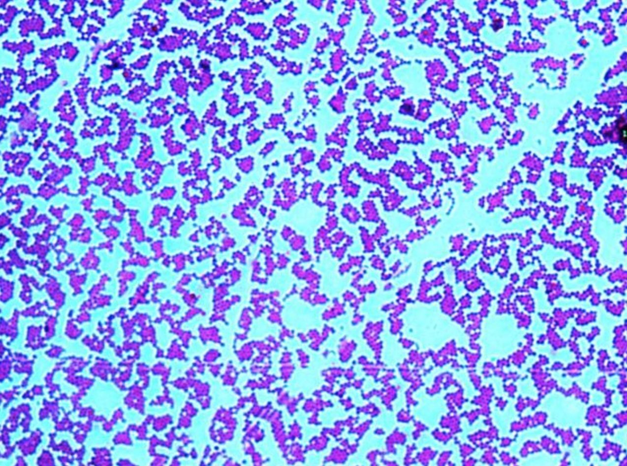
- 58—Streptococcus, methylene blue, pure culture
Simple staining
Fixed smears are stained using neutral and basic aniline dyes.
Only one dye is used, for example, methylene blue, safranin, etc.
All bacteria will stain the same colour, however it will be possible to evaluate the shape, size and arrangement of bacteria.
The slide is placed on a stand over the container and flooded with methylene blue. The dye should be left on for 5 minutes. When using safranin, the dye should be left on for 4 minutes.
Afterwards the slide is washed with water and dried. Only dry slides are examined under oil immersion.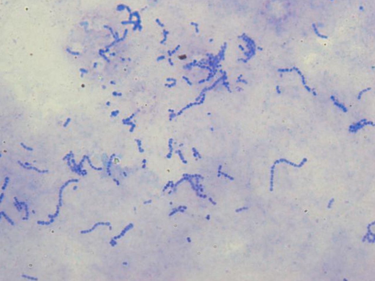
- 59—Streptococcus, methylene blue, pure culture
Simple staining
Fixed smears are stained using neutral and basic aniline dyes.
Only one dye is used, for example, methylene blue, safranin, etc.
All bacteria will stain the same colour, however it will be possible to evaluate the shape, size and arrangement of bacteria.
The slide is placed on a stand over the container and flooded with methylene blue. The dye should be left on for 5 minutes. When using safranin, the dye should be left on for 4 minutes.
Afterwards the slide is washed with water and dried. Only dry slides are examined under oil immersion.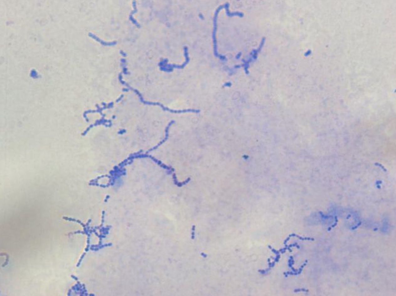
- 60—Streptococcus, methylene blue, pure culture
Simple staining
Fixed smears are stained using neutral and basic aniline dyes.
Only one dye is used, for example, methylene blue, safranin, etc.
All bacteria will stain the same colour, however it will be possible to evaluate the shape, size and arrangement of bacteria.
The slide is placed on a stand over the container and flooded with methylene blue. The dye should be left on for 5 minutes. When using safranin, the dye should be left on for 4 minutes.
Afterwards the slide is washed with water and dried. Only dry slides are examined under oil immersion.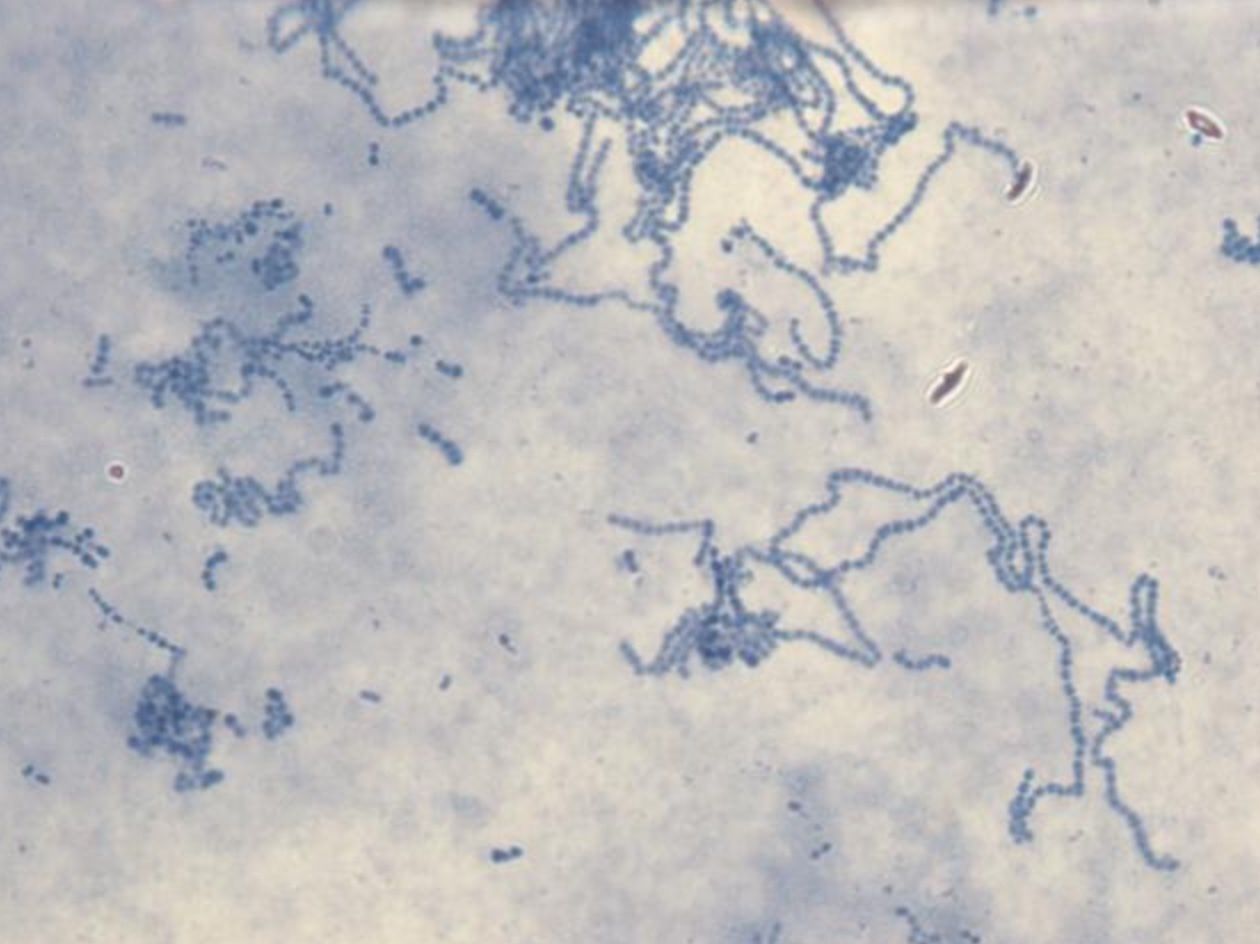
- 61—T.pallidum, negative staining, patient's sample
It differs from the other techniques in that the background is stained rather than the actual microbial cells. This technique is used to stain microorganisms that contain a high concentration of lipids and do not bind aniline dyes. If India ink is used, then the background stains black whereas if colargol solution is used, the background stains yellowish brown. The microbial cells remain uncoloured.
1. A small drop of the ink or 2% colargol solution is placed near one end of the slide.
2. The bacterial culture is then dispersed in the drop with an inoculation loop.
3. The smear is prepared by using another clean slide.
4. The smear is dried and examined under oil immersion without fixation.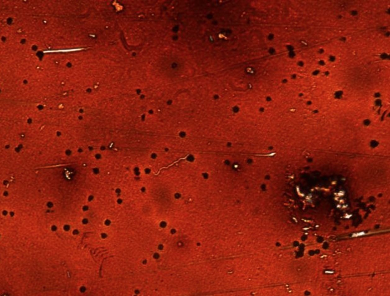
- 62—Treponema pallidum, negative staining, patient's sample
It differs from the other techniques in that the background is stained rather than the actual microbial cells. This technique is used to stain microorganisms that contain a high concentration of lipids and do not bind aniline dyes. If India ink is used, then the background stains black whereas if colargol solution is used, the background stains yellowish brown. The microbial cells remain uncoloured.
1. A small drop of the ink or 2% colargol solution is placed near one end of the slide.
2. The bacterial culture is then dispersed in the drop with an inoculation loop.
3. The smear is prepared by using another clean slide.
4. The smear is dried and examined under oil immersion without fixation.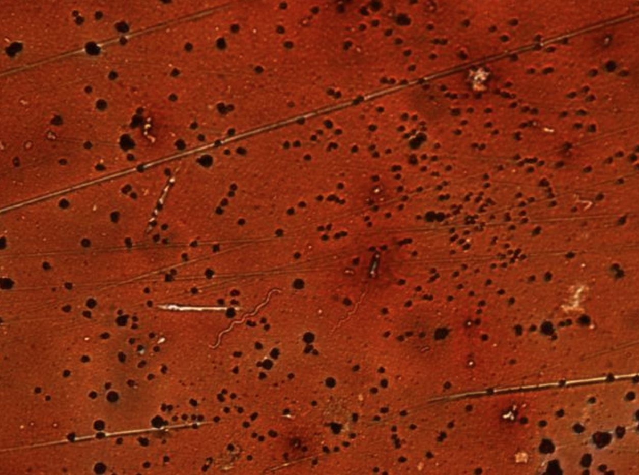
- 63—T.pallidum, negative staining, patient's sample
It differs from the other techniques in that the background is stained rather than the actual microbial cells. This technique is used to stain microorganisms that contain a high concentration of lipids and do not bind aniline dyes. If India ink is used, then the background stains black whereas if colargol solution is used, the background stains yellowish brown. The microbial cells remain uncoloured.
1. A small drop of the ink or 2% colargol solution is placed near one end of the slide.
2. The bacterial culture is then dispersed in the drop with an inoculation loop.
3. The smear is prepared by using another clean slide.
4. The smear is dried and examined under oil immersion without fixation.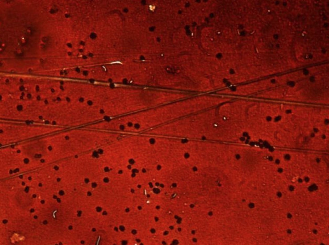
- 64—T.pallidum, negative staining, patient's sample
It differs from the other techniques in that the background is stained rather than the actual microbial cells. This technique is used to stain microorganisms that contain a high concentration of lipids and do not bind aniline dyes. If India ink is used, then the background stains black whereas if colargol solution is used, the background stains yellowish brown. The microbial cells remain uncoloured.
1. A small drop of the ink or 2% colargol solution is placed near one end of the slide.
2. The bacterial culture is then dispersed in the drop with an inoculation loop.
3. The smear is prepared by using another clean slide.
4. The smear is dried and examined under oil immersion without fixation.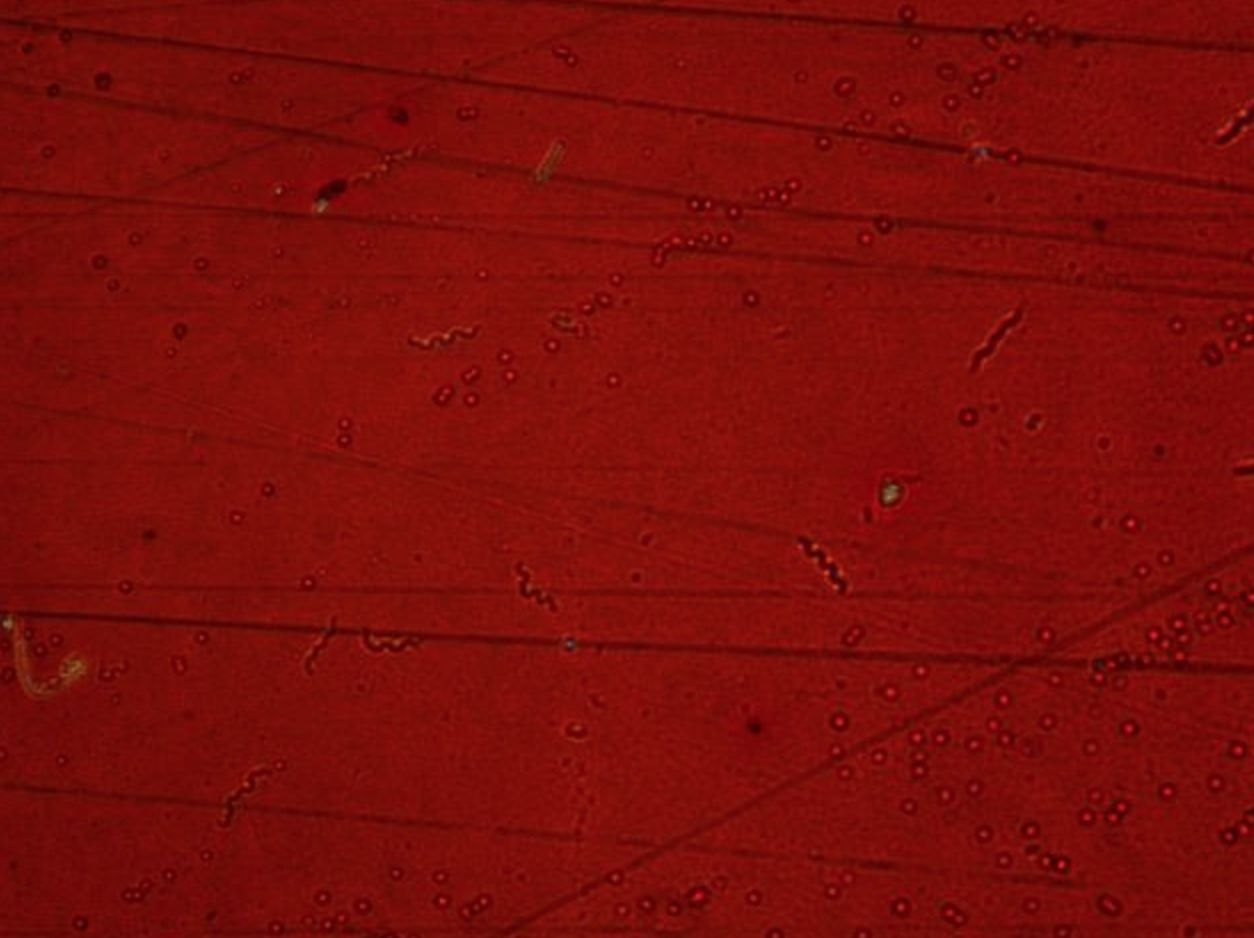
- 65—T.pallidum, negative staining, patient's sample
It differs from the other techniques in that the background is stained rather than the actual microbial cells. This technique is used to stain microorganisms that contain a high concentration of lipids and do not bind aniline dyes. If India ink is used, then the background stains black whereas if colargol solution is used, the background stains yellowish brown. The microbial cells remain uncoloured.
1. A small drop of the ink or 2% colargol solution is placed near one end of the slide.
2. The bacterial culture is then dispersed in the drop with an inoculation loop.
3. The smear is prepared by using another clean slide.
4. The smear is dried and examined under oil immersion without fixation.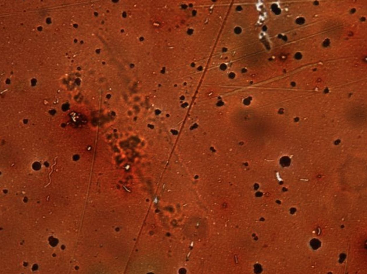
- 66—T.pallidum, negative staining, patient's sample
It differs from the other techniques in that the background is stained rather than the actual microbial cells. This technique is used to stain microorganisms that contain a high concentration of lipids and do not bind aniline dyes. If India ink is used, then the background stains black whereas if colargol solution is used, the background stains yellowish brown. The microbial cells remain uncoloured.
1. A small drop of the ink or 2% colargol solution is placed near one end of the slide.
2. The bacterial culture is then dispersed in the drop with an inoculation loop.
3. The smear is prepared by using another clean slide.
4. The smear is dried and examined under oil immersion without fixation. - 67—Vibrio et spirillum, negative staining, patient's sample
It differs from the other techniques in that the background is stained rather than the actual microbial cells. This technique is used to stain microorganisms that contain a high concentration of lipids and do not bind aniline dyes. If India ink is used, then the background stains black whereas if colargol solution is used, the background stains yellowish brown. The microbial cells remain uncoloured.
1. A small drop of the ink or 2% colargol solution is placed near one end of the slide.
2. The bacterial culture is then dispersed in the drop with an inoculation loop.
3. The smear is prepared by using another clean slide.
4. The smear is dried and examined under oil immersion without fixation.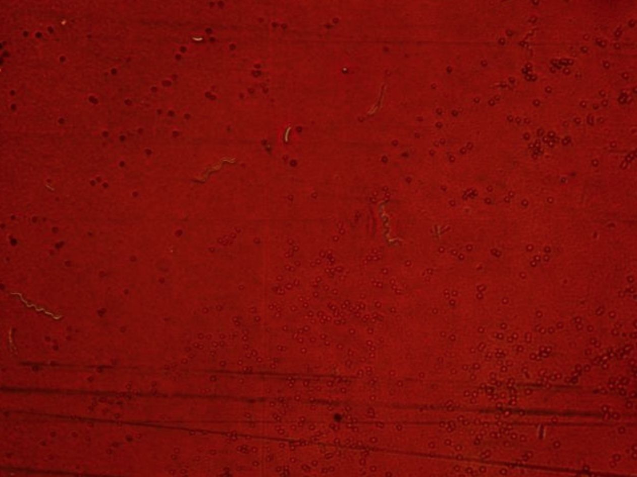
- 68—Vibrio et spirillum, negative staining, patient's sample
It differs from the other techniques in that the background is stained rather than the actual microbial cells. This technique is used to stain microorganisms that contain a high concentration of lipids and do not bind aniline dyes. If India ink is used, then the background stains black whereas if colargol solution is used, the background stains yellowish brown. The microbial cells remain uncoloured.
1. A small drop of the ink or 2% colargol solution is placed near one end of the slide.
2. The bacterial culture is then dispersed in the drop with an inoculation loop.
3. The smear is prepared by using another clean slide.
4. The smear is dried and examined under oil immersion without fixation.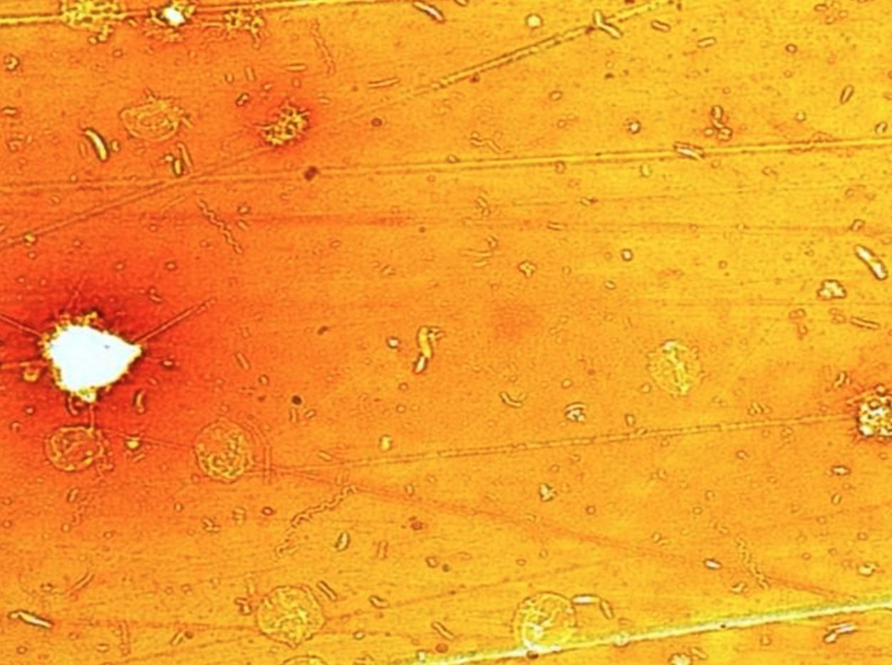
- 69—Vibrio et spirillum, negative staining, patient's sample
It differs from the other techniques in that the background is stained rather than the actual microbial cells. This technique is used to stain microorganisms that contain a high concentration of lipids and do not bind aniline dyes. If India ink is used, then the background stains black whereas if colargol solution is used, the background stains yellowish brown. The microbial cells remain uncoloured.
1. A small drop of the ink or 2% colargol solution is placed near one end of the slide.
2. The bacterial culture is then dispersed in the drop with an inoculation loop.
3. The smear is prepared by using another clean slide.
4. The smear is dried and examined under oil immersion without fixation.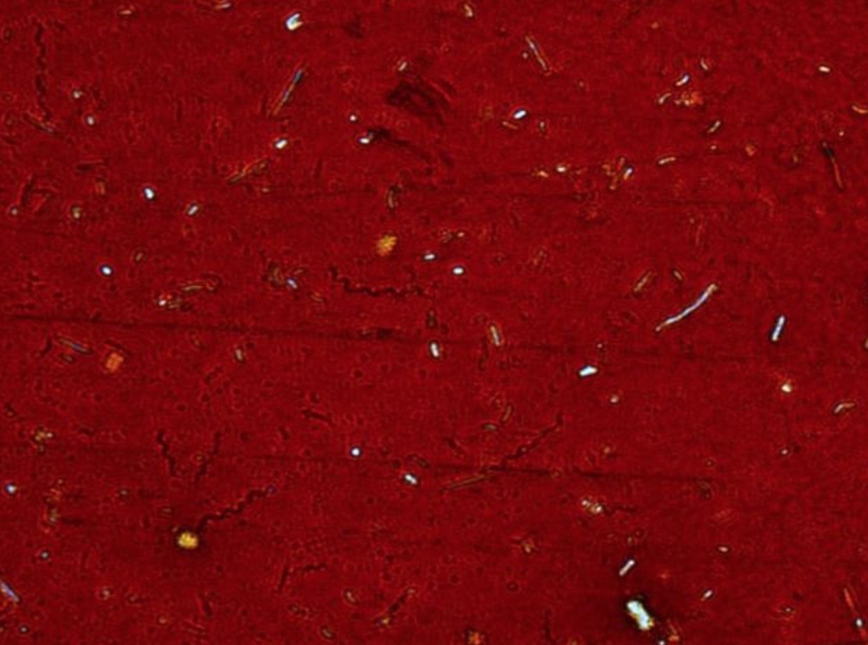
- 70—Vibrio et spirillum, negative staining, patient's sample
It differs from the other techniques in that the background is stained rather than the actual microbial cells. This technique is used to stain microorganisms that contain a high concentration of lipids and do not bind aniline dyes. If India ink is used, then the background stains black whereas if colargol solution is used, the background stains yellowish brown. The microbial cells remain uncoloured.
1. A small drop of the ink or 2% colargol solution is placed near one end of the slide.
2. The bacterial culture is then dispersed in the drop with an inoculation loop.
3. The smear is prepared by using another clean slide.
4. The smear is dried and examined under oil immersion without fixation.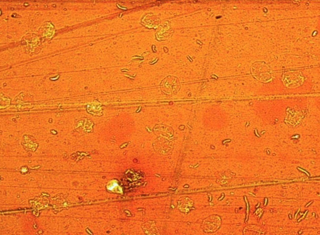
- 71—Vibrio et spirillum, negative staining, patient's sample
It differs from the other techniques in that the background is stained rather than the actual microbial cells. This technique is used to stain microorganisms that contain a high concentration of lipids and do not bind aniline dyes. If India ink is used, then the background stains black whereas if colargol solution is used, the background stains yellowish brown. The microbial cells remain uncoloured.
1. A small drop of the ink or 2% colargol solution is placed near one end of the slide.
2. The bacterial culture is then dispersed in the drop with an inoculation loop.
3. The smear is prepared by using another clean slide.
4. The smear is dried and examined under oil immersion without fixation.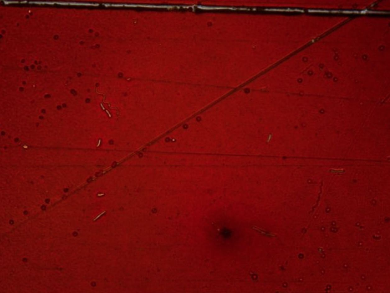
- 72—Staphylococcus, Grams staining, pure culture
Gram stain technique
1. The heat fixed bacterial smear is flooded with crystal violet and left on for 2 minutes.
2. Then it is flooded with Lugol’s iodine (mordant) which is left on for 1 minute.
3. The smear is then decolourized with ethanol (96%) for 30 seconds up to one minute.
4. Afterwards it is flooded with safranin which is left on for 2-3 minutes.
5. Finally it is washed with water, dried and examined under oil immersion.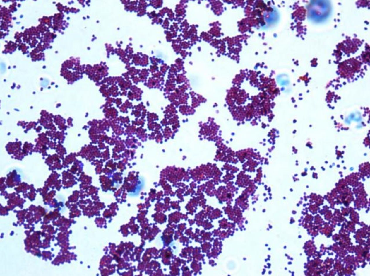
Information
- Description
- Medicine Flashcards on micro, created by Kerim Yildirim on 22/03/2018.
- Created by
- Kerim Yildirim
- Views
- 0
- Rating