
Flowchart
Untitled
Flowchart content
3.3.4 Mass transport
Tissue Fluid
Heart & Cardiac cycle
Haemglobin
Plants: Translocation and Transpiration
Formed by: Hydrostatic pressure & Oncotic pressure.
-Hydrostatic pressure is high at arteriole end of capillary causing net movement of fluid
- fluid secretes out of leaky endothelium cells into the surrounding tissue.
-Platlets, proteins are too big to diffuse out.
- returns to venous end.
Blood flow in Arterioles
Capillary bed Fluid secreted out in tissues and cells
Tissue exits through the venule down vein
Diffusion and active uptake in gas exchange of nurtrients
fluid enters through lymphatic system and turns to lymph as it passes lymph nodes
Lymph is returned to the blood system through the subclavian vein
Artery from heart: BP- very high. S-Small lumen, elastin fibres,smooth muscle collagen
Arterioles to heart: BP- high. S- Blood flow is restrcited at lumen contracts. small lumen
.Capillary: BP- low. S- cell is squamous very small lumen leaky endothelium
Veins: BP- lowest. S-opening/closing valves prevent backflow.
Venule: BP- lower. S- Elastic tissue. Thin collagen.
Transpiration: XYLEM
Translocation: PHLEOM
1) H+ ions are pumped out of companion cells actively using ATP.
2) H+ ions enter back to companion cells passively with sucrose through co-transport proteins
1) water evaporates from mesophyll layer and out stomata generating a water vapour gradient
2) Caspirian strips stop apoplast pathway. water moves through symplast
2) sucrose, H+ enter the sieve tube elements of phloem lowering water potential, water moves in through osmosis maintaining a hydrostatic pressure gradient. Mass flow generated
3) ion pumps pump ions lowering water potential cause water to move in along the cortex to the medulla through osmosis
Investigating plant transport
(tracer Experiment) Autoradiography: Isotope carbon 14 generates carbon 12 Makes CO2 for photosynthesis. Photographic film used to detect the flow of sugars
(Ring Experiment) Wood: Bark of wood with phloem is removed. Top of log is swelling bottom part is dying. No transportation of sugars as phloem was removed
Blood flow in Heart
Right atrium
Right ventricle
Pulmonary artery
lungs
pulmonary vein
left atrium
Lungs, kidneys, intestines,
Arota
Left ventricle
Vena Cava
Heart rate
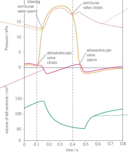
1) Electrical impulse travel through SLV through atria. Increasing its pressure. Atrium systole
2) Electrical impulse travel through bundle of His and through Purkinje fibres. High pressure. Ventricle systole
Ventricle Pressure
Arotic Pressure
Atrial pressure: Lower pressure excerted due to thin walls
Ventricle Volume
Information
- Description
- Flowchart on Untitled, created by joanna maranon on 05/06/2017.
- Created by
- joanna maranon
- Views
- 0
- History
-
- Created by joanna maranon · Jun 5, 2017
- Copied by joanna maranon · Jun 5, 2017
- Copied by joanna maranon · Jun 5, 2017
- Rating