
Mind map
Chapter 10 Muscular tissue
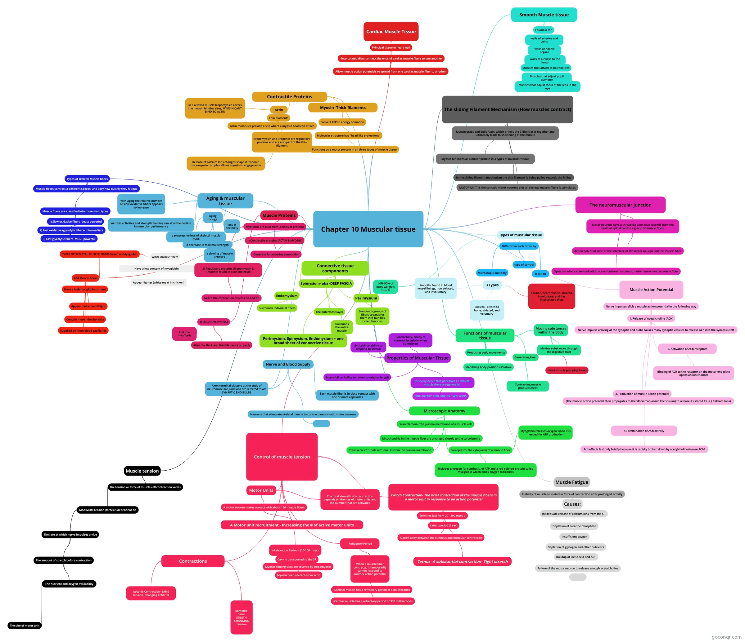
Mind map: Muscular tissue
Map content
Chapter 10 Muscular tissue
Types of muscular tissue
- 3 Types
- Skeletal- attach to bone, striated, and voluntary
- Cardiac- heart muscle, striated, involuntary, and has intercalated discs
- Smooth- Found in blood vessel linings, non striated, and involuntary
- Differ from each other by
- Microscopic anatomy
- location
- type of control
- 40%-50% of body weigh is muscle
- 3 Types
Functions of muscular tissue
- Producing body movements
- Stabilizing body positions- Posture
- Moving substances within the Body
- Heart muscle pumping blood
- Moving substances through the digestive tract
- Generating Heat
- Contracting muscle produces heat
Properties of Muscular Tissue
- for every nerve that penetrates a skeletal muscle there are generally :
- ONE ARTERY AND ONE OR TWO VEINS
- Excitability- ability to respond to stimuli
- Contractility- ability to contract forcefully when stimulated
- Extensibility- Ability to return to original length
- for every nerve that penetrates a skeletal muscle there are generally :
Connective tissue components
- Epimysium- aka -DEEP FASCIA
- The outermost layer
- surrounds the entire muscle
- Perimysium
- Surrounds groups of fibers separating them into bundles called fascicles
- Endomysium
- surrounds individual fibers
- Perimysium, Epimysium, Endomysium = one broad sheet of connective tissue
- Epimysium- aka -DEEP FASCIA
Axon terminal clusters at the ends of neuromuscular junctions are referred to as: SYNAPTIC END BULBS
- Nerve and Blood Supply
- Neurons that stimulate skeletal muscle to contract are somatic motor neurons
- Each muscle fiber is in close contact with one or more capillaries
- Neurons that stimulate skeletal muscle to contract are somatic motor neurons
- Nerve and Blood Supply
Microscopic Anatomy
- Scarcolemma- The plasma membrane of a muscle cell
- Mitochondria in the muscle fiber are arranged closely to the sarcolemma
- Transverse (T tubules) -Tunnel in from the plasma membrane
- Sarcoplasm- the cytoplasm of a muscle fiber
- includes glycogen for synthesis, of ATP and a red-colored protein called myoglobin which binds oxygen molecules
- Myoglobin releases oxygen when it is needed for ATP production
- Scarcolemma- The plasma membrane of a muscle cell
Muscle Proteins
- Myofibrils are built from 3 kinds of proteins
- 1) Contractile proteins (ACTIN & MYOSIN)
- -Generate force during contraction
- 2) Regulatory proteins (Tropomyosin & Troponin found in actin molecule
- switch the contraction process on and off
- 1) Contractile proteins (ACTIN & MYOSIN)
- 3) Structural Proteins
- Align the thick and thin filaments properly
- Link the myofibrils
- Myofibrils are built from 3 kinds of proteins
Contractile Proteins
- Myosin- Thick filaments
- convert ATP to energy of motion
- Molecular structure has "head like projections"
- Functions as a motor protein in all three types of muscle tissue
- Molecular structure has "head like projections"
- convert ATP to energy of motion
- Actin
- Thin filaments
- Actin molecules provide a site where a myosin head can attach
- Tropomyosin and Troponin are regulatory proteins and are also part of the thin filament
- In a relaxed muscle tropomyosin covers the myosin binding sites, MYOSIN CANT BIND TO ACTIN
- Release of calcium Ions changes shape if troponin tropomyosin complex allows myosin to engage actin
- Thin filaments
- Myosin- Thick filaments
The sliding Filament Mechanism (How muscles contract)
- Myosin grabs and pulls Actin, which bring s the Z-disc closer together and ultimately leads to shortening of the muscle
- Myosin functions as a motor protein in 3 types of muscular tissue
- In the sliding filament mechanism the thin filament is being pulled towards the M-line
- MOTOR UNIT- is the somatic motor neurons plus all skeletal muscle fibers it stimulates
The neuromuscular junction
- Motor neurons have a threadlike axon that extends from the brain or spinal cord to a group of muscle fibers
- Action potential arise at the interface of the motor neuron and the muscle fiber
- Synapse- where communication occurs between a somatic motor neuron and a muscle fiber
- Motor neurons have a threadlike axon that extends from the brain or spinal cord to a group of muscle fibers
Muscle Action Potential
- Nerve Impulses elicit a muscle action potential in the following way
- 1. Release of Acetylcholine (ACH)
- Nerve impulse arriving at the synaptic end bulbs causes many synaptic vesicles to release ACh into the synaptic cleft
- 2. Activation of ACh receptors
- Binding of ACh to the receptor on the motor end plate opens an Ion channel
- 1. Release of Acetylcholine (ACH)
- 3. Production of muscle action potential
- (The muscle action potential then propagates to the SR (Sarcoplasmic Recticulum) to release its stored Ca++ ( Calcium Ions)
- 4.) Termination of ACh activity
- Ach effects last only briefly because it is rapidly broken down by acetylcholinesterase AChE
- Nerve Impulses elicit a muscle action potential in the following way
Muscle Fatigue
- Inability of muscle to maintain force of contraction after prolonged activity
- Causes:
- Inadequate release of calcium ions from the SR
- Depletion of creatine phosphate
- Insufficient oxygen
- Depletion of glycogen and other nutrients
- Buildup of lactic acid and ADP
- Failure of the motor neuron to release enough acetylcholine
- Failure of the motor neuron to release enough acetylcholine
- Buildup of lactic acid and ADP
- Depletion of glycogen and other nutrients
- Insufficient oxygen
- Depletion of creatine phosphate
- Inadequate release of calcium ions from the SR
- Causes:
- Inability of muscle to maintain force of contraction after prolonged activity
Muscle tension
- the tension or force of muscle cell contraction varies
- MAXIMUM tension (force) is dependent on
- The rate at which nerve impulses arrive
- The amount of stretch before contraction
- The nutrient and oxygen availability
- The size of motor unit
- The nutrient and oxygen availability
- The amount of stretch before contraction
- The rate at which nerve impulses arrive
- MAXIMUM tension (force) is dependent on
- the tension or force of muscle cell contraction varies
Control of muscle tension
- Motor Units
- A motor neuron makes contact with about 150 muscle fibers
- A Motor unit recruitment - Increasing the # of active motor units
- The total strength of a contraction depends on the size of motor units and the number that are activated
- A motor neuron makes contact with about 150 muscle fibers
- Twitch Contraction- The brief contraction of the muscle fibers in a motor unit in response to an action potential
- Twitches last from 20 - 200 msec L
- Latent period (2 sec)
- A brief delay between the stimulus and muscular contraction
- Latent period (2 sec)
- Tetnus- A substantial contraction- Tight stretch
- Twitches last from 20 - 200 msec L
- --Relaxation Period-- (10-100 msec)
- Ca++ is transported to the SR
- Myosin binding sites are covered by tropomyosin
- Myosin heads detach from actin
- Myosin binding sites are covered by tropomyosin
- Ca++ is transported to the SR
- --Refractory Period-
- When a muscle fiber contracts, it temporarily cannot respond to another action potential
- -Skeletal muscle has a refractory period of 5 milliseconds
- -Cardiac muscle has a refractory period of 300 milliseconds
- -Skeletal muscle has a refractory period of 5 milliseconds
- When a muscle fiber contracts, it temporarily cannot respond to another action potential
- Contractions
- Isotonic Contraction- SAME tension, Changing LENGTH
- Isometric- Same LENGTH, CHANGING tension
- Motor Units
Red Muscle fibers
- TYPES OF SKELETAL MUSCLE FIBERS based on Myoglobin
- White muscle fibers
- Have a low content of myoglobin
- Appear lighter (white meat In chicken)
- White muscle fibers
- Have a high myoglobin content
- Appear darker and thighs
- Contain more mitochondria
- Supplied by more blood capillaries
- TYPES OF SKELETAL MUSCLE FIBERS based on Myoglobin
Types of skeletal Muscle fibers
- Muscle fibers contract a different speeds, and vary how quickly they fatigue
- Muscle fibers are classified into three main types
- 1) Slow oxidative fibers -Least powerful
- 2) Fast oxidative -glycolytic fibers- Intermediate
- 3) Fast glycolytic fibers- MOST powerful
- 2) Fast oxidative -glycolytic fibers- Intermediate
- 1) Slow oxidative fibers -Least powerful
Cardiac Muscle Tissue
- Principal tissue in heart wall
- -intercalated discs connect the ends of cardiac muscle fibers to one another
- Allow muscle action potentials to spread from one cardiac muscle fiber to another
- -intercalated discs connect the ends of cardiac muscle fibers to one another
- Principal tissue in heart wall
Smooth Muscle tissue
- Found in the
- walls of arteries and veins
- walls of hollow organs
- walls of airways to the lungs
- Muscles that attach to hair follicles
- Muscles that adjust pupil diameter
- Muscles that adjust focus of the lens in the eye
- Muscles that adjust pupil diameter
- Muscles that attach to hair follicles
- walls of airways to the lungs
- walls of hollow organs
- walls of arteries and veins
- Found in the
Aging & muscular tissue
- Aging brings
- a progressive loss of skeletal muscle mass
- a decrease in maximal strength
- a slowing of muscle reflexes
- loss of flexibility
- with aging the relative number of slow oxidative fibers appears to increase
- Aerobic activities and strength training can slow the decline in muscular performance
- Aging brings
Information
- Description
- Mind Map on Chapter 10 Muscular tissue, created by April Brown on 19/09/2021.
- Created by
- April Brown
- Views
- 0
- Rating