
Mind map
Demyelinating disorders
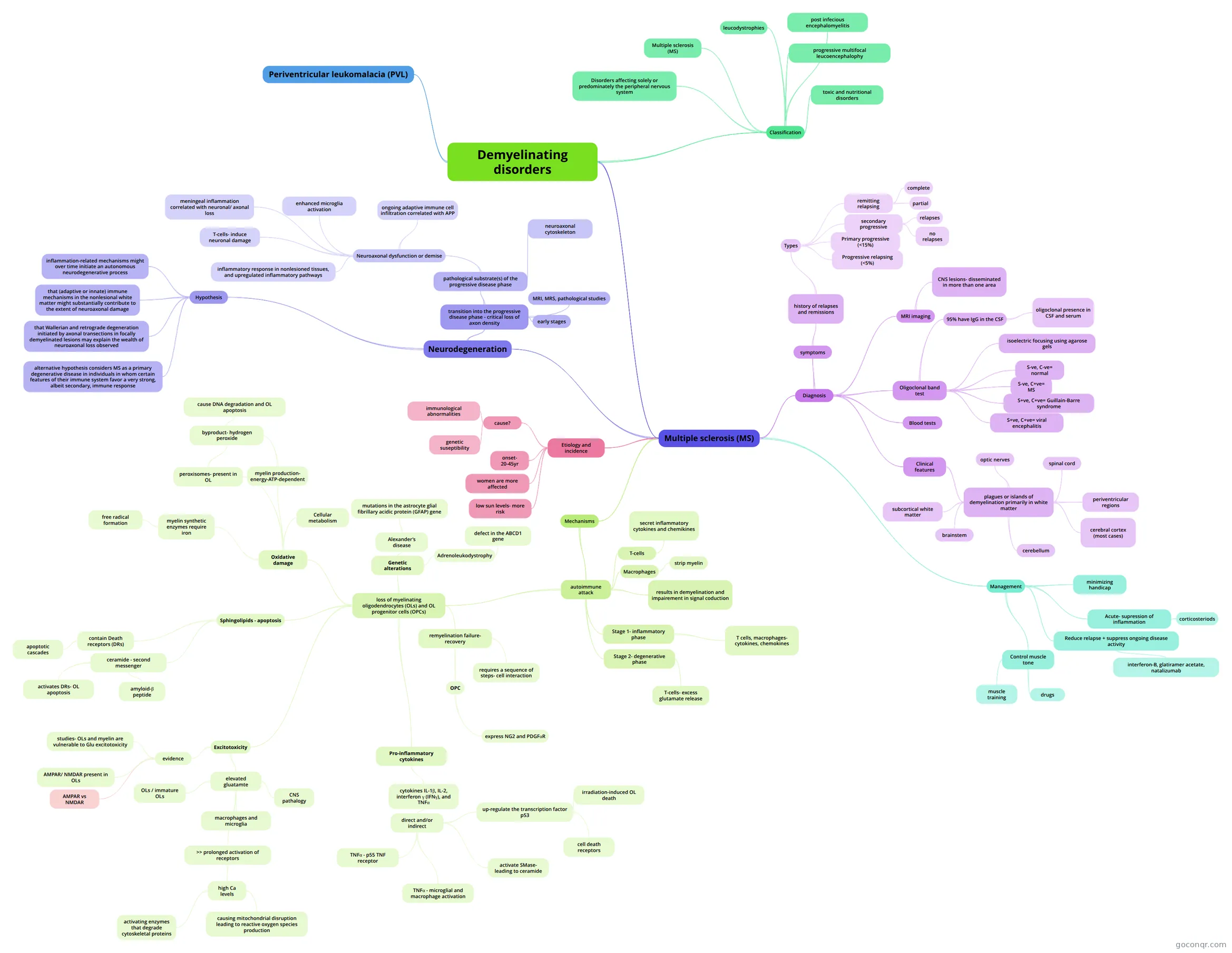
Mind map: demyelinating disorders
Map content
Demyelinating disorders
Classification
- A group of disease which involve damage and destruction of myelin sheet that surround nerve fibres. The axons of these fibres may be damages as well. The authiology of most demyelinating disorders is unknown.
- Multiple sclerosis (MS)
- post infecious encephalomyelitis
- progressive multifocal leucoencephalophy
- toxic and nutritional disorders
- leucodystrophies
- Disorders affecting solely or predominately the peripheral nervous system
Multiple sclerosis (MS)
- slowly progressive CNS disease characterised by disseminated patches of demyelination in the brain and spinal cord, resulting in multiple and varied neurological symptoms and signs usually with remissions and exacerbations
- Etiology and incidence
- onset- 20-45yr
- women are more affected
- cause?
- immunological abnormalities
- genetic suseptibility
- low sun levels- more risk
- Mechanisms
- autoimmune attack
- T-cells
- secret inflammatory cytokines and chemikines
- IFN, interferon, IL-2, interleukin-2, TNFa
- secret inflammatory cytokines and chemikines
- Macrophages
- strip myelin
- results in demyelination and impairement in signal coduction
- Stage 1- inflammatory phase
- T cells, macrophages- cytokines, chemokines
- Stage 2- degenerative phase
- T-cells- excess glutamate release
- excitotoxicity hypothesis- Ca permeable receptors- over-reaction may lead to cell death
- T-cells- excess glutamate release
- loss of myelinating oligodendrocytes (OLs) and OL progenitor cells (OPCs)
- remyelination failure- recovery
- Remylenation process- proliferation, migration, differentiation, mylenation>> once this occurs the symptoms disappear
- This repair process is mediated by a population of cells located throughout the gray and white matter that has often been referred to as adult OPCs
- requires a sequence of steps- cell interaction
- misscomunication between different cells may lead to impairement in remyelinition
- OPC
- express NG2 and PDGFαR
- these molecules have been used to identify and characterize OPCs in vivo
- express NG2 and PDGFαR
- Oxidative damage
- Thus, the combination of high metabolism, numerous peroxisomes, lipid byproducts, and iron all make OLs particularly vulnerable to oxidative damage
- myelin production- energy-ATP-dependent
- myelin production is energy dependent, large amounts of ATP are consumed in the process, which translates into increased ATP production and significant oxygen consumption
- byproduct- hydrogen peroxide
- cause DNA degradation and OL apoptosis
- if not metabolized, has been shown in vitro to cause DNA degradation and OL apoptosis
- cause DNA degradation and OL apoptosis
- peroxisomes- present in OL
- byproduct- hydrogen peroxide
- Cellular metabolism
- Cellular metabolism also creates reactive oxygen species which are highly toxic and induce lipid peroxidation and DNA damage
- myelin synthetic enzymes require iron
- OLs and OL progenitors have the largest intracellular stores of iron in the adult brain (20-fold greater than astrocytes under baseline culture conditions)
- free radical formation
- While iron is necessary for myelin production, it is also highly reactive and can evoke free radical formation and lipid peroxidation. For example, iron catalyzes the conversion of hydrogen peroxide into hydroxyl radicals, which directly damage intracellular compartments
- Excitotoxicity
- elevated gluatamte
- results from traumatic injury, stroke, AD, Parkinson’s disease, amyotrophic lateral sclerosis, and even normal aging
- OLs / immature OLs
- glutamate release induced by anoxia or compression was due to reversal of glutamate transport, likely from axons and OLs (Li et al. 1999). Immature OLs can also release glutamate via reverse glutamate transport, which can then feedback and damage the cells through calcium influx
- CNS pathalogy
- macrophages and microglia
- >> prolonged activation of receptors
- high Ca levels
- activating enzymes that degrade cytoskeletal proteins
- causing mitochondrial disruption leading to reactive oxygen species production
- high Ca levels
- evidence
- studies- OLs and myelin are vulnerable to Glu excitotoxicity
- AMPAR/ NMDAR present in OLs
- AMPAR vs NMDAR
- Data from these studies indicate that the receptors are functional as NMDA-mediated currents and Ca2+entry into the cytoplasm were detected. This work also provided evidence that the NMDA receptors contribute to OL toxicity during ischemia, which was recently confirmed by Bakiri et al. (2008). These results are in contrast, however, to those of Tekkok et al. (2007) who used a similar in vitro optic nerve preparation and showed that AMPA/kainate but not NMDA antagonists prevented white matter damage.
- The reason for contrasting reports is not clear but may relate to such differences in experimental design as recording temperatures (33° vs. 37°), stimulus intensities (‘low’ vs. ‘supramaximal’) and/or antagonist concentrations
- elevated gluatamte
- Pro-inflammatory cytokines
- Pro-inflammatory cytokines are often present at sites of CNS injury or disease and are thought to contribute to OL pathology.
- cytokines IL-1β, IL-2, interferon γ (IFNγ), and TNFα
- promote OL death in vitro
- direct and/or indirect
- TNFα - p55 TNF receptor
- TNFα can directly kill OLs by binding to the p55 TNF receptor, which induces apoptosis-inducing factor to translocate from the cytoplasm to the nucleus where it evokes DNA degradation and caspase-independent apoptosis
- TNFα - microglial and macrophage activation
- microglial and macrophage activation by TNFα and IFNγ leads to production of free radicals which in turn can kill OLs through oxidative methods described above
- activate SMase- leading to ceramide
- up-regulate the transcription factor p53
- cell death receptors
- when over-expressed in cultured adult human OLs, caused an up-regulation of the death receptors FAS, DR4 and DR5 and led to caspase-mediated apoptosis (Wosik et al. 2003). Analysis of MS tissue revealed that p53 was present in OLs within active lesions, suggesting that p53-mediated mechanisms may contribute to OL loss in this disease (Wosiket al. 2003).
- irradiation-induced OL death
- Based on work from knock-out mice, p53 also contributes to irradiation-induced OL death, which can deplete the OL cell population by invoking OL apoptosis and/or by elevating TNFα and IFNγ production
- cell death receptors
- TNFα - p55 TNF receptor
- Genetic alterations
- Alexander’s disease
- Dysmyelination or demyelination is the primary clinical pathology in Alexander’s disease
- mutations in the astrocyte glial fibrillary acidic protein (GFAP) gene
- It is hypothesized that OL pathology results from poor/aberrant astrocyte and OL communication (gap junction disruption), lack of astrocytic glutamate uptake or cytotoxic molecules generated by astrocytes
- Adrenoleukodystrophy
- Adrenoleukodystrophy, another genetic myelin disorder with documented OL loss
- defect in the ABCD1 gene
- accumulation of very long chain fatty acids; these molecules can alter cell membrane function and render OLs vulnerable to cell death via inflammatory mediators
- Alexander’s disease
- Sphingolipids - apoptosis
- The brain is enriched in sphingolipids, which are the major lipid components of plasma membranes and comprise up to 20% of the dry weight of myelin. These molecules were long thought to be important for structural support but to have no role in cellular signaling
- contain Death receptors (DRs)
- apoptotic cascades
- ceramide - second messenger
- released intracellularly by enzymatic cleavage of membrane sphingolipids
- activates DRs- OL apoptosis
- amyloid-β peptide
- increased intracellular ceramide levels and OL apoptosis
- remyelination failure- recovery
- T-cells
- autoimmune attack
- Diagnosis
- quite good diagnosis for MS
- Clinical features
- plagues or islands of demyelination primarily in white matter
- with destruction of oligodendroglya and perivascular inflammation are disseminated through the CNS
- periventricular regions
- deep white matter- euphoria, poor memory and concentration, dementia, acute psychriatric disorders
- optic nerves
- subacute unilateral loss of vision, blind spot in the centre vision, pain when the eye is moved
- spinal cord
- weakness, heaviness or stiffness of both legs or all four limbs and sometimes just one limb, sensory loss in the legs that spreads up into the trunk over a few days, altered sensation, bladder and bowel dysfunction, sexual dysfunction
- subcortical white matter
- cerebral cortex (most cases)
- brainstem
- double vision, slurred speech, unsteady gait, incoordination of the hands, numbness of the face, unilateral facial weakness, difficulty swallowing
- cerebellum
- slured speech, insteady gait, nystagmus
- plagues or islands of demyelination primarily in white matter
- MRI imaging
- CNS lesions- disseminated in more than one area
- Oligoclonal band test
- 95% have IgG in the CSF
- IgG= immunoglobin
- oligoclonal presence in CSF and serum
- isoelectric focusing using agarose gels
- S-ve, C-ve= normal
- S-ve, C+ve= MS
- S+ve, C+ve= Guillain-Barre syndrome
- S+ve, C+ve= viral encephalitis
- 95% have IgG in the CSF
- symptoms
- - paresthesias in one or more extremities, in the trunk or on one side of the face - weakness or clumsiness of a leg or a hand - visual disturbances (partial blindness and pain in one eye; retrobulbar optic neuritis) - apathy - emotional lability is common - scanning speech - deep reflexes (e.g.: knee and ankle jerk) are generally increased - superficial reflexes (e.g.: abdominal) are diminished or absent. - intention tremor (cerebellar lesion) - slight incontinence, sexual impotence, genital anesthesia (spinal cord involvement)
- history of relapses and remissions
- Types
- remitting relapsing
- Patients experience a series of attacks followed by complete or partial disappearance of the symptoms (remitting) until another attack occurs (relapse). It may be weeks to years between relapses.
- complete
- partial
- secondary progressive
- relapses
- no relapses
- Primary progressive (<15%)
- Progressive relapsing (<5%)
- remitting relapsing
- Types
- Blood tests
- Management
- Acute- supression of inflammation
- Methylprednisolone (Corticosteroids reduce the duration of the relapse, but do not influence the long-term outcome)
- corticosteriods
- minimizing handicap
- (physiotherapy, occupational therapy, adaptations to work and home environments) Increase strength, endurance, movement strategies assistive devices
- Reduce relapse + suppress ongoing disease activity
- Interferon-b, glatiramer acetate and natalizumab reduce the appearance of new MRI abnormalities and relaps rate and may have an effect on disease progression in both relapsing-remitting and secondary progressive disease. (Costs!) Interferon may also reduce the rate at which patients with a single episode of CNS demyelination progress to clinically definite MS. Azothioprine (intravenous IgG) may reduce relapse rates
- interferon-B, glatiramer acetate, natalizumab
- Control muscle tone
- muscle training
- drugs
- Acute- supression of inflammation
- Neurodegeneration
- Most commonly, the term neurodegeneration in the context of MS is used to describe the full extent of neuroaxonal damage, be it related or not to focal lesion formation. In contrast to classic neurodegenerative diseases, most authors currently do not favor a central nervous system (CNS) autonomous neurodegenerative process, but rather consider inflammation central to its pathogenesis
- transition into the progressive disease phase - critical loss of axon density
- MRI, MRS, pathological studies
- early stages
- axonal loss and dysfunction, respectively, occur very early in the disease course
- pathological substrate(s) of the progressive disease phase
- Neuroaxonal dysfunction or demise
- reduction in neuroaxonal amino acid
- meningeal inflammation correlated with neuronal/ axonal loss
- ongoing adaptive immune cell infiltration correlated with APP
- amyloid precursor protein >> accumulations in axons, indicating acute axonal damage
- T-cells- induce neuronal damage
- enhanced microglia activation
- disrupted nodes of Ranvier in brain diseases with enhanced microglia activation, exemplified by MS and Parkinson's disease
- inflammatory response in nonlesioned tissues, and upregulated inflammatory pathways
- Molecular studies of normal-appearing and periplaque white matter indicate a subtle inflammatory response also in nonlesioned brain tissue; however, in addition, they point to the fact that anti-inflammatory pathways are upregulated, which may indicate the activation of endogenous protective mechanisms
- neuroaxonal cytoskeleton
- Neuroaxonal dysfunction or demise
- Hypothesis
- evidence has led to these hypothesis- not mutually exclusive
- inflammation-related mechanisms might over time initiate an autonomous neurodegenerative process
- similar to that observed in neurodegenerative diseases such as Alzheimer's disease, Parkinson's disease, or amyotrophic lateral sclerosis
- that (adaptive or innate) immune mechanisms in the nonlesional white matter might substantially contribute to the extent of neuroaxonal damage
- that Wallerian and retrograde degeneration initiated by axonal transections in focally demyelinated lesions may explain the wealth of neuroaxonal loss observed
- alternative hypothesis considers MS as a primary degenerative disease in individuals in whom certain features of their immune system favor a very strong, albeit secondary, immune response
Periventricular leukomalacia (PVL)
Information
- Description
- Neurological and Psychiatric disorders Mind Map on Demyelinating disorders, created by Cher Bachar on 24/05/2013.
- Created by
- Cher Bachar
- Views
- 141
- Tags
- Rating