
Mind map
MICROSCOPY TECHNIQUES_1
Map content
MICROSCOPY TECHNIQUES_1
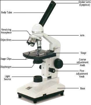
MICROSCOPE PARTS
- Place where you look through
- One hand should grasp this part when carrying the unit
- To be place the slide
- For focusing in low and scanning power
- For focusing in high power
- To be placed at the flat surface
- Control the light intensity
- Control the amount of light passing through the specimen
- To hold the slide
- Adjust from low to high power
- Holds the objective lenses and revolves to switch the objective lens
- Connect eyepiece to objective lenses
- Holds the objective lenses and revolves to switch the objective lens
- Adjust from low to high power
- To hold the slide
- Control the amount of light passing through the specimen
- Control the light intensity
- To be placed at the flat surface
- For focusing in high power
- For focusing in low and scanning power
- To be place the slide
- One hand should grasp this part when carrying the unit
- Place where you look through
HOW TO FOCUS THE MICROSCOPE
- 1. place the slide on the stage between the stage clips
- 2. Objective lens at low power
- 3. Looking at the slide - adjust the lowest power lens down close to the specimen without touching it.
- 4. Look through eyepiece - focus upward until the image is sharp (use coarse adjustment knob)
- 5. Change to high power objective lens - fine objective knob
- 4. Look through eyepiece - focus upward until the image is sharp (use coarse adjustment knob)
- 3. Looking at the slide - adjust the lowest power lens down close to the specimen without touching it.
- 2. Objective lens at low power
- 1. place the slide on the stage between the stage clips
OIL IMMERSION
- To improve the resolution power (ability to differentiate between 2 dots) by focusing more light to the specimen
- Use at high power objective lens (100x)
- To improve the resolution power (ability to differentiate between 2 dots) by focusing more light to the specimen
MICROSCOPES
- Bright Field
- Dark Field
- Phase Contrast
- Enhances contrast of transparent or colourless by influencing the optical path of light
- Microbial motility Determining the shape of the living cell Detecting bacteria component
- Enhances contrast of transparent or colourless by influencing the optical path of light
- Fluorescene
- Image itself emits light - after excitation with fluorescene stain called fluorochrome
- Electron Microscope
- Transmisson Electron Microscope
- Analysing internal structure - works like a slide projector
- Scanning Electron Microscopy
- Analysing surface structure
- Transmisson Electron Microscope
NEW TECHNIQUE IN MICROSCOPY
- Confocal Microscope
- Uses laser beam to illuminate specimen Provide reconstruction of images
- Scanning Probe Microscopy
- Uses electrons and application of small voltage to the specimen
- Atomic Force Microscopy
- Forcing electron on surface at short distance without using voltage
- Confocal Microscope
Images and files in the map
Information
- Description
- Mind Map on MICROSCOPY TECHNIQUES_1, created by munirahadibah on 02/03/2014.
- Created by
- munirahadibah · Mar 2014
- Updated
- Feb 19, 2016
- Views
- 11
- History
-
- Created by munirahadibah · Mar 2, 2014
- Copied by munirahadibah · Mar 2, 2014
- Rating