
Fichas
Biol 251 Lab practical
Fichas del conjunto (199)
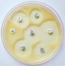
- 1What test is this and what do the results determine?Kirby-Bauer method; it tells us how sensitive or resistant a pathogenic bacteria is to various antibiotics
- 2What are the standardized variables for a Kirby-Bauer test?1. Medium: Mueller-Hinton II agar
2. pH: 7.2-7.4
3. Agar thickness: 4mm in a petri dish
4. Bacterial growth: grown in broth to a density standard of 10^7
5. Bacteria are applied in a lawn with a cotton swab
6. Discs with antibiotics are applied & incubate for 16-18 hrs - 3What is the zone of inhibition (Kirby-Bauer)?The area around the disk where no bacteria grow
- 4How is the susceptibility of a bacterium to a particular antibiotic determined? (Kirby-Bauer)The diameter of the zone of inhibition is measured in mm and compared to a standardized chart to determine the susceptibility rating.
- 5Natural AntibioticAntimicrobial compounds (antibiotics) produced by microbes in nature that inhibit growth of competing microbes; gives the producer an environmental advantage
- 6Semi-synthetic AntibioticsAntimicrobial compounds that are altered in a lab by modifying the R group while keeping the active moiety (shape) in tact.
- 7Synthetic AntibioticsAntimicrobial compounds that are made entirely in a lab; no part of it is naturally made by a microbe.
- 8PenicillinFirst antibiotic used; natural antibiotic
- 9Semi-synthetic penicillinAntibiotics derived from penicillin that retain the beta-lactam moiety, but change the R group to prevent the cleaving of the beta-lactam ring by resistant microbes.
- 10BactericidalAntibiotics that kill bacteria outright
- 11BacteriostaticAntibiotics that inhibit microbial growth but do not kill them; work together with the immune system to eliminate the pathogen
- 12Narrow Spectrum AntibioticsAntibiotics that target single, specific groups of pathogens (vancomycin v. Gram positive, polymyxin B v. Gram neg)
- 13Broad Spectrum AntibioticsTarget more than one group of bacteria (tetracycline v. gram pos, gram neg, chlamydias)
- 14What is an advantage and disadvantage of broad spectrum antibiotics?They target & inhibit many types of bacteria - by doing so, they remove normal microbiota and can lead to superinfections.
- 15What are the targets of antibiotics?1. Cell wall synthesis (penicillin)
2. Protein synthesis - change the bacterial ribosome so they cannot make necessary proteins
3. Disruption of the cell membrane (polymyxin B - highly toxic for humans since we too have a cell membrane that can be disrupted)
4. Inhibit DNA & RNA synthesis by inhibiting replication/transcription enzymes (DNA gyrase, RNA polymerase) - can be toxic to humans - 16High selective toxicityDrug targets the bacteria and NOT the human
- 17Low selective toxicityDrugs that cause more serious side effects in humans because it is not able to target specific pathogen components without affecting human processes
- 18Sensitive bacteriaA bacterium that is susceptible to an antibiotic and is either killed or has it's growth inhibited
- 19Resistant bacteriaBacterium that is not inhibited by the drug (no effect on growth)
- 20Intermediate BacteriaBacteria may be gaining resistance to the drug, but the drug can be used at a different concentration
- 21What test is this & what is is used to determine?E- Test
It is used to determine the antimicrobial susceptibility of a microbe to an antibiotic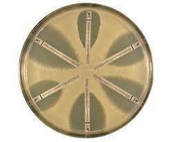
- 22What is the minimum inhibitory concentration (MIC) & why is it important?The lowest concentration of an antimicrobial that will inhibit visible growth of a microbe
It is important to determine the lowest concentration of an antibiotic that will prevent the growth of microbes. It is important so we use the lowest effective dose to reduce side effects in the patient. - 23What is the MIC of this antibiotic?.25ug/ml
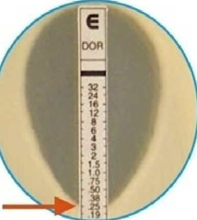
- 24How is the MIC read?The point of the elliptical zone (lower number) intersects the strip; the number it touches is the MIC.
- 25What is UV radiation?Nonionizing, short wavelength, high energy radiation
- 26Longest wavelengthUVA
- 27280-315 nmUVB
- 28100-280 nm; most damaging wavelengthUVC
- 29What UV wavelength results in DNA damage?254-260nm (UVC)
- 30How is DNA damaged by prolonged exposure to UV radiation?UV damage causes pyrimidine dimers - covalent bonds between pyrimidines which causes "kinks" in the DNA. Polymerase cannot replicate the DNA resulting in mutations/cell death
- 31What is the most common pyrimidine dimer?Thymine-Thymine dimers
- 32How do bacterial cells repair DNA damage from UV exposure?Via the SOS system which is activated when large amounts of DNA damage occurs. The dimer is cut out and replaced with new pyrimidine molecules, fixing the damage.
- 33What are the variables for UV lethality?1. Time of exposure - increased time of continuous exposure eventually results in lethal effects
2. Wavelength - 260nm is most damaging/lethal
3. Distance from UV source - the closer to the source, the more damaging
4. Barriers - Nonionizing UV cannot penetrate barriers of any kind; anything in the way will not allow the waves to pass through and no damage occurs
5. Endospores - bound with acid soluble proteins that protect if from damage as well as enzymatic repair of damaged DNA
6. Biofilms - provide some UV protection - 34What medium is used in the UV radiation experiment?TSA (Tryptic Soy Agar)
- 35Conjugation, transformation, transduction & transposons are what kind of gene transfer?Horizontal gene transfer
- 36ConjugationTransferring genetic material (usually via plasmids) from a donor cell to a recipient cell by a sex pilus.
- 37What is a plasmid?A small, circular, double-stranded DNA molecule that is not part of the chromosome and do not carry essential genes. Plasmids carry genes that may provide antibiotic resistance
- 38Is conjugation more or less common in biofilms?More common
- 39What type of gene was transferred to a recipient bacterium by conjugation in lab?Resistance gene for streptomycin resistance
- 40What medium is used for the conjugation test?LB Broth
- 41What microbe was used for the conjugation experiment?E. Coli
- 42How does vertical gene transfer differ from horizontal?Vertical gene transfer is done via sexual reproduction (parent to offspring), while horizontal transfer is the transfer of genes to cells of the same generation via a direct link (sex pili)
- 43Do bacteria reproduce sexually?No
- 44How do biofilms increase horizontal gene transfer?Biofilms promote plasmid stability and increase the range of mobile genetic elements that are transferred. Since microbes are close together in a biofilm, transfer is easier and can reach a greater number of cells.
- 45Why is conjugation important in the healthcare setting?Conjugation can spread antibiotic resistance; patients in the hospital are already immunocompromised and are on antibiotics, allowing a higher rate of selection for resistance.
- 46What is transformation?Insertion of a gene into an organism that changes the recombinant organism's traits.
- 47Are bacterial cells usually naturally competent?No
- 48How do competent cells take up free or naked DNA?Competent cells have DNA receptors on their surface, allowing them to bring the DNA into the cell.
- 49How can we induce competence in a bacterial cell?By using heat shock and CaCl2 (calcium chloride). Heat shock creates a pressure difference between the inside & outside of the cell, inducing pore formation that the DNA can pass through.
- 50In lab, E. Coli was transformed with a plasmid containing what gene?A gene that encodes for the enzyme Beta-lactamase (bla), which breaks down ampicillin.
- 51How were bacteria containing the bla gene selected for in lab?Ampicillin was placed in the growth medium, allowing only resistant cells to grow
- 52Why are most prokaryotic cells unable to perform endocytosis?Due to their small size and rigid cell walls.
- 53What do prokaryotic cells do instead of endocytosis to bring molecules inside the cell?These cells will secrete exoenzymes into the environment and
digest large molecules into its base components and then bring it
into the cell - 54What are the steps of transformation?1. Competent cells express DNA binding proteins on their surface that bind free DNA from the environment.
2. Nuclease domain in the complex above will digest one of the strands of DNA bound to the complex
3. Undigested strand (ssDNA) is transported into the cell intact
4. ssDNA undergoes homologous recombination (recombines with the bacterial chromosome) resulting in new traits - 55How is a plasmid incorporated into a cell via transformation?1. DNA circularizes
2. Complementary strand is synthesized
3. Newly formed dsDNA exists extra-chromosomally - 56What are the differential media used to detect unknown enteric bacteria testing for?Enzymatic activity by using pH indicators.
- 57Enterobacteriaceae1. Gram Negative rods
2. Ferment glucose
3. Normal GI microbiota, opportunists (E. Coli), and pathogens (Salmonella, Shigella) - 58How are bacteria identified based on metabolic/enzymatic activity?By the use of differential media, which shows color changes as the pH of the media changes. These changes show us the presence (or lack of) enzymatic activity.
- 59Sulfide, Indole, Motility (SIM)Combined differential Media that tests for:
1. Sulfur reduction to hydrogen sulfide (H2S)
2. Indole production from the amino acid tryptophan
3. Motility - 60Motility in SIMA needle is used to inoculate SIM with a stab. If the bacteria swim away from the line, they are motile (flagella). If bacteria grow only on the line, they are non-motile.
- 61Sulfur reduction (H2S production) in SIM mediaBacterial enzymes remove the sulfur from the amino acid cysteine, or reduce sulfate directly; both produce H2S.
The H2S combines with ferrous sulfate in the media and turns black. - 62Indole production in SIM-Tryptophanase hydrolyzes tryptophan, producing indole & pyruvate.
-Indole is detected by adding Kovac's reagent. Reagent combines with indole to make rosindole
-Rosindole turns the pH indicator bright cherry red - 63Tripe Sugar Iron (TSI) AgarDifferentiates bacteria based on sugar fermentation & sulfur reduction
- 64What are the 3 sugars in TSI?1. Glucose
2. Sucrose
3. Lactose - 65How is TSI used to determine sugar fermentation?Sugar fermented: pH is acidic; medium turns yellow
No fermentation: Sugar was not fermented so peptones were used; medium turns red due to alkaline pH
Bubbles or cracks in the medium indicate fermentation due to the production of CO2 - 66How does TSI detect sulfur reduction?Media contains ferrous sulfate, turning black in the presence of H2S from sulfur reduction
- 67What is the pH indicator in TSI agar?Phenol red
- 68Simmon's Citrate Agar (SCA)Differential media that allows us to differentiate between gram negative bacteria based on citric acid utilization.
- 69What is the sole carbon source in SCA?Sodium citrate
- 70What is the nitrogen source in SCA?Ammonium phosphate
- 71What is the pH indicator in SCA?bromthymol blue
- 72What does the enzyme citrase do?It allows bacteria to use sodium citrate for energy, converting ammonium phosphate to ammonia turning the media blue
- 73Differentiates bacteria based on the presence of the enzyme ureaseUrea Agar
- 74What does urease do?It hydrolyzes certain amino acids and urea to make ammonia
- 75What is the pH indicator in Urea Agar?Phenol red
- 76What medium is this and what are the results?Triple Sugar Iron (TSI) Agar
Yellow slant & butt: bacteria fermented the glucose (butt; no oxygen required) and either sucrose or lactose (or both) (slant; oxygen required).
Fermentation produces acidic byproducts, turning the medium yellow (acidic pH)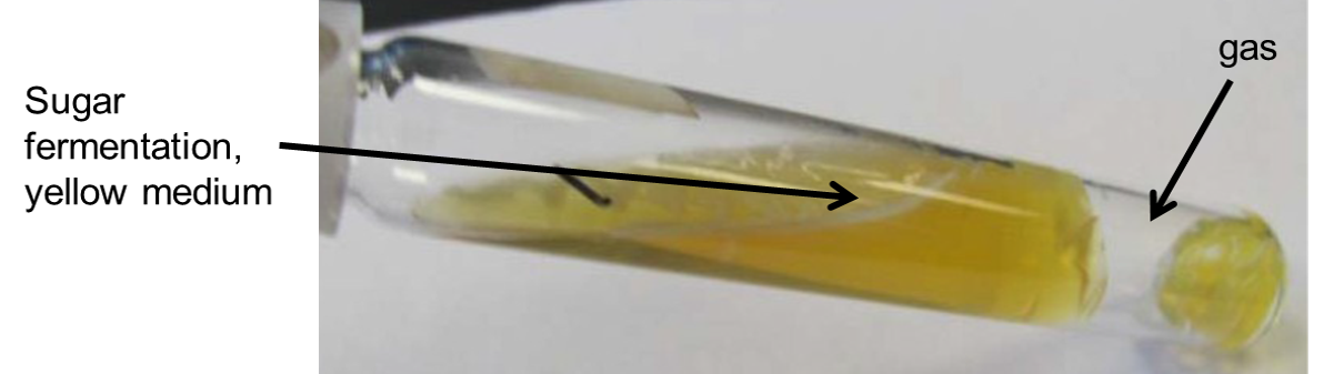
- 77What medium is this & what are the results?Triple sugar iron (TSI) agar
Red butt & slant (K/K): the bacteria did not ferment any of the sugars; only peptones were used. Peptones break down into ammonia, making the media alkaline.
Bacteria are NOT enterobacteriaceae or alkaline reversion/conversion occured.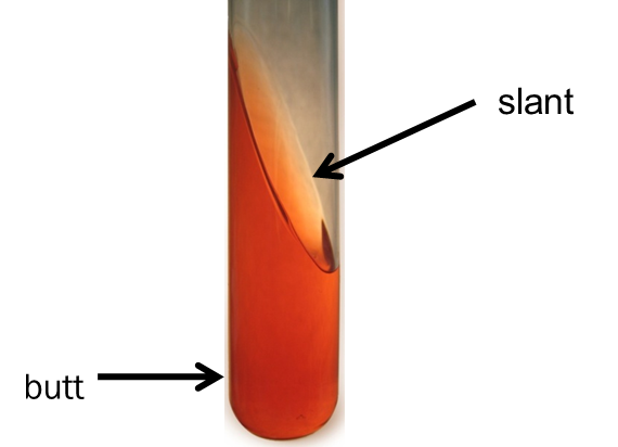
- 78What medium is this & what are the results?TSI
Yellow butt (acidic): bacteria fermented the glucose but we unable to use the other sugars. Acidic byproducts of fermentation cause the pH to become acidic
Pink slant (alkaline): The bacteria used the amino acids for food after the glucose (peptonization) producing ammonia; ammonia is alkaline, turning the media pink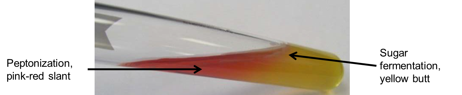
- 79What is this medium & what are the results?TSI
Black butt: bacteria reduced sulfate (anaerobic respiration) and produced H2S gas. Ferrous sulfate turns black in the presence of H2S.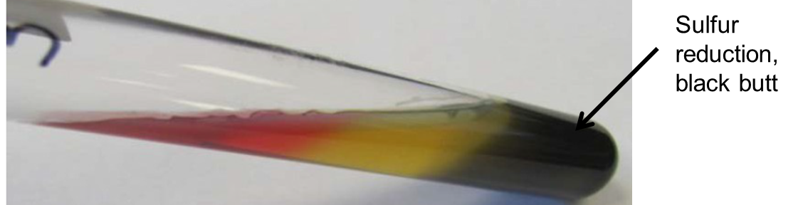
- 80What is alkaline reversion/conversion?Bacteria initially rapidly ferment all the sugars, turning the media yellow due to the acidic products of fermentation.
They then use amino acids for food and produce ammonia, turning the media red (alkaline). - 81What medium is this & what is this testing for?Sulfide Indole Motility (SIM) medium
Tests for sulfur reduction, indole production & motility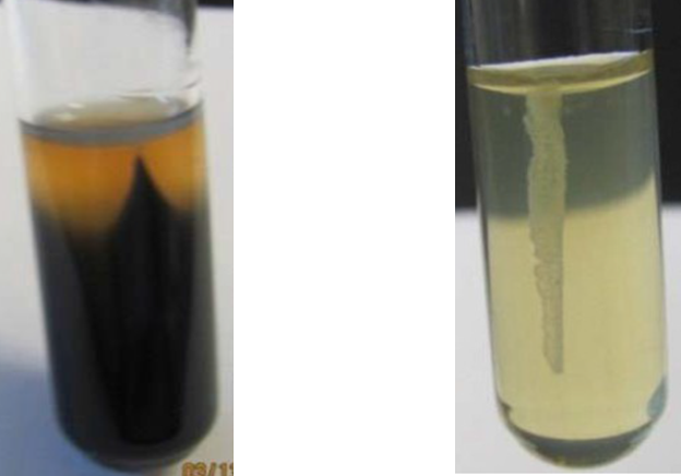
- 82What do the results on the left indicate (SIM)?Black indicates the bacteria reduced sulfur and produced H2S.
- 83What do the results on the right indicate (SIM)?Clear medium: The bacteria did not reduce sulfur
- 84What medium is this & what is it testing for?SIM
This is testing for the presence of Indole.
Tryptophanase hydrolyzes tryptophan, producing indole & pyruvate. Indole reacts with DMABA in Kovac's reagent to make rosindole, which turns the media pink.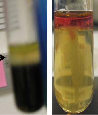
- 85What do the results on the left indicate? (SIM)Bacteria did not break down tryptophan, no indole production.
- 86What do the results on the right indicate? (SIM)Red top: DMABA in Kovac's Reagent reacted with indole to form rosindole, turning it pink.
This indicates the bacteria metabolized tryptophan & produced indole. - 87What medium is this & what is being tested?SIM
Testing for motility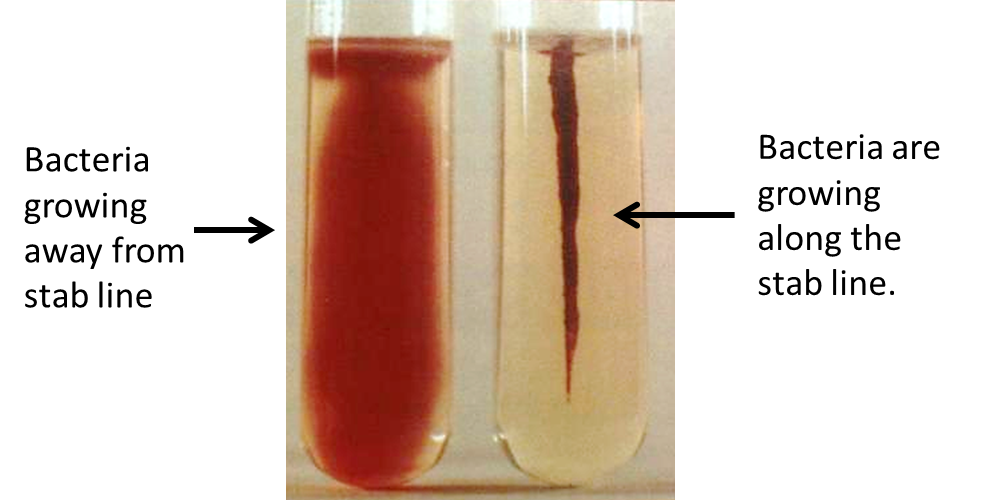
- 88What do the results on the left indicate? (SIM)Motile
Cloudiness extending from the stab line indicates the bacteria are able to move through the solid media - 89What do the results on the right indicate? (SIM)Not motile
Growth is only seen along the stab line, bacteria were not able to move through the media - 90What medium is this and what is it testing for?Simmon's Citrate Agar (SCA)
Detects the presence of the enzyme citrase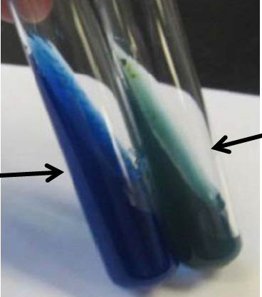
- 91What do the results on the left indicate?Positive for citrase activity
Bacteria with citrase enzyme were able to use sodium citrate & the nitrogen to make ammonia, which changes the pH to alkaline/ turns the media blue (bromthymol blue) - 92What do the results on the right indicate?Negative for citrase activity
Bacteria do not have citrase, and cannot use citrate as food, leaving the color unchanged. - 93What is the medium and what enzyme is being detected?Urea Agar
Used to detect the presence of urease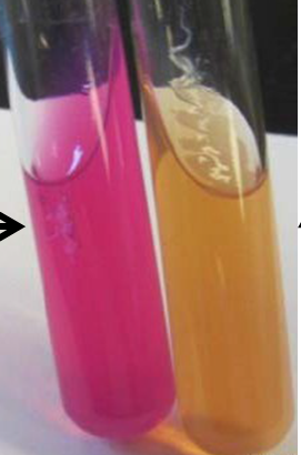
- 94What do the results on the left indicate?Urea Agar
Positive for urease activity
Bacteria that have urease hydrolyze urea to ammonia, turning the pH alkaline.
Alkaline pH: bright pink (phenol red) - 95What do the results on the right indicate?Urea Agar
Negative for urease activity
Bacteria do not have the urease enzyme so they cannot use urea; the medium stays orange - 96What are coliform bacteria?1. Gram Negative bacillus
2. Facultative anaerobes
3. Ferment lactose producing acid and gas
4. Only live in the GI tract of animals - 97What are the differential mediums used in the identification of unknown enteric bacteria?1. Triple Sugar Iron (TSI) Agar
2. Sulfide-Indole-Motility (SIM) medium
3. Simmon's Citrate Agar (SCA)
4. Urea Agar - 98What does the presence of coliform bacteria in a water sample indicate?Indicates fecal contamination of water supply and the POSSIBLE presence of GI pathogens.
- 99What selective-differential mediums are used to screen water to detect coliforms?1. Rapid'E.coli 2 Agar
2. Eosin Methylene Blue
3. Hektoen Enteric Agar
4. MacConkey Agar - 100What do the selective-differential mediums select for and differentiate in water testing?1. Selects for the growth of gram-neg bacteria while inhibiting growth of gram positive (Coliforms are gram-neg)
2. Allows differentiation between lactose fermenters (coliforms) and non-lactose fermenters (non-coliform) - 101What 2 dyes are in Eosin Methylene Blue (EMB)?1. eosin Y
2. methylene blue - 102What do the dyes in EMB do?Inhibit the growth of Gram-pos bacteria & act as an indicator that reacts with the acid produced during lactose fermentation
- 103If coliforms turn EMB pink/dark purple, what does this indicate and why?Coliforms are lactose fermenters and produce acid as a byproduct of fermentation. The acidic byproducts turn the medium pink/dark purple as a result of the media becoming acidic.
- 104If EMB remains red, what does this indicate and why?If EMB stays red, there was no lactose fermentation so the pH does not change.
This means no coliforms are present, but there ARE non-coliforms growing. - 105If EMB turns pink/dark purple and the bacteria are a metallic green, what does this indicate and why?E. Coli are present- the metallic green color is from excessive acid production (rapid fermentation) and the media turns pink in response to an acidic pH change from the acidic byproducts of fermentation.
- 106What does Hektoen Enteric Agar (HEA) contain to inhibit the growth of gram-postive bacteria?Bile salts
- 107What 3 sugars does HEA contain?1. Lactose
2. Sucrose
3. Salicin - 108How do the 3 sugars (lactose, sucrose and salicin) differentiate between microbes?It is used to differentiate the presence of coliforms and Salmonella & Shigella from other enterics.
- 109If HEA turns yellow-pinkish orange, what does this indicate?Bacteria are fermenting any of the 3 sugars & produce acidic byproducts, turning the media yellow-pinkish orange in response to the acidic media.
This indicates coliforms are present. - 110If HEA is blue-green, what does this indicate and why?No fermentation has occured, no coliforms present BUT there are non-coliforms growing.
- 111What does HEA contain to detect H2S production?1. Sodium thiosulfate (sulfur)
2. Ferric Ammonium Citrate - 112If HEA produces black spots, what does this indicate and why?Black spots indicate the bacteria used the sulfur as food & produced H2S gas.
H2S reacts with the ferric ammonium citrate to produce ferrous sulfide, turning the media black.
Salmonella present, non-coliform - 113What 2 things does MCA have to inhibit growth of gram-positive microbes?1. Bile salts
2. Crystal Violet - 114What pH indicator does MCA use?neutral red indicator
Due turns pink when lactose fermentation occurs (acidic; coliforms present) - 115If MCA turns pink, what does this indicate and why?Lactose fermentation produced acidic byproducts that reacts with the neutral red indicator, turning the media pink due to the acidic pH.
- 116If MCA remains colorless, what does this indicate?No lactose fermentation occured, no coliforms present BUT there and non-coliforms growing
- 117What type of medium is Rapid'E. coli 2 Agar and what is it detecting?Selective chromogenic medium
Used to detect E. coli and other coliforms - 118What 2 enzymatic activities are detected simultaneously in Rapid'E. coli 2 Agar?1. Galactosidase (GAL)
2. Glucuronidase (GLUC) - 119What 2 chromogenic substances are used in Rapid'E. coli 2 Agar?1. 1 specific to GAL, creates blue colored colonies if GAL is present
2. 1 specific to GLUC, creates pink colored colonies if GLUC is present. - 120What forms blue/green colonies in Rapid'E. coli 2 Agar?Coliforms
GAL +
GLUC - - 121What forms violet/pink colonies on Rapid'E. coli 2 Agar?E. coli
GAL/GLUC + - 122If GLUC is detected (violet/pink colonies), what microbe is most likely present and why?E. coli, as they are one of the only species of enterobacteriaceae to possess the GLUC enzyme.
There are a few strains of Salmonella & Shigella that are GLUC positive as well. - 123What is this medium and what do the results on the left indicate?Eosin Methylene Blue (EMB)
Metallic green colonies are from E. coli rapidly fermenting lactose
The media turned pink due to the acidic byproducts of lactose fermentation, indicating the presence of coliforms.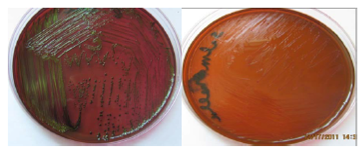
- 124What is the medium and what do the results on the right indicate?EMB
Agar remained red, no lactic acid fermentation occured so no coliforms are present BUT non-coliforms are growing. - 125What is the medium and what do the results on the left indicate?Hektoen Enteric Agar (HEA)
Bacteria (coliforms) have fermented any of the 3 sugars. Fermentation produces acidic byproducts, turning the media yellow-pinkish orange due to the acidic pH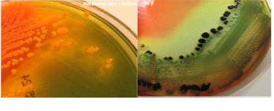
- 126What is the medium and what do the results on the right indicate?HEA
Blue-green media indicates no sugar fermentation occured (no coliforms present) BUT non-coliforms are growing.
Black colonies indicate the bacteria used sulfur to produce H2S gas. H2S reacts with the ferric ammonium citrate to produce ferrous sulfide (black color) - 127What is the medium and what do the results on the left indicate?MacConkey Agar (MCA)
Lactose fermentation has occured producing acidic byproducts. Acidic byproducts react with the neutral red indicator, turning the media pink
Coliforms present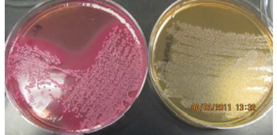
- 128What is the medium and what do the results on the left indicate?MCA
Clear agar means no fermentation occured, so there is no pH change.
Coliforms not present BUT non-coliforms are growing. - 129What medium is this and what do these results indicate/what enzyme is detected?Rapid'E. coli 2 Agar
Blue/Green colonies indicate GAL enzyme is detected
Coliforms present (GAL+/GLUC-)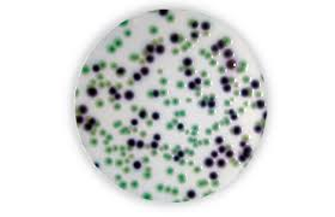
- 130What medium is this & what do the results indicate/ what enzyme is detected?Rapid'E. coli 2 Agar
Violet/pink colonies indicate the detection of GLUC enzyme.
GLUC detection is most likely E.Coli (GLUC & GAL+)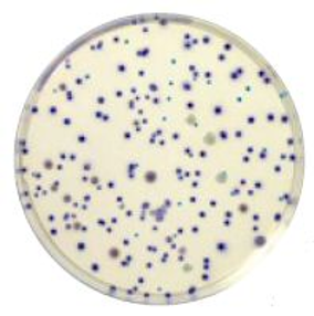
- 131What is a Bacteriophage?Viruses that infect and replicate inside bacteria
- 132What are the steps of lytic infection in bacteria from a bacteriophage?1. Phage attaches to host cell by the tail fiber
2. Phage penetrates host cell and injects it's DNA
3. Phage DNA directs synthesis of viral components by the host cell
4. Viral components are assembled into virions
5. Host cell lyses releasing mature virions - 133How does lysogenic infection differ from lytic infection (bacteriophage)?Lytic results in lysing the cell to exit.
Lysogenic infection incorporates phage DNA into the bacteria's chromosome becoming a dormant prophage. Bacteria reproduces normally, replicating phage DNA with bacterial DNA. This cycle continues until some event causes the prophage to be released, where it can enter a lytic cycle. - 134In both lytic & lysogenic cycles, what must phage DNA do once injected into the bacterial cell?After the phage attaches to the host cell and injects it's DNA, the phage DNA must circularize. It can then enter a lytic or lysogenic stage.
- 135What are the steps of transduction?1. Phage attaches to host cell & injects phage DNA.
2. Phage proteins & DNA are made, and the bacterial DNA is "chopped up" to make sure the host cannot fight back.
3. A mistake is made, and pieces of the bacterial DNA are packaged in the phage capsid.
4. Host cell lyses, releasing phage particles containing bacterial DNA
5. Phage carrying bacterial DNA injects a new host cell, where the new DNA can recombine producing a recombinant cell with a different genotype from both donor and recipient cells.
This can result in antibiotic resistance being transferred - 136Staphylococcus aureusGram positive Cocci
Pyogenic (pus-forming) causing abscesses & can invade tissues
Halotolerant
Ferment mannitol (sugar)
Hemolytic
Coagulase positive - 137Staphylococcus saprophyticusGram positive Cocci
May be normal flora in the vaginal tract; associated with UTI
Halotolerant
Ferment mannitol (sugar)
Non-hemolytic
Coagulase negative - 138Staphylococcus epidermidisGram positive Cocci
Common skin flora, but may cause blood infections (nosocomial) when IV is administered
Halotolerant
Do not ferment sugar
Non-hemolytic
Coagulase negative - 139Enterococcus species can cause what kind of infections?Blood stream infections & can be resistant to antibiotics (vancomysin resistant Enterococcus [VRE])
- 140How is Hydrogen Peroxide (H2O2) used to detect catalase enzyme in Staph species?Staph live in oxygen rich environments and must be able to break down reactive oxygen species (ROS) and superoxide radicals; toxic metabolic byproducts of aerobic cellular respiration.
Hydrogen peroxide is a ROS. Staph are catalase-positive, and the enzyme breaks down hydrogen peroxide into water and oxygen.
Bubbling indicates the presence of catalase. - 141If the catalase test is positive, what would you expect to see and what type of bacteria is it?Oxygen bubbles forming after mixing bacteria with hydrogen peroxide, which tells us the bacteria is most likely Staph (catalase positive)
- 142If the catalase test is negative, what results would you expect to see and what bacteria is present?No bubbles mean no catalase is present; bacteria is not Staph.
- 143What 3 tests are used AFTER the catalase test to determine the species of Staph?1. Mannitol Salt Agar
2. Hemolysin
3. Coagulase - 144What does Mannitol Salt Agar contain?1. Mannitol (sugar)
2. 7.5% salt - 145What does Mannitol Salt Agar differentiate?1. The salt will inhibit the growth of non-halotolerant bacteria, while halotolerant bacteria (Staph) are able to grow.
2. S. aureus and S. saprophyticus can ferment mannitol and produce acid, turning the medium yellow. S. epidermidid cannot ferment mannitol, so the agar stays red. - 146How do we differentiate between S. aureus & S. epidermidis & S. saprophyticus?1. Hemolysin: S. aureus is hemolytic (Beta) and break down RBC which can be seen on the blood agar. S. epidermidis & sapro are non-hemolytic and will show gamma hemolysis.
2. Coagulase: S. aureus is the only species of Staph with the coagulase enzyme which results in clotting of plasma by converting fibrin to fibrinogen. - 147EpidemiologyThe study of diseases in a population
- 148OutbreakAn occurrence of a disease in a population
- 149Point source outbreakEveryone affected got it from a common source (food poisoning from a restaurant)
- 150Propagated outbreakDisease is spread between people or animals. The number of cases build over time, like the flu or cold.
- 151Index case/ Patient 0The individual was was originally infected and spreads the infection throughout a community
- 152Epidemicdiseases/disorders that occur in a particular community at higher than expected rates
- 153PandemicEpidemic that spreads to more than 1 continent, infecting a large number of people.
- 154What are the 3 types of diseases?1. Communicable
2. Noncommunicable
3. Zoonosis - 155Communicable diseaseDisease that is spread between people, causing a propagated outbreak.
- 156Noncommunicable diseaseDisease that is spread by a vector (Lyme's disease)
- 157Zoonotic diseaseDisease that is spread from an animal to a person (rabies)
- 158What are the 3 types of immunity?1. Innate
2. Acquired
3. Herd - 159What is Innate Immunity?Non-specific immunity, naturally present at birth. Does not depend on prior experience.
- 160What is Acquired Immunity?Immunity developed as a result of exposure to a specific organism
- 161Herd ImmunityThe resistance of a group to the invasion or spread of an infectious organism.
- 162How is Herd Immunity acquired?By vaccinating the majority of a population from the infection
- 163How is DNA Fingerprinting used to ID unknown microorganisms?The DNA is cut into pieces (fragments) and are separated by size. The unknown fingerprint is compared to known fingerprints. Same fingerprint=same or similar microbe as the known. Different fingerprint=organisms are not the same or could be a mutation of the known microbe.
- 164What enzymes are used to cut DNA?Restriction enzymes
- 165How do restriction enzymes cut DNA into fragments?Restriction enzymes recognize specific nucleotide sequences, bind to the DNA and cleave the DNA between the bases.
- 166How are DNA fragments separated from each other?By using agarose gel electrophoresis which separates the (-) charged DNA fragments using and electric current.
DNA moves from the (-) pole to the (+) pole and is separated by size - 167What medium is used in the DNA fingerprinting and what does it do?Agarose Gel
It is porous and allows DNA to move through it when an electric current is applied. - 168What 2 stains are used in DNA fingerprinting?1. Ethidium bromide
3. GelGreen - 169How do the stains in DNA fingerprinting allow us to visualize DNA?They bind to DNA; the DNA-dye complexes emit a light under UV that can be visualized.
- 170What is Serology?The study of antigen-antibody reactions
- 171Antigenmacromolecules or fragments of molecules that may trigger an immune response
- 172What are 3 examples of antigens attached to cells?1. Lipopolysaccharides (O-antigens)
2. Flagellar proteins (H-antigens)
3. Capsule (K-antigens) - 173What is an example of a soluble antigen?Toxins found in serum
- 174AntibodyLarge, Y-shaped proteins produced by B cells that can recognize specific antigens.
- 175What does the variable region of an antibody bind to?Antigenic binding site of the variable region binds to the specific antigen at small structures (epitopes) or antigenic determinants
- 176What is an antigen-antibody reaction?The binding of an antibody to the epitope of an antigen
- 177What can an antigen-antibody reaction result in?1. Precipitation
2. Agglutination
3. Neutralization of toxins/viruses
4. Complement fixation
5. Opsonization - 178PrecipitationAntigen-antibody reaction that occurs between soluble antigens and their specific antibody
- 179How is precipitation studied?Gel immunodiffusion; antiserum is placed in one well and an antigen is placed in the other. The two diffuse out toward one another (double diffusion)
- 180What medium does gel immunodiffusion use and how does it work?Porous agarose medium that allows antibodies and antigens to diffuse through easily.
- 181What does a white preciptin line indicate?A specific antibody recognises and binds to the soluble antigen. The white preciptin line forms as their diffusion paths cross.
- 182What is agglutination?A reaction between antibodies & antigens that results in clumping of bacterial cells.
- 183What is the purpose of latex beads in the Slide Agglutination Test?Beads with specific antibodies adsorbed (attached) to the latex.
Enhances the observation of clumps in agglutination. - 184What causes crosslinking in the Slide Agglutination test?The antibodies on the latex beads contact the epitope of it's matching antigen & bind together. The crosslinking between antigens allow agglutination clumps to be seen easily.
- 185What is slide agglutination used for?Rapid, specific and definitive identification of bacteria
- 186What is an essential trace element for all living organisms?Iron
- 187How do our cells protect our iron from invading bacteria?Iron is sequestered -bound to carrier molecules & stored inside cells)
- 188How do bacteria steal iron from host cells?Via the hemolytic toxin "Hemolysin" which binds to & lyse RBCs, releasing hemoglobin.
Siderophores (bacterial iron binding proteins) pull iron from hemoglobin & bring it into the bacterial cell. - 189What is a titer?A ratio used to determine the functional amount of a compound in a soultion.
- 190How are serial dilutions used in the titer test?The solution is diluted until the compound no longer functions.
- 191How is the titer calculated?The titer is the reciprocal of the last dilution showing function
- 192How are titers used in healthcare?They are used to determine the effectiveness of a vaccine or exposure to a pathogen.
- 193What test is this and what do the results on the left indicate?Slide agglutination
Positive
latex agglutination, visible clumps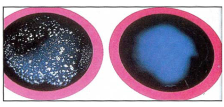
- 194What test is this and what do the results on the right indicate?Slide agglutination
Negative
No agglutination, no visible clumps - 195What medium is this and what do the results on the left indicate?Mannitol Salt Agar (MSA)
No acid production, so mannitol was not fermented. pH remains the same; agar does not change.
Since there is growth, it is halotolerant S. epidermidis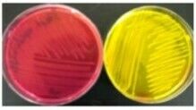
- 196What is the medium & what do the results on the right indicate?Mannitol Salt Agar (MSA)
Mannitol was fermented producing acidic byproducts. The acid changes the agar's pH to acidic, turning the indicator yellow.
The bacteria is halotolerant; it could be either S. aureus or S. saprophyticus - 197What test is this & what do the results on the top indicate?Coagulase test
The tube on the top is coagulase positive, causing the blood plasma in the tube to clot and solidify.
This is most likely S. aureus, since S. saprophyticus is coagulase neg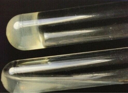
- 198What is this test & what do the results on the bottom indicate?Coagulase test
No coagulation occured, so the microbe is coagulase negative.
S. saprophyticus - 199What test is this & what do the results on the top indicate?Hemolysis (Blood Agar)
Beta hemolysis; complete lysis of RBC
S. aureus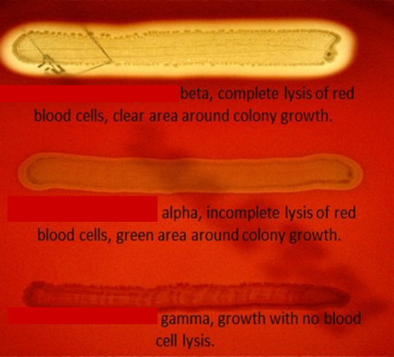
Información
- Descripción
- Biology Fichas sobre Biol 251 Lab practical , creado por Olivia Poston el 18/04/2023.
- Creado por
- Olivia Poston
- Visitas
- 7
- Valoración