
Diagrama de flujo
DNA Sequencing- SANGER Method
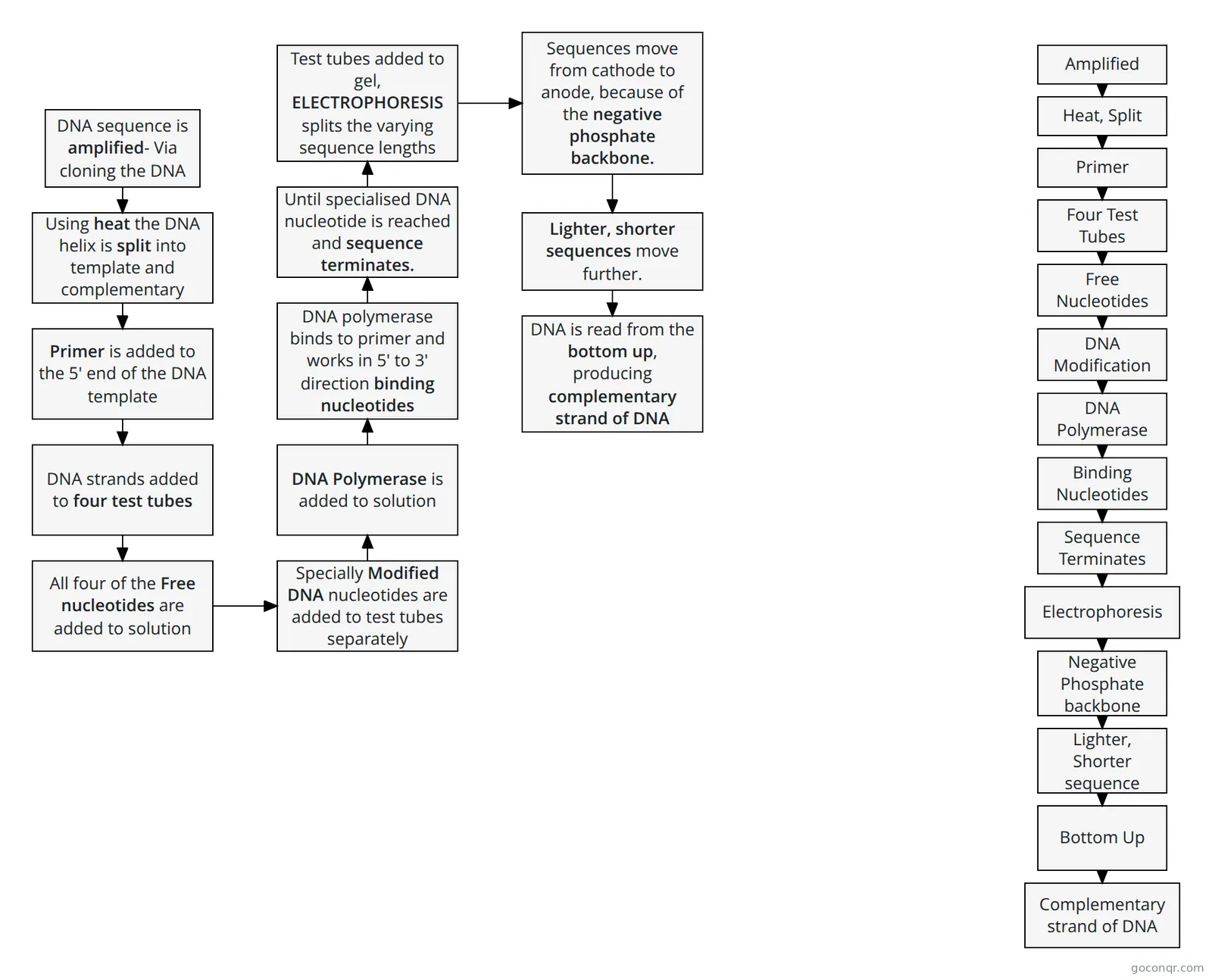
Flowchart: DNA sequencing Sanger method
Contenido del diagrama
DNA sequence is amplified- Via cloning the DNA
Using heat the DNA helix is split into template and complementary strand
Primer is added to the 5' end of the DNA template
DNA strands added to four test tubes
All four of the Free nucleotides are added to solution
Specially Modified DNA nucleotides are added to test tubes separately
DNA Polymerase is added to solution
DNA polymerase binds to primer and works in 5' to 3' direction binding nucleotides
Until specialised DNA nucleotide is reached and sequence terminates.
Test tubes added to gel, ELECTROPHORESIS splits the varying sequence lengths
Sequences move from cathode to anode, because of the negative phosphate backbone.
Lighter, shorter sequences move further.
DNA is read from the bottom up, producing complementary strand of DNA
Amplified
Heat, Split
Primer
Four Test Tubes
Free Nucleotides
DNA Modification
DNA Polymerase
Binding Nucleotides
Sequence Terminates
Electrophoresis
Negative Phosphate backbone
Lighter, Shorter sequence
Bottom Up
Complementary strand of DNA
Información
- Descripción
- AS level (Module 6- Genetics and Ecosystems) Biology Diagrama sobre DNA Sequencing- SANGER Method, creado por 007842-Stuart Denton el 30/05/2017.
- Creado por
- 007842-Stuart Denton
- Visitas
- 20
- Valoración