
Mapa mental
Microscopy and Staining
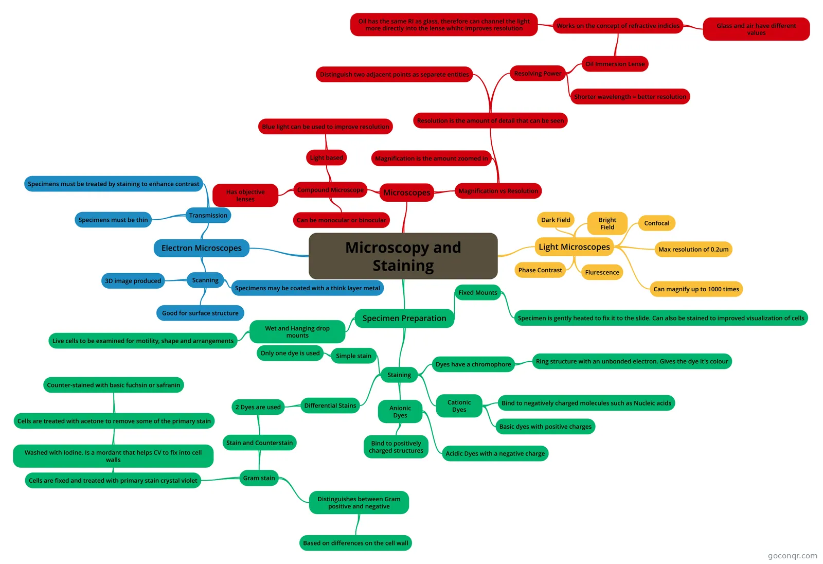
Mind map: Microscopy and staining
Contenido del mapa
Microscopy and Staining
Microscopes
- Compound Microscope
- Can be monocular or binocular
- Has objective lenses
- Light based
- Blue light can be used to improve resolution
- Magnification vs Resolution
- Magnification is the amount zoomed in
- Resolution is the amount of detail that can be seen
- Distinguish two adjacent points as separete entities
- Resolving Power
- Shorter wavelength = better resolution
- Oil Immersion Lense
- Works on the concept of refractive indicies
- Glass and air have different values
- Oil has the same RI as glass, therefore can channel the light more directly into the lense whihc improves resolution
- Works on the concept of refractive indicies
- Compound Microscope
Light Microscopes
- Bright Field
- Dark Field
- Phase Contrast
- Flurescence
- Confocal
- Can magnify up to 1000 times
- Max resolution of 0.2um
Electron Microscopes
- Transmission
- Specimens must be thin
- Specimens must be treated by staining to enhance contrast
- Scanning
- 3D image produced
- Specimens may be coated with a think layer metal
- Good for surface structure
- Transmission
Specimen Preparation
- Wet and Hanging drop mounts
- Live cells to be examined for motility, shape and arrangements
- Fixed Mounts
- Specimen is gently heated to fix it to the slide. Can also be stained to improved visualization of cells
- Staining
- Cationic Dyes
- Basic dyes with positive charges
- Bind to negatively charged molecules such as Nucleic acids
- Anionic Dyes
- Acidic Dyes with a negative charge
- Bind to positively charged structures
- Dyes have a chromophore
- Ring structure with an unbonded electron. Gives the dye it's colour
- Simple stain
- Only one dye is used
- Differential Stains
- 2 Dyes are used
- Stain and Counterstain
- Gram stain
- Distinguishes between Gram positive and negative
- Based on differences on the cell wall
- Cells are fixed and treated with primary stain crystal violet
- Washed with Iodine. Is a mordant that helps CV to fix into cell walls
- Cells are treated with acetone to remove some of the primary stain
- Counter-stained with basic fuchsin or safranin
- Cells are treated with acetone to remove some of the primary stain
- Washed with Iodine. Is a mordant that helps CV to fix into cell walls
- Distinguishes between Gram positive and negative
- Gram stain
- Stain and Counterstain
- 2 Dyes are used
- Cationic Dyes
- Wet and Hanging drop mounts
Información
- Descripción
- 2 Biology Mapa Mental sobre Microscopy and Staining, creado por Danny Que el 15/03/2016.
- Creado por
- Danny Que
- Visitas
- 15
- Valoración