
Mapa mental
Pattern Recognition
Contenido del mapa
Pattern Recognition
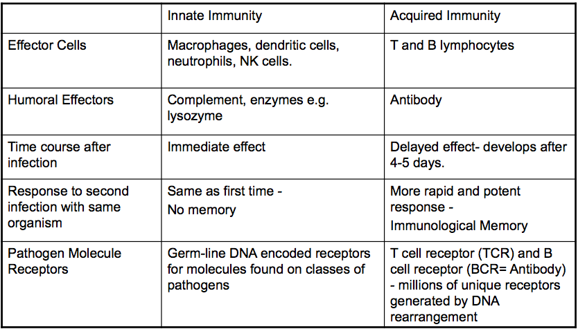
Innate vs Acquired Immunity
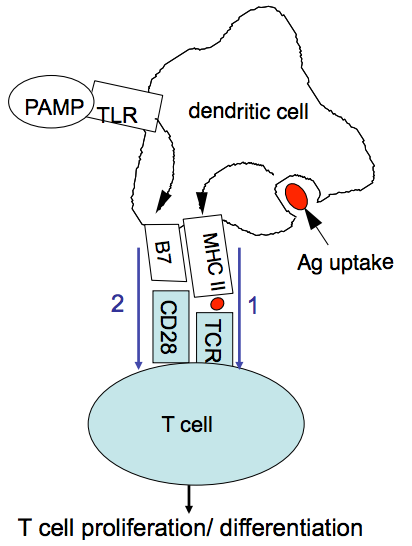
T cell activation by DCs requires 2 signals
- 1) MHC-peptide:TCR 2) cosstimulatory signal (e.g. B7 on DC: CD28 on T cell
- activation of an antigen specific T cell is determined by the presence of costimulatory molecules on the DC (not present in resting DCs).
- costimulatory molecules can be induced by recognition of PAMPs or DAMPs by the DC through pathogen-specific receptors such as TLRs
- 1) MHC-peptide:TCR 2) cosstimulatory signal (e.g. B7 on DC: CD28 on T cell
Self/Non-self Discrimination
- Self/non-self hypothesis
- Burnet and Medawar 1959
- Burnet = australian virologist/immunologist, developed theory immune tolerance. Director of Walter and Eliza Hall Institute (1944-1965). Won a prize in 1960
- Ag specific cells which recognise self molecules are eliminated in life, leading to tolerance of self.
- this leaves only cells which recognise foreign antigens. In this model, specific responses to foreign molecules is just dependent on T and B cells, and the process of tolerance
- Inconsistent with this hypothesis are:
- requirement for activated DC to present Ag to T cells
- requirement for adjuvant in vaccination
- vaccination with pure protein has little effect bc the acquired immune system does not recognise the antigen as foreign.
- Adjuvants: aluminium salts, LPS, bacterial DNA + polysaccharides, Freunds adjuvant (emulsion of killed mycobacteria)
- act as innate immune stimulants
- Adjuvants: aluminium salts, LPS, bacterial DNA + polysaccharides, Freunds adjuvant (emulsion of killed mycobacteria)
- vaccination with pure protein has little effect bc the acquired immune system does not recognise the antigen as foreign.
- not rejected, but built upon in later hypotheses
- Burnet and Medawar 1959
- Infectious non-self hypothesis
- distinction between self and foreign relies on the recognition of characteristic microbial molecules (PAMPs) by the innate immune system (PAMP=pathogen associated molecular pattern)
- PAMPs recognised by PRRs/TLRs on innate cells
- activation of DCs by PAMPs upregulates the costimulatory molecules required for T cell activation
- known that PAMPs are not the only route to DC activation (DAMPS)
- distinction between self and foreign relies on the recognition of characteristic microbial molecules (PAMPs) by the innate immune system (PAMP=pathogen associated molecular pattern)
- Danger hypothesis
- distinction relies on release of cellular molecules which indicate stress, damage, or danger to the cell
- in support: acquired immunity response to tumours and rejection of grafts in the absence of infectious agents
- self molecules called DAMPs (danger associated molecular patterns) can act through PRRs that recognise PAMPs
- Evidence for DAMPs: Monosodium urate crystals
- cytoplasmic extract behaved as an adjuvant
- it activated DCs by increasing B7 (CD86) and generated specific CTL against the antigen
- cytoplasmic extract was fractionated to find an active component - uric acid, active as monosodium urate crystals - note these will only form when outside the cell (indicating cell lysis) due to the elevated Na concentration outside the cell
- uric acid is normally present at 40-60micrograms/mL in blood, close to the limit of solubility in body fluids (70micrograms/mL). cells have uric acid concentrations between 1-4mg/mL which increase if cell is dying due to DNA and RNA breakdown (uric acid is a purine derivative. therefore when cells lyse, uric acid will form crystals with Na - an abundant ion
- cell damage can be caused by injury or infection
- uric acid is normally present at 40-60micrograms/mL in blood, close to the limit of solubility in body fluids (70micrograms/mL). cells have uric acid concentrations between 1-4mg/mL which increase if cell is dying due to DNA and RNA breakdown (uric acid is a purine derivative. therefore when cells lyse, uric acid will form crystals with Na - an abundant ion
- cytoplasmic extract was fractionated to find an active component - uric acid, active as monosodium urate crystals - note these will only form when outside the cell (indicating cell lysis) due to the elevated Na concentration outside the cell
- it activated DCs by increasing B7 (CD86) and generated specific CTL against the antigen
- cytoplasmic extract behaved as an adjuvant
- Examples:
- Small molecules: monosodium urate, extracellular ATP
- Intracellular proteins: HMGB1 (high mobility group protein B1)
- Mitochondrial molecules (similar to bacterial due to endosymbiotic theory of evolution: DNA from mitochondria (not methylated on cytosine), and N-formyl peptides from mitochondria
- Evidence for DAMPs: Monosodium urate crystals
- PAMPs:
- Bacterial Cell wall/surface molecules
- (GRAM -VE) LPS, lipoprotein, peptidoglycan, (GRAM+VE) lipoteichoic acid, peptidoglycan
- Flagellin
- yeast cell wall: "zymosan" beta 1-3 linked glucose
- DNA: bacterial and viral DNA has unmelthylated CG dinucleotides (170-80% of our CGs are methylated (5-methyl cytosine) and we have lower than random CG frequency)
- ssRNA: wide range of base modifications in pathogen RNA
- dsRNA: long dsRNA is foreign to vertebrates + is an intermediate in viral life cycle, may result from txn off both DNA strands of DNA viruses
- Bacterial Cell wall/surface molecules
- Self/non-self hypothesis
PRRs:
- named because they recognised repeating patterns characteristic of PAMPs
- also recognise DAMPs
- mediate
- phagocytosis (macrophage mannose receptor in MBL pathway)
- Chemoattraction (formyl peptide receptors)
- Production of chemokines and cytokines (inflammation)
- DC activation, leading to activation of the acquired immune system
- Examples
- Localisation
- Recognition of PAMPs in the cytosol tell the cell that it itself is infected
- important for viral detection and for bacteria such as Listeria which can live and or inject molecules into the cytoplasm out of the endosome
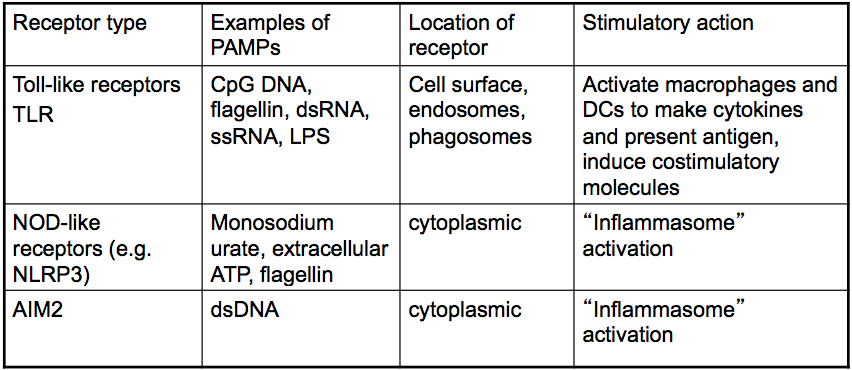
- performed by cytosolic TLRs to recognise nucleic acids
- TLRs on endosome = engulfed microbe (local extracellular infection)
- TLRs on cell surface = extracellular infection (local)
- Recognition of PAMPs in the cytosol tell the cell that it itself is infected
- Discoveries
- 1st discovered in Drosophila as a gene affecting embryonic development (strange morphologies formed) and defence against fungal and bacterial infection as its pathways induces antimicrobial peptides
- Mammalian TLR
- shown to stimulate cytokine production
- a cytoplasmic TLR domain was fused with an extracellular domain that dimerised upon binding. this lead to a protein cascade in the cytoplasm to produce inflammatory molecules
- Types:
- Extracellular: Cell membrane
- TLR5: flagellin
- TLR6+TLR2: diacyl lipopeptides
- TLR1+TLR2: triacyl lipopeptides
- TLR 4: LPS
- any ligands have been proposed fro TLR2 and 4
- Intracellular: inside Phagosome/endosome which is required to break down foreign DNA or RNA from the pathogen
- TLR3: dsDNA
- TLR7: ssRNA
- TLR9: unmethylated CG dinucleotides of DNA
- Bacterial and vertebrate DNA look very similar overall, so specific innate recognition of bacterial DNA was initially very surprising
- unmethylated CG DNA = CpG DNA, has sequences like AACGTT
- acts on macrophages, DCs and B cells
- induces inflammatory cytokines, promotes Ag presentation and induces co-stimulatory molecules on DCs
- short CpG oligonucleiotides are powerful immune stimulants and are in clinical trials as vaccine adjuvants, treatments for infectious disease and cancer, and as immunomodulators for allergy
- Extracellular: Cell membrane
- TLR structure: 2 important domains
- Leucine rich repeat (LRR) domain involved in ligand recognition
- has horseshoe shape
- LRR of TLR3 binds dsRNA directly
- Toll/interleukin-1 receptor (TIR) domain: cytoplasmic signalling
- this component is brought together in dimerisation to initiate a signal cascade
- Leucine rich repeat (LRR) domain involved in ligand recognition
Nod-like receptors, AIM2 and the inflammasome
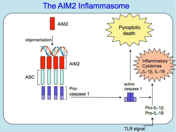
- the inflammasome = a large cytoplasmic protein which recruits and activates the protease caspase 1
- caspase 1 cleaves pro-IL1beta and pro-IL18 into active inflammatory cytokines
- IL-1beta
- causes vasodilation, fever, hypotension, infiltration if inflammatory cells, chemokine and cytokine release, activation of APCs
- role in inflammatory diseases
- major sources are macrophages, DCs and monocytes
- IL-18
- promotes T cell activation
- CASPASE-1
- initially called IL-1beta converting enzyme
- an inflammatory caspase (others are involved in apoptosis)
- caspases 8,9 +12 cleave caspases 3 and 7 to induce apoptosis
- caspases 4 and 5 are also inflammatory and are involved in pyroptosis
- its pro-domain in what is recruited to the inflammasome, where it is activated and induced to dimerise
- IL-1beta
- activation also leads to inflammatory lytic cell death termed pyroptosis
- activation initiated by a range of PAMPs and DAMPs causing oligomerisation of NLRs (particularly NLRP3 aka NALP3) and/or cytoplasmic DNA recognition by AIM2
- AIM2 binds DNA, it recruits ASC, ASC recruits procaspase 1, cleaves to caspase 1 and close proximity results in caspase1 dimerisation to form active caspase 1
- AIM2 - a cytoplasmic DNA receptor
- introduction of DNA into the cytoplasm of macrophages activates the inflammasome to increase ILs to cause pyroptosis
- ASC
- clusters in inflammasome formation. when tagged with GFP AIM2 and ASC are shown to be spread out in cytoplasm until DNA is added, in which case clustering occurs
- AIM2 - a cytoplasmic DNA receptor
- AIM2 binds DNA, it recruits ASC, ASC recruits procaspase 1, cleaves to caspase 1 and close proximity results in caspase1 dimerisation to form active caspase 1
- activation initiated by a range of PAMPs and DAMPs causing oligomerisation of NLRs (particularly NLRP3 aka NALP3) and/or cytoplasmic DNA recognition by AIM2
- caspase 1 cleaves pro-IL1beta and pro-IL18 into active inflammatory cytokines
- What activates the inflammasome?
- DAMPs
- extracellular ATP: binds P2X7 receptor to increase the potassium influx into the cell, resulting in a change in conformation of the NLRP3 so it can bind ASC to cluster it
- monosodium urate crystals (also particulate stimulus): macrophages engulf crystals, disrupts phagosome, inflammasome is activated, causing IL-1beta release and continuous inflammation, IL-1 receptor antagonist is a potentially successful treatment (NLRP3
- present in high levels in gout
- Particulate Stimuli
- monosodium urate (NLRP3)
- alum (used as vaccine adjuvant) (NLRP3)
- asbestos (NLRP3)
- silica (NLP3)
- beta-amyloid (NLRP3)
- cholesterol crystals (NLRP3)
- PAMPS
- Flagellin (NLRC4)
- cytoplasmic DNA (AIM2)
- bacterial pore-forming toxins, anthrax lethal toxins (NLRP3)
- Associated diseases include gout, Alzheimer's, atherosclerosis, diabetes
- DAMPs
- AIM2 inflammasome and Francisella tularensis (FT) innate immure recognition (PAPER)
- 1. prove that AIM2 is a receptor for DNA using macrophages from gene knockout mice
- primed cells with LPS to induce the need for an inflammatory event
- when ASC absent: no inflammatory response, so it is essential for IL release
- when AIM2 absent: DNA stimuli have no effect, but ATP and flagella have IL release, therefore AIM2 is essential for responses to DNA, so it is a receptor for DNA
- when ASC absent: no inflammatory response, so it is essential for IL release
- primed cells with LPS to induce the need for an inflammatory event
- 2. Prove AIM2 is a receptor that activates caspase 1
- performed western blot with AIM2 knockout cells
- found that pro-caspase-1 is found in all cells, but active caspase-1 is only present in WT cells (not AIM2 knockouts)
- procedure:1. run protein extracts on SDS-PAGE, 2. transfer proteins io nitrocellulose membrane, 3. incubate with soecific Ab to bind protein of interest (active caspase1- and pro-caspase-1), 4. use secondary Ab that binds first Ab. Second Ab will have an enzyme attached that emits light to allow the position of the antibody to be dtermined
- performed western blot with AIM2 knockout cells
- 3. prove AIM2 is involved in recognition of a cytosilic bacterium
- AIM2 knockouts had no inflammatory response when infected with FT
- 4. showed that AIM2 was co-localised to bacterial DNA in infected cells
- 5. AIM2 + caspase-1 knokouts has the same result, with increased FT growth comparted to WT
- therefore AIM2 inflammasone decreases bacterial growth and proliferation, as does caspase-1
- SO, "absent in melanoma 2" =AIM2 is required for innate imune recognition of Francisella tularensis
- 1. prove that AIM2 is a receptor for DNA using macrophages from gene knockout mice
- the inflammasome = a large cytoplasmic protein which recruits and activates the protease caspase 1
Imágenes y archivos del mapa
Información
- Descripción
- 3rd Year Immunology Mapa Mental sobre Pattern Recognition, creado por Amy Allen el 07/04/2016.
- Creado por
- Amy Allen
- Visitas
- 0
- Valoración