
Mapa mental
Embryology
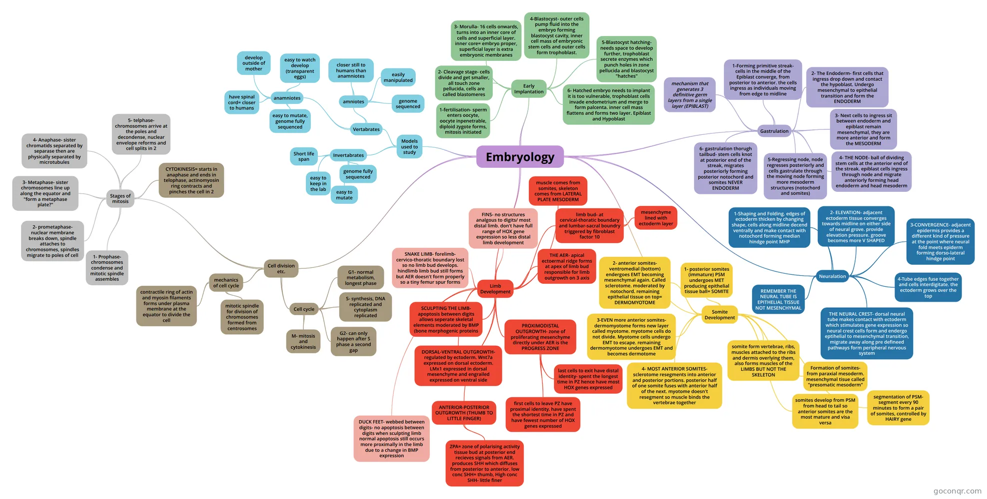
Mind map: Embryology
Contenido del mapa
Embryology
Models used to study
- Invertabrates
- Short life span
- easy to keep in the lab
- genome fully sequenced
- easy to mutate
- Vertabrates
- anamniotes
- have spinal cord= closer to humans
- develop outside of mother
- easy to watch develop (transparent eggs)
- easy to mutate, genome fully sequenced
- amniotes
- closer still to humans than anamniotes
- easily manipulated
- genome sequenced
- anamniotes
- Invertabrates
Early Implantation
- 1-fertilisation- sperm enters oocyte, oocyte inpenetrable, diploid zygote forms, mitosis initiated
- 2- Cleavage stage- cells divide and get smaller, all touch zone pellucida, cells are called blastomeres
- 3- Morulla- 16 cells onwards, turns into an inner core of cells and superficial layer. inner core= embryo proper, superficial layer is extra embryonic membranes
- 4-Blastocyst- outer cells pump fluid into the embryo forming blastocyst cavity, inner cell mass of embryonic stem cells and outer cells form trophoblast.
- 5-Blastocyst hatching- needs space to develop further, trophoblast secrete enzymes which punch holes in zone pellucida and blastocyst "hatches"
- 6- Hatched embryo needs to implant it is too vulnerable, trophoblast cells invade endometrium and merge to form palcenta. inner cell mass flattens and forms two layer. Epiblast and Hypoblast
Gastrulation
- mechanism that generates 3 definitive germ layers from a single layer (EPIBLAST)
- 1-Forming primitive streak- cells in the middle of the Epiblast converge, from posterior to anterior. the cells ingress as individuals moving from edge to midline
- 2- The Endoderm- first cells that ingress drop down and contact the hypoblast. Undergo mesenchymal to epithelial transition and form the ENDODERM
- 3- Next cells to ingress sit between endoderm and epiblast remain mesenchymal, they are more anterior and form the MESODERM
- 4- THE NODE- ball of dividing stem cells at the anterior end of the streak. epiblast cells ingress through node and migrate anteriorly forming head endoderm and head mesoderm
- 5-Regressing node, node regresses posteriorly and cells gastrulate through the moving node forming more mesoderm structures (notochord and somites)
- 6- gastrulation thorugh tailbud- stem cells knot at posterior end of the streak, migrates posteriorly forming posterior notochord and somites NEVER ENDODERM
Neuralation
- FORMATION OF NEURAL TUBE FROM ECTODERM FORMS CNS BRAIN AND SPINAL CHORD
- 1-Shaping and Folding. edges of ectoderm thicken by changing shape, cells along midline decend ventrally and make contact with notochord forming median hindge point MHP
- plate now reffered to as neural grove
- 2- ELEVATION- adjacent ectoderm tissue converges towards midline on either side of neural grove. provide elevation pressure. groove becomes more V SHAPED
- 3-CONVERGENCE- adjacent epidermis provides a different kind of pressure at the point where neural fold meets epiderm forming dorso-lateral hindge point
- 4-Tube edges fuse together and cells interdigitate. the ectoderm grows over the top
- REMEMBER THE NEURAL TUBE IS EPITHELIAL TISSUE NOT MESENCHYMAL
- THE NEURAL CREST- dorsal neural tube makes contact with ectoderm which stimulates gene expression so neural crest cells form and endergo eptihelial to mesenchymal transition, migrate away along pre defineed pathways form peripheral nervous system
Somite Development
- somite form vertebrae, ribs, muscles attached to the ribs and dermis overlying them, also forms muscles of the LIMBS BUT NOT THE SKELETON
- Formation of somites- from paraxial mesoderm. mesenchymal tisue called "presomatic mesoderm"
- segmentation of PSM- segment every 90 minutes to form a pair of somites, controlled by HAIRY gene
- somites develop from PSM from head to tail so anterior somites are the most mature and visa versa
- 1- posterior somites (immature) PSM undergoes MET producing epithelial tissue ball= SOMITE
- 2- anterior somites- ventromedial (bottom) endergoes EMT becoming mesenchymal again. Called sclerotome. moderated by notochord. remaining epithelial tissue on top= DERMOMYOTOME
- 3-EVEN more anterior somites- dermomyotome forms new layer called myotome. myotome cells do not divide. Myotome cells undergo EMT to escape. remaining dermomyotome undergoes EMT and becomes dermotome
- all mesenchymal tissue again
- 4- MOST ANTERIOR SOMITES- sclerotome resegments into anterior and posterior portions. posterior half of one somite fuses with anterior half of the next. myotome doesn't resegment so muscle binds the vertebrae together
Limb Development
- muscle comes from somites, skeleton comes from LATERAL PLATE MESODERM
- limb bud- at cervical-thoratic boundary and lumbar-sacral boundry triggered by fibroblast factor 10
- mesenchyme lined with ectoderm layer
- THE AER- apical ectoermal ridge forms at apex of limb bud responsible for limb outgrowth on 3 axis
- proximodistal anterior-posterior dorsal ventral -dorsal is back of hand
- PROXIMODISTAL OUTGROWTH- zone of proliferating mesenchyme directly under AER is the PROGRESS ZONE
- first cells to leave PZ have proximal identity. have spent the shortest time in PZ and have fewest number of HOX genes expressed
- last cells to exit have distal identity- spent the longest time in PZ hence have most HOX genes expressed
- ANTERIOR-POSTERIOR OUTGROWTH (THUMB TO LITTLE FINGER)
- ZPA= zone of polarising activity tissue bud at posterior end recieves signals from AER. produces SHH which diffuses from posterior to anterior. low conc SHH= thumb, High conc SHH- little finer
- DORSAL-VENTRAL OUTGROWTH- regulated by ectoderm. Wnt7a expressed on dorsal ectoderm. LMx1 expressed in dorsal mesenchyme and engrailed expressed on ventral side
- FINS- no structures analgous to digits/ most distal limb. don't have full range of HOX gene expression so less distal limb development
- DUCK FEET- webbed between digits- no apoptosis between digits when sculpting limb normal apoptosis still occurs more proximally in the limb due to a change in BMP expression
- SCULPTING THE LIMB- apoptosis between digits allows seperate skeletal elements moderated by BMP (bone morphogenic proteins
- SNAKE LIMB- forelimb- cervico-thoratic boundary lost so no limb bud develops. hindlimb limb bud still forms but AER doesn't form properly so a tiny femur spur forms
Cell division etc.
- Cell cycle
- G1- normal metabolism, longest phase
- S- synthesis, DNA replicated and cytoplasm replicated
- G2- can only happen after S phase a second gap
- M- mitosis and cytokinesis
- mechanics of cell cycle
- mitotic spindle for division of chromosomes formed from centrosomes
- contractile ring of actin and myosin filaments forms under plasma membrane at the equator to divide the cell
- Stages of mitosis
- 1- Prophase- chromosomes condense and mitotic spindle assembles
- 2- prometaphase- nuclear membrane breaks down, spindle attaches to chromosomes, spindles migrate to poles of cell
- 3- Metaphase- sister chromosomes line up along the equator and "form a metaphase plate?"
- 4- Anaphase- sister chromatids separated by separase then are physically separated by microtubules
- 5- telphase- chromosomes arrive at the poles and decondense, nuclear envelope reforms and cell splits in 2
- CYTOKINESIS= starts in anaphase and ends in telophase, actinomyosin ring contracts and pinches the cell in 2
- Cell cycle
Información
- Descripción
- (Developmental Biology Strand) Year 1 Mapa Mental sobre Embryology, creado por Clodagh Mullins el 05/11/2016.
- Creado por
- Clodagh Mullins
- Visitas
- 9
- Valoración