8. Specialised Cells: CD8+ T Cells
Diapositivas del conjunto (24)
-
- The properties of CD8+ T cells/CTL
- Three cell activation model and IL-2 requirement
- Restriction to MHC class I
- Divergent need for co-stimulation depending on activation status; naïve versus effector
- The process of CTL killing
- Performed cytolytic granules, generation of new granules via Th1 help
- Importance of pro-and anti-apoptotic molecules in stability of mitochondria
- Cytochrome c release and formation of apoptosome
- Triggering of initiator and effector caspases leading to apoptosis of cell
- Clearance of apoptotic bodies by macrophage
- Fas-mediated fratricide
-
Cytotoxic T Cell Killers (CTLs)
- CD8 cells transform into cytotoxic T cell killer (CTLs) and kill cells that express peptide-MHC complexes specific for TCR on CD8 T cells
- Properties of CTL killing:
- Rapid
- Highly specific- important for non-renewing cells eg neurons
- Forms immunological synapse with infected cell
- Can serially kill many target cells
- At site of infection, do not need co-stimulatory signal
-
- Apoptosis:
- Physiological
- Cellular condensation
- Nuclear fragmentation
- Rapid phagocytosis
- Lack of inflammation
- Internal components remain encapsulated, wont create response to own tissue
- Necrosis:
- Pathological
- Organelles swell
- Membrane rupture
- Leakage of cell contents - taken up by innate immune cells and cause marked inflammation
- Consequence: pathogenic bystander activation for inducing autoimmunity
- Immune response initially to pathogen subsequently spreads to host tissue (epitope spreading)
-
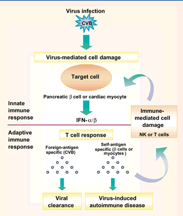
- Viral Infection:
- Innate immune system activated to infection, produce IFNs that dampen virus ability to replicate
- IFNs damage surrounding healthy cells-release internal components of healthy cells that can be picked up by APC in context of inflammation
- Present healthy cell internal components to a T cell recognising self
- Autoimmunity
-
CTLs Kill Targets via Apoptosis
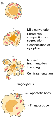
- Programmed cell death
- Governed by series of biochemical events and activation of serine proteases (caspases) lead to:
- Nuclear blebbing
- Chromatin condensation, DNA fragmentation
- Shedding of membrane vesicles
-
CTLs Efficient Killing Machines
- Contain preformed cytoxic granules:
- Perforin- delivers contents of granules to target cell cytosol
- Granzymes- serine proteases that activate apoptosis once in cytoplasm
- Granulysins- antimicrobial activity
-
CD4-APC-CD8 Interaction: CD8 Activation
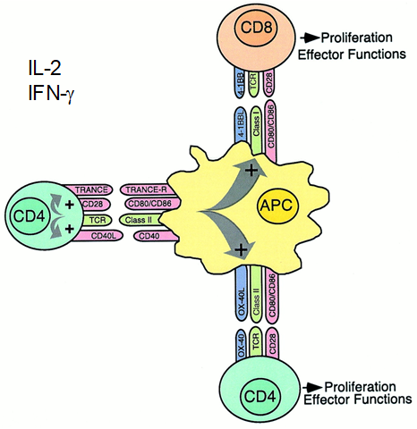
- APC presents molecules to both CD8(MHC-I) and CD4(MHC-II)
- Brings CD4 and CD8 into close proximity
- CD4 co stimulatory molecule CD40L binds CD40 on APC-> upregulation of MHC-peptide complexes and costimulatory molecules (for CD8)
- CD8+ T cell co-stimulatory 4-1BB binds 4-1BBL on APC and provides survival signal for activated CD8 T cell
- CTL replenishes granules with weapons in by receiving signals from Th1 cell by IFNϒ
- IL-2 drives proliferation
- IFNϒ drives differentiation to effector status cytotoxic T cell (CTL)
-
CD4-APC-CD8 Interaction: CD8 Activation
-
Granzyme B + Perforin Expression
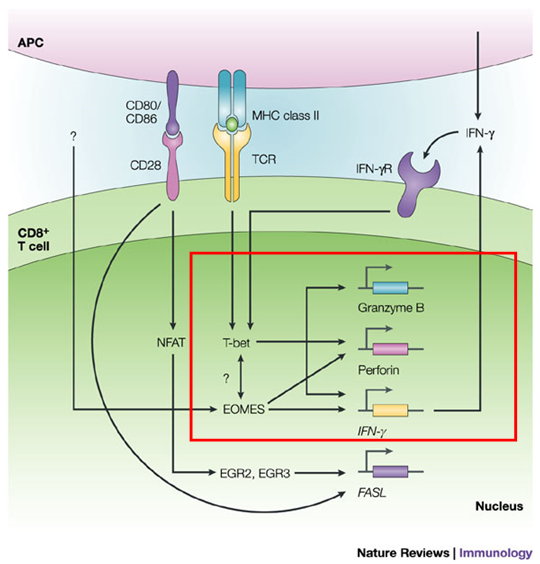
- Granzyme B and Perforin mRNA expression is controlled by TFs T-bet and Eomes:
- IFNϒ produced by Th1 cell binds IFNϒR on effector CD8 T cell
- Combined signals via immunological synapse +IFNϒRs trigger TF T-bet
- T-bet activates transcription of granzyme B and perforin
- In absence of T-bet, Eomes can substitute
-
Granzyme B + Perforin Expression
-
- Whilst CD8 T cell activation in LN, at site of infection:
- Innate cells are producing interferons-> modify proteosome inside infective tissue
- Replace certain catalytic subunits
- Peptides produced that are more receptive to binding MHC-I= more stable immunologcial synapse with T cells
- IFNgam modifications of top and bottom of immunoproteasome increases rate of peptides released- widens
- Innate cells produce cytokines that change epithelial cell lining
- Change adhesion molecules specific for T cells
- Once T cell activated in LN, downregulates chemokine R keeping it in LN, upregulate chemokine receptors specific for epithelial cells: entry of activated cells only at site of infection
-
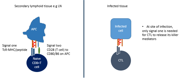
- Need 2 signals to generate effector CD8 T cell in LN
- In infected tissue, only need 1 signal to make immunological synapse and release weapons
-
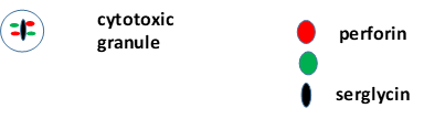
- Perforin and granzymes work together to kill target cell: form multimeric complex stabilised by scaffold molecule serglycin
-
- Formation of immunological synapse vital to prevent unwarranted death of healthy cells:
- Reorganisation of Golgi apparatus and MTs at strongest point of contact in immunological synapse
- Tight binding to target cell via TCR and MHC-I-peptide
- Signals into CTL to polarise its granules in direction of target cell
- Accumulation of granzyme and perforin molecules at site of immunological synapse
- Tubule extends into target cell and delivers molecules (undirectional)
-
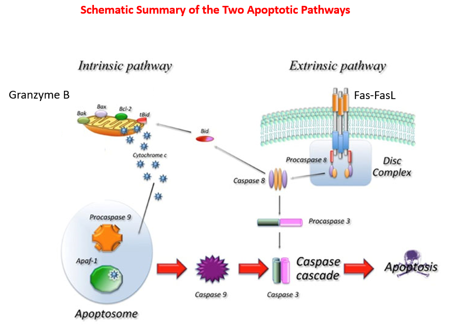
Intrinsic Pathway of Apoptosis
- Involves release of cytochrome c from mitochondria
- Granzyme B acts on molecules that prevent release of cytochrome c
- Release of cyt c helps formation of complexes necessary to destroy DNA in cell
- Apoptosome triggers activation of caspases -> destruction of DNA
-
Intrinsic Pathway of Apoptosis
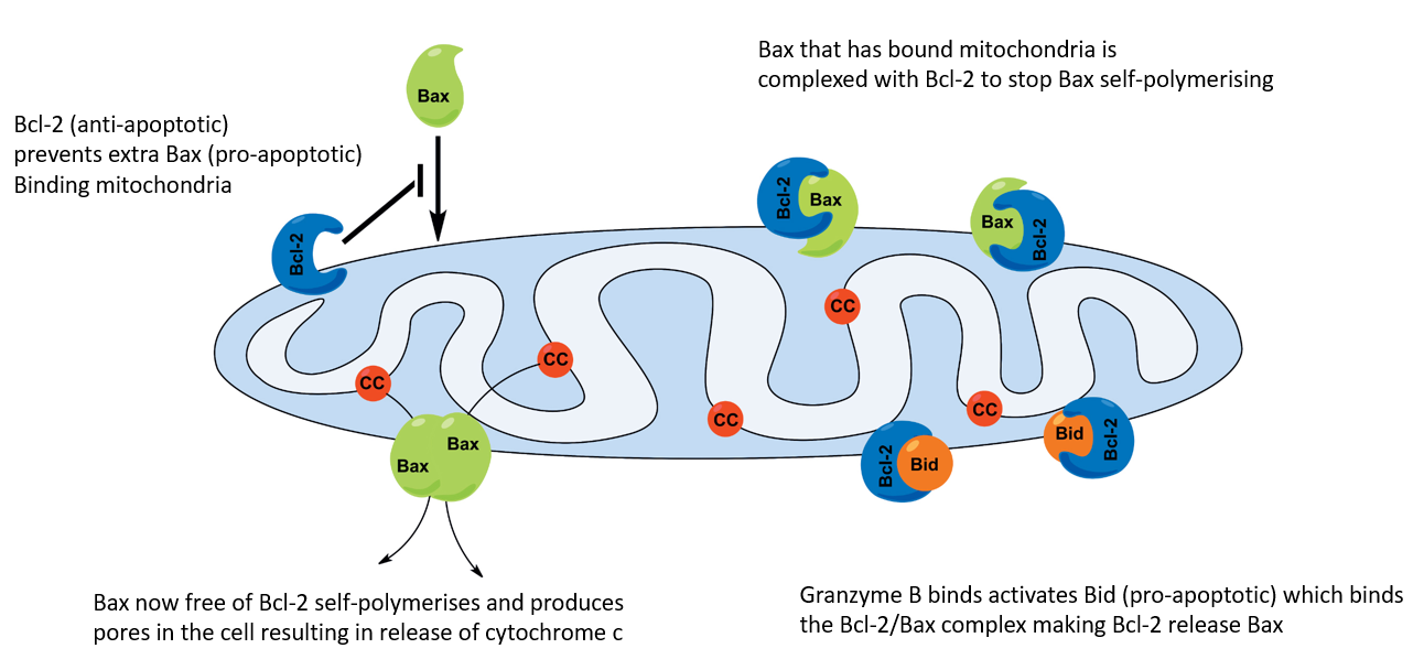
- The Bcl family molecules= key regulators of apoptosis
- Bcl-2= anti-apoptotic molecule
- Bax= pro-apoptotic molecule- binds and forms pore with mitochondria walls to release cyt c
- Bcl-2 binds Bax preventing Bax polymerising to form pore
- When infected:
- Granzyme B activates Bid (or Bad)
- Bid binds Bcl-2/Bax complex-> releases Bcl-2
- Bax can now self-polymerise and cause pore in mitochondria wall
-
Intrinsic Pathway of Apoptosis
-
Intrinsic Pathway of Apoptosis
- Death by caspases:
- Caspase= cysteine proteases that cleave substrates on C- side of aspartate residues
- Synthesised as inactive zymogens (proenzymes), require proteolytic modification to be activated
- Classified as:
- Initiators- activate procaspases by cleaving them (these are 8,9)
- Effectors- executioners, once activated they kill (these are 3,7 )
- Apoptosome formation:
- Upon cyt c release, it complexes with Apaf-1= apoptosome (apoptotic protease activating factor-1)
- Apoptosome recruits procaspase-9 (initiator) which also complexes = complete apoptosome
- Upon recruitment, procaspase 9 cleaved to active caspase 9
-
Intrinsic Pathway of Apoptosis
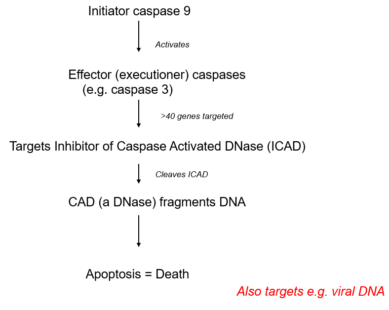
- How caspases lead to cell death:
- Active caspase 9, cleaves and activates effector caspase 3
- Effector caspase targets and cleaves upto 40 genes, including the inhibitor of caspase activated DNase (ICAD)
- CAD DNase now active, targets and fragments the DNA in the cell- doesnt discriminate between your DNA and viral DNA
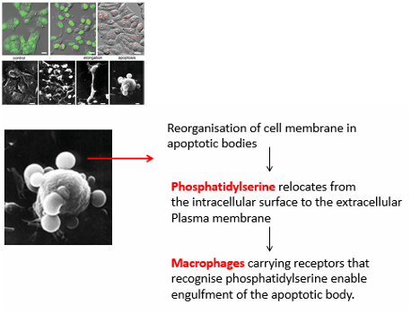
- Combined mitochondrial and DNA damage -> cell shrinkage and 'blebbing'= apoptotic bodies
- Encapsulation into apoptotic bodies is CRUCIAL to self-contain own and virus molecules
- Phosphatidylserine relocates from intraceullular surface to extracellular PM of apoptotic body
- Macrophages receptor recognises phosphatidylserine and engulf body- phagocytosis
- Rapid removal of apoptotic bodies to prevent tissue autoimmunity , would get high levels of inflammation
-
Intrinsic Pathway of Apoptosis
-
Extrinsic Pathway of Apoptosis
- Cytotoxic T cells can also kill cells using non-cytotoxic granule-mediated mechanisms (instead receptor-mediated Fas-FasL pathway):
- CTL upregulates FsL on its surface that binds to Fas on target cell
- Similar to action of granules, Fas-FasL interaction triggers caspase cascade -> fragmentation of target cell DNA and death by apoptosis
-
Extrinsic Pathway of Apoptosis
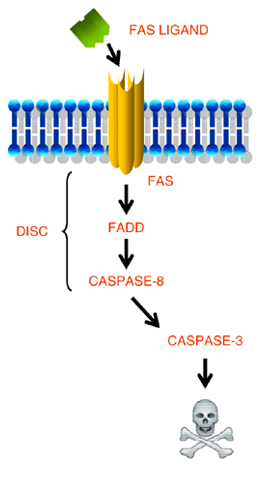
- FasL on CTL binds Fas on target cell
- Triggers Fas-Associated protein Death Domain (FADD)
- Acts as adaptor protein with procaspase 8 and activates it = Death Inducing Signal Complex (DISC)
- Caspase 8 activates executioner caspase 3
-
Intrinsic vs Extrinsic Pathway
-
Killing CTLs once Pathogen killed
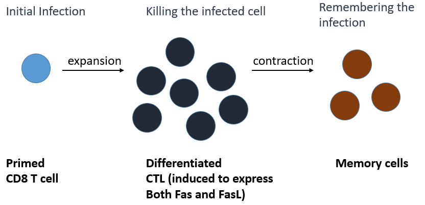
- Fas-FasL interactions are important at killing CTL once pathogenic threat is cleared= Fratricide (CTL cells kill eacother)
- Leave small pool of memory cells that survey body for re-infection