
Flashcards
CBL MSK
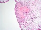
Cards in this deck (54)
- 1Cardinal feature of osteoarthritisPAIN
- first aggravated by movement and relieved by rest
- as time goes by, relief is less complete
- aching character
- poorly localised
- late stages, pain may awaken patient from sleep
- muscle spasm around joint may add to pain - 2Other symptoms of OAStiffness
- waking in morning or after periods of inactivity such as sitting
- as joint is limbered up, patient feels better
Limitation of Motion
- eg. in the hip, patients lose ABduction as well as rotation
- may be flexion contracture masked by postural adjustment of the pelvis and lumbar spine
- Joint deformity
- Wasting of associated muscles
- joint swelling
- localised tenderness
- palapable osteophytes and crepitus
- limp or antalgic gait - 3Investigations to confirm diagnosis of OAX ray and blood tests
- 4What abnormalities would be seen on OA x-ray?- loss of joint space
- subchondral sclerosis
- bone cysts
- osteophytes at lateral margin of joint - 5What blood tests would you do for OA and why? - secondary OA?FBC – normal
•ESR, CRP – normal
•Rheumatoid factor, Anti nuclear factor – negative
•Calcium, phosphate, alkaline phosphatase – normal
Blood tests should all be within normal limits. Patients may have laboratory abnormalities in secondary OA such as high levels of uric acid in gout or high levels of alkaline phosphatase in Paget’s disease - 6Management of patient with OAPain relief – analgesics and anti inflammatory agents
•Increasing mobilisation – joint movement and muscle tone may be improved by physiotherapy
•Reducing load – weight loss, walking stick
•Surgical options – osteotomy, arthrodesis, joint replacement, excision arthroplasty - 7Surgical options for OA1. A realignment osteotomy can relieve pain. It may change the mechanical axis of the joint to allow weight bearing on a part of the articular surface which is not affected.
2. Arthrodesis is best performed when adjacent joints are relatively unaffected, because after a joint is fused, more demand is placed on them. Arthrodesis may be considered for younger patients in whom a joint replacement would be expected to fail after a relatively short time because their demands are higher.
3. Joint replacement remains the best treatment for established OA of the hip and knee. The patient and surgeon must weigh up the benefits of pain relief and improved function and the risks of major surgery.
4. Sometimes an excision arthroplasty can be performed where the affected joint is excised and the resulting space is allowed to fill with fibrous tissue. - 86 characterisitic features of synovial joints1. the parts of the bones which are in contact with each other are covered by smooth articular cartilage
2. the joints are surrounded by a connective tissue capsule
3. there is a joint cavity
4. the inner surface of the capsule and the non articular surfaces of the bones are covered by a synovial membrane
5. the capsule is reinforced by ligaments
6. the joint is capable of movement - 9Macroscopic pathological features in OAloss of articular cartilage with eburnation (polishing) of surface
•subarticular cyst formation
•osteophyte formation at the joint margins
•sclerosis (thickening) of subchondral bone
- Cartilage destruction may first appear as softening of the joint surface. This may be followed by fissuring, flaking or complete loss of articular cartilage with exposed subchondral bone.
- These processes usually start where the load across the joint is greatest, for example, the medial compartment of the knee.
- raised intra-articular pressure is a cause of subarticular cyst formation
- Osteophytes are bony outgrowths which occur peripherally or centrally - 10Microscopic pathological features in OAfissuring, flaking then full thickness loss of articular cartilage
•subarticular cysts
•nodules of reactive bone and cartilage (osteophytes) at the joint margins
•Sclerosis of subchondral bone
Osteoarthritic cartilage is characterised by increased water content, alterations in proteoglycans, collagen abnormalities and binding of proteins to hyaluronic acid. In addition, the rate of synthesis of DNA, collagen and proteoglycans is increased. - 11Normal articular cartilage histologynormal articular cartilage is made up of chondrocytes which manufacture and lie in a matrix made of collagens, proteoglycans, and non-collagenous proteins.
- 12—This is a detritus synovitis – in OA the synovium is secondarily involved, with flakes of bone and cartilage broken off from the damaged joint embedding in the synovium and causing mild villous hyperplasia and chronic inflammation
•You can see villous hyperplasia and chronic inflammation around blood vessels as well as fragments of cartilage detritus - 13What are the causes of osteoarthritis?Primary osteoarthritis
•Secondary osteoarthritis –
- intra articular fracture,
- high intensity impact,
- ligament injuries,
- avascular necrosis,
- acromegaly,
- Paget’s disease,
- previous joint sepsis,
- haemophilia,
- haemochromatosis,
- ochronosis,
- pseudogout,
- Charcot’s joint - 14Presentation of RA– joints: hands, wrists, feet, knees
– sore to the touch, swollen, feel warm
- limited range of movement
- subcutaneous nodules on elbows - 15Blood tests in RAHaematology
• 1. FBC
• 2. Blood film
• 3. Haematinics
• Liver function tests
Immunology
• 1. autoantibodies (antinuclear antibodies, rheumatoid factor, complement studies)
• 2. immunoglobulins
• 3. serum protein electrophoresis
• 4. acute phase proteins – CRP - 16What would haematology blood tests be expected to be like in RA?1. FBC low haemoglobin, normal white cell count, normal platelets, elevated ESR
2. Blood film - Anaemia which is a normochromic normocytic anaemia of chronic disease. Patients who take NSAIDs can also develop iron deficiency anaemia from upper GI bleeding. Patients with Felty’s syndrome (RA + splenomegaly) can develop haemolytic anaemia.
3. Haematinics – will depend on the underlying form of anaemia present - 17LFTs in RANormal liver function tests except a mildly elevated alkaline phosphatase and possibly gamma GT.
An elevated alkaline phosphatase and gamma GT are commonly raised during acute flares of joint disease - 18Expected immunology results in RA1. autoantibodies
• antinuclear antibodies – negative (used to exclude lupus but may be positive in 20% of RA patients)
• rheumatoid factor - positive
• complement studies - normal (used to exclude lupus and vasculitis)
2. immunoglobulins – increased total globulins and IgG and IgA with normal IgM
3. serum protein electrophoresis - normal
4. acute phase proteins – CRP – high (increased due to acute phase response)
Rheumatoid factor is positive in 70% of all cases of RA, but in 100% of cases with systemic manifestations.
Anti CCP antibodies (cyclic citrullinated protein) are more specific and sensitive for RA but are only available to rheumatologists. - 19What is rheumatoid factor?Rheumatoid factor is an autoantibody, IgM directed against Fc portion of IgG.
- 20What abnormalities are present on this x-ray of hands?– periarticular osteopaenia
- periarticular erosions
- soft tissue swelling around metacarpo-phalangeal joints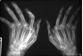
- 21Histological findings in RA• Villous architecture (hyperplasia)
• Synovial lining cell hyperplasia
• Fibrin exudation
• Marked chronic inflammation with lymphoid aggregates and plasma cells - 22What pathological changes can you see?This is a knee joint in which yellow pannus (tissue made up of rather vascular fibrous connective tissue) can be seen destroying the articular cartilage from the edge.
The upper histology image shows this pannus destroying the cartilage and the lower image shows villiform synovium typical of RA.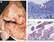
- 23Pathogenesis of RA- joint swelling due to active synovitis
- in RA the synovium is full of macrophages, T lymphocytes and plasma cells (which produce rheumatoid factor)
- macrophages are activated and produce pro-inflammatory cytokines such as TNF, IL1 and IL6 which activate enzymes in the synovium
- this leads to formation of a pannus which destroys the cartilage and bone - 24Which other organ systems can be affected in RA?• CVS – pericarditis, myocarditis, vasculitis
• Resp – pleuritis, pulmonary fibrosis, rheumatoid nodules
• Skin – rheumatoid nodules
• Eyes- Sjogren’s, scleromalacia perforans
• Haem – anaemia, splenomegaly (Felty’s syndrome)
Amyloidosis - 25Pharmacotherapy for RA• NSAIDs – reduce and inflammation by interfering with prostaglandin synthesis.
• Second line agents, DMARDs (disease modifying anti rheumatic drugs)– salazopyrin, hydroxychloroquine, methotrexate
• corticosteroids – intra-articular, intramuscular or oral, often used as bridging therapy until DMARDs have taken effect (2-3 months) - 26Biologics for RAanti TNF agents e.g. infliximab.
70% of patients respond to therapy with improvements in joint disease, systemic disease, fatigue and quality of life. They are expensive.
Rituximab which causes B lymphocyte depletion and an anti IL-6 therapy called tociluzimab are relatively new to the market for RA treatment - 27Surgical interventions for RA• tendon repair – ruptured tendons need to be repaired
• synovectomy – effective for resistant synovitis
• joint replacement – knee replacement in particular is beneficial
• joint fusion – useful in ankles and wrists as a last resort - 28Role of allied health professionals in RA - physiotherapy - occupational therapy - podiatry/surgical appliancesA multi disciplinary approach is essential in RA consisting of both in patient and out patient management.
Physiotherapy
– this helps maintain movement and prevents muscle atrophy
Occupational therapy
– appropriate adaptations to the patients’ lifestyle and housing can improve the long-term outlook for patients
Podiatry/surgical appliances
- the feet are almost always involved in RA. Appropriate alteration to shoe wear and maintenance of good foot hygiene are imperative for the disease. - 29When to start DMARDs?Early DMARD therapy reduces joint damage. Therefore DMARDs should be ideally used within 3 months of
disease onset, with an attempt at aggressive control of inflammation, and consequently symptom control and
radiological damage - 30Examples of: - anti TNF - anti B cell - inhibition of interleukinsanti-TNF
- etanercept
- adalumimab
- infliximab
anti b cell
- rituximab
inhibition of interleukins
- tocilizumab - IL6 receptor
- Anakinra IL1 - 31general manifestations of RAmalaise, weight loss, fever
- 32vasculitis in RAsmall vessel inflammation leads to micro infarction in peri-ungual area.
In fingers and toes can produce rheumatoid ulcers in lower limbs. - 33Haematological manifestations of RA- Anaemia, usually of chronic disease
- B12 deficiency may occur with pernicious anaemia.
- Folic acid deficiency may occur with poor diet.
- Iron deficiency may be dietary or secondary to blood loss - 34skin manifestations of RAsubcutaneous nodules in 20-30% of patients
Occur on elbows, finger joints, Achilles tendon, bony prominences. More common in smokers - 35Felty's syndrome in RALymphadenopathy, splenomegaly, leucopenia, thrombocytopenia.
Increased risk of infection and lympho-proliferative disease - 36pulmonary involvement in RA- Asymptomatic involvement found in 50% of patients at post mortem.
- Effusions are exudates with lymphocytes.
- Rheumatoid nodules can mimic bronchial Ca on X-ray.
- Nodules in coal-miners (Caplan’s syndrome).
- Pulmonary fibrosis with progressive alveolar scarring.
- Pulmonary artery hypertension can follow pulmonary fibrosis - 37Amyloidosis in RARare.
Complication of longstanding poorly controlled arthropathy. Deposition in the kidney produces proteinurea and in
the heart cardiac failure. Amyloid may also involve liver, spleen, intestine and skin - 38Ocular involvement in RAKerato-conjunctivitis sicca (secondary Sjogren’s syndrome) affects
35% of patients
More common with longstanding RA.
Scleromalacia, - thinning of sclera.
Scleritis - acute pain and redness, normal visual acuity. - 39Cardiac and Neurological and renal involvement in RACardiac involvement: Nodule formation, amyloidosis, pericarditis and valve fibrosis
Neurological involvement: Vasculitis of vasa-nervorum causing peripheral neuropathy If many nerves are involved - mononeuritis multiplex
Renal involvement: Has tended to be a complication of older therapies or of amyloidosis in long-standing disease - 40Blood tests in RA in consideration of alternative diagnosesUrate
• Vitamin D
• CK
• TFTs
• HLA B27
• ANA - 41DAS28Composite measure of swollen joints, tender joints, ESR and patient visual analogue score.
• DAS28 <3.2 represents low disease activity - 42OA on examinationLook: deformity, muscle wasting, swelling nodules
Feel: tender, swollen
Move: range of motion, pain, crepitus
Special tests: Thomas, Trendelenberg
Gait – antalgic, Trendelenberg, short leg - 43Risks of surgery for OAMedical complications – cardiorespiratory, DVT / PE
Periprosthetic infection – osteolysis, joint loosening and failure
Dislocation (especially hip), fracture,
Failure of fixation – loosening and migration - 44composition of prosthetic jointsMetal on high density polyethylene (HDPE) bearings for most joints
Newer bearing surfaces for hips – metal on metal, ceramic on ceramic
10,15,20 years survival of prostheses - 45gene in RAHLA DW4 linked
- 46epidemiology of RAany age but most commonly 35 – 45 years
Women are affected 2 – 3 times more commonly than men - 47RA examination - hand - wrists - feet - cervical spine - large jointsHand: symmetrical; PIP, MCP laxity, characteristic deformities, tendon rupture, nodules
Wrists: carpal subluxation, distal radioulnar joint disruption, median nerve compression
Feet: MTP subluxation, loss of longditudinal arch,
Cervical spine: atlantoaxial instability
Large joints: cartilage loss leads to loss of hip / knee / shoulder function - 48Pain control for RA - ladder - nerve root - non pharmacological1. non opoid +/- adjuvant
2. opioid for mild to moderate pain +/- non-opioid or adjuvant
3. opioid for moderate to severe pain +/- non-opioid or adjuvant
- Consider nerve root pain, use e.g. carbamazepine, amitriptyline
or gabapentin.
- TENS machines are an effective and non toxic means of relieving pain in many patients.
• Non-pharmacological measures such as acupuncture can
be employed by physiotherapists to aid pain relief.
• Remember that pain may have a psychological component. - 49NSAIDs actionNSAIDs inhibit an enzyme called cyclo-oxygenase, of which two isoforms are known, COX-1 and COX-2. COX-2 is
known to be more involved in joint inflammation, and selectively inhibiting this enzyme reduces GI bleeds and ulceration.
Other GI toxicity and particularly cardiovascular and renal toxicity is still significant however. - 50Patients at risk of gastric ulcer disease>65yr of age, previous ulceration, on other medications increasing the
risk of ulceration (e.g.e steroids), should also be prescribed a proton pump inhibitor or H2 receptor antagonist - 51side effects of steroidsAdrenal suppresion
• Risk of infection including opportunistic infection
• Osteoporosis
• Diabetes mellitis
• Cataracts
• • Bruising/thinning of skin - 52histology of rheumatoid nodulemacrophages giant cells and T cells surrounding an area of
necrosis. They are thought to represent a local reaction to an
underlying area of vasculitis - 53non-medical management of RA- education
- psychosocial (may need social worker, DLA, earning capacity)
- diet (calcium; weight reduction)
- depression
- co-morbidity (CVS risks as RA is an independent risk factor for CVD. osteoporosis)
- fertility (methotrexate is teratogenic, some DMARDs impair male fertility)
- self-help groups - 54Rheumatology nurse inputFollow up and monitoring of especially vulnerable/frail patients.
» Patient advice for flares/ DMARD queries/ other issues
» Education of patients/ relatives
» Exercise group coordinator.
» Research
» Advice on wound dressing/ community follow up of patients with vasculitic ulceration or other wounds related to RA
» Home visits
» Help with allowances ( via social work dept.)
» Specialised nursing of patients with rheumatic diseases and a knowledge of their complications
Information
- Description
- Musculoskeletal Flashcards on CBL MSK, created by Ellie Britt on 09/12/2017.
- Created by
- Ellie Britt
- Views
- 4
- Rating