
Flashcards
The Endocrine System
Cards in this deck (70)
- 1What are the 8 components of the endocrine system?1. Pituitary gland
2. Thyroid gland
3. Parathyroids
4. Pancreas
5. Suprarenals (adrenals)
6. Gonads - ovaries & testes
7. Thymus
8. Pineal gland - 2What is the difference between endocrine & exocrine glands?ENDOCRINE glands are ductless glands & secrete directly into the bloodstream.
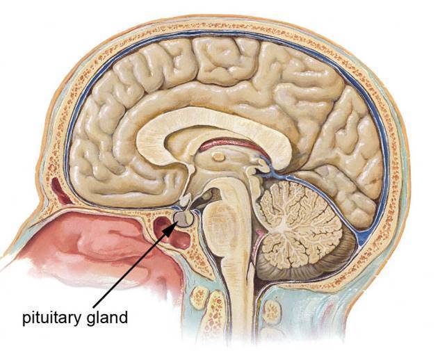
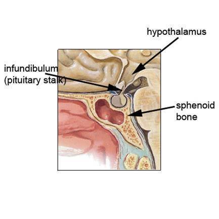
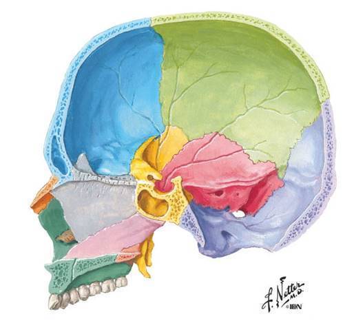
Whereas, EXOCRINE glands have ducts & secrete directly into/onto an organ/tissue. For example: salivary glands, sweat glands, pancreas (digestive juices) - 3Describe where the pituitary gland is placed in the body?The pituitary gland is attached to the under-surface of the brain (hypothalamus) by the infundibulum or the pituitary stalk.
And it located in the sella turcica of the sphenoid bone. - 4Label the following: - hypothalamus - infundibulum / pituitary stalk - sphenoid bone
- 5Define hypo/hyperpituitarism.HYPOPITUITARISM = not enough growth hormone released, resulting in growth deficiency.
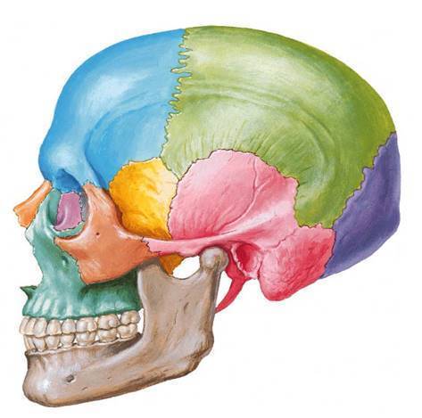
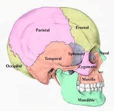
HYPERPITUITARISM = over-production of the growth hormone. - 6Label the following: - frontal bone - nasal bone - maxillary bone - mandible - temporal bone - occipital bone - parietal bone - sphenoid bone
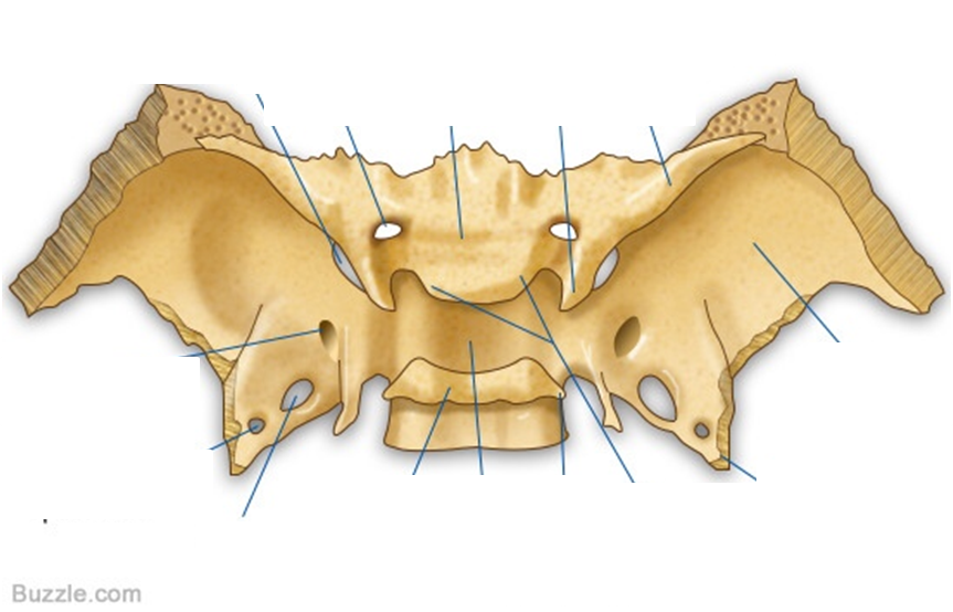
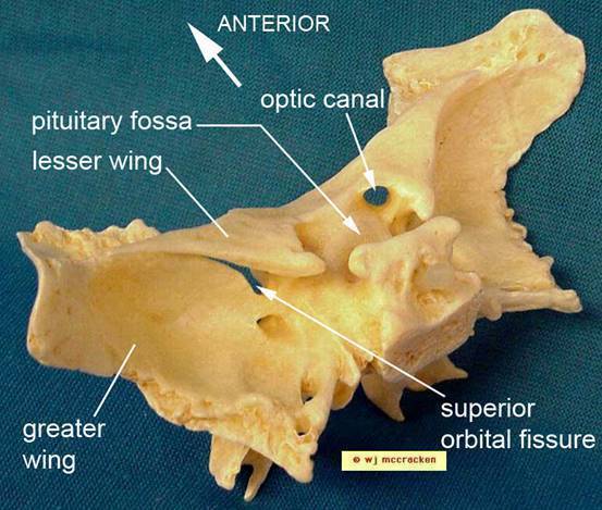
- 7Where does the pituitary gland sit?In the circular part of the sphenoid bone known as the pituitary fossa.
- 8Label the following: - pituitary fossa / sella turcica - optic canal - superior orbital fissure - the body - greater wing - lesser wing
- 9Where is the thyroid gland positioned in the body?The thyroid gland is positioned:
- on the trachea, anterolateral to the trachea & the larynx.
- lies on the top of the 2nd & 3rd tracheal rings anteriorly
- extends from the vertebral levels of C5 - T1
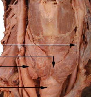
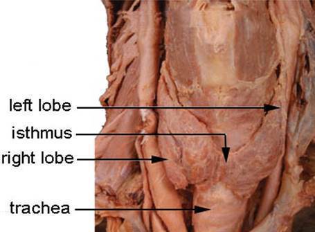
- laterally (sideways) related to cricoid & laryngeal cartilages - 10What is the structure of the thyroid gland?- consists of left & right lobes united by a narrow isthmus.
- it can have a pyramidal lobe is present in 50% of the population, usually on the left side. - 11What is the pyramidal lobe?the pyramidal lobe arises from the isthmus upwards along the midline.
- 12Label the following: - right lobe -left lobe - isthmus - trachea
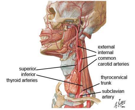
- 13What is the arterial supply of the thyroid gland?The thyroid gland has 2 arteries: SUPERIOR & INFERIOR thyroid arteries.
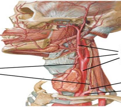
The SUPERIOR thyroid artery is the 1st branch of the external common carotid artery. (left or right?)
The INFERIOR thyroid gland is a branch of the thyrocervical trunk, which is a brach of the subclavian artery. (left or right?) - 14Label the following: - external + internal common carotid arteries - thyrocervical trunk - subclavian artery - superior + inferior thyroid arteries
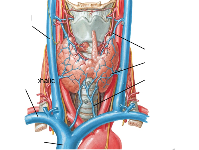
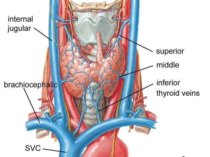
- 15What is the venous drainage of the thyroid gland?There are 3 thyroid veins that are involved in its drainage:
- superior
- middle
- inferior
SUPERIOR + MIDDLE drain into the internal jugular veins.
INFERIOR drains into the brachiocephalic vein.
INTERNAL JUGULAR VEIN + BRACHIOCEPHALIC merge into the SVC. - 16Label the following: - superior+middle+inferior thyroid veins - SVC - brachiocephalic vein - internal jugular vein
- 17What is the thyroid gland covered by?The thyroid gland is covered by the infra-hyoid strap muscles. There are 4 different types of strap muscles.
- 18Can the thyroid gland be palpated?You cannot palpate the thyroid gland, unless the gland is enlarged.
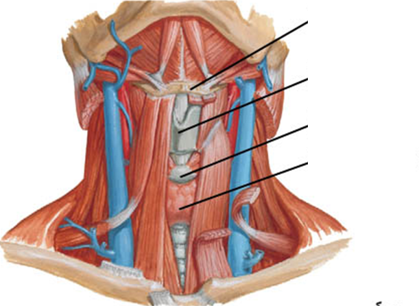
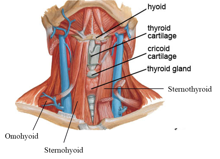
Enlarged thyroid gland due to iodine deficiency = GOITRE - 19What are the 4 different types of infra-hyoid strap muscles?The following strap muscles cover the thyroid gland:
- omohyoid (lateral)
- sternohyoid (medial)
- thyrohoid
- sternothyroid (deep) - 20Label the following: - hyoid bone - omohyoid - sternohyoid - sternothyroid - thyroid cartilage - cricoid cartilage - thyroid gland
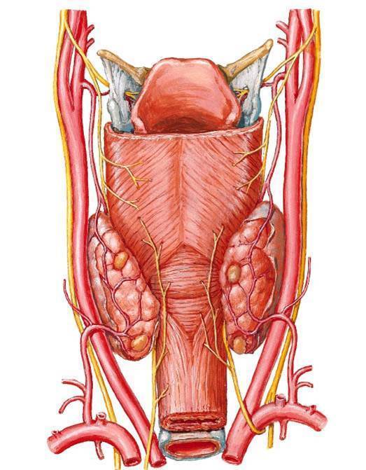
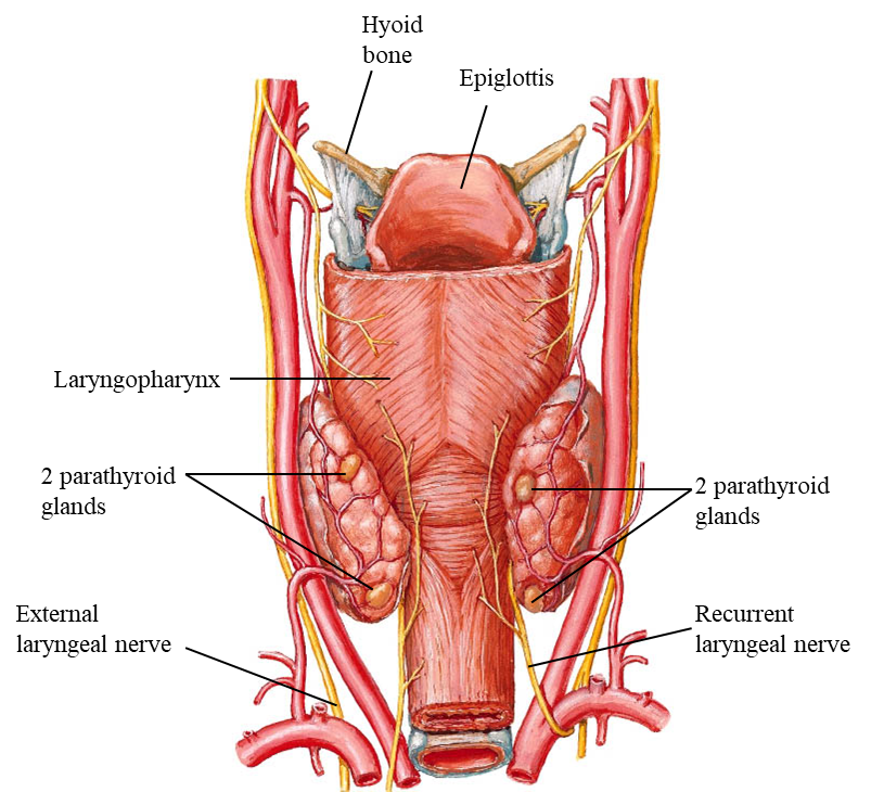
- 21What are the parathryoid glands?- four tiny glands (2 pairs)
- embedded in the deep surface of they thyroid gland
- blood supplied by the inferior thyroid arteries
- produces parathyroid hormone
- it is responsible to regulate the body's calcium levels. - 22Label the following: - hyoid bone - epiglottis - laryngopharyx - 4 parathyroid glands - recurrent laryngeal nerve - external laryngeal nerve
- 23Where is the pancreas located in the body?- located on the posterior abdominal wall
- the head is enclosed by the loop of the duodenum (first part of the small intestine)
- the tail is located in the hilum of the spleen
- its head is located on the transpyloric plane (L1)
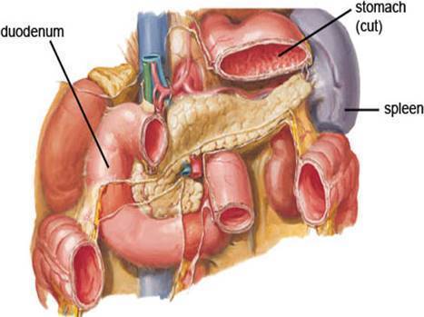
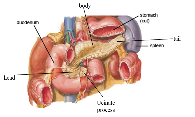
- the body crosses the abdominal aorta & left renal vein. - 24Describe the pancreas.- Has a head, neck, body, tail & uncinate process.
- A mixed endo & exocrine gland
- Insulin is produced in the islets of Langerhans.
- located behind the stomach. - 25Label the following: - body - head - ucinate process - tail
- 26Label the following in the abdominal aorta: - coeliac trunk - inferior mesenteric artery - superior mesenteric artery
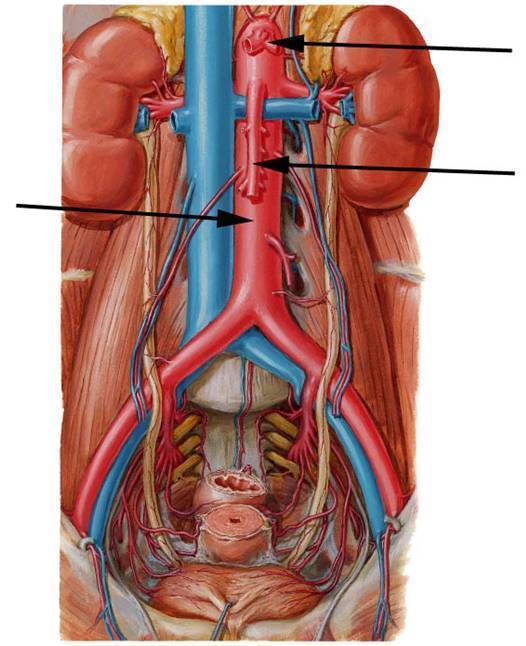
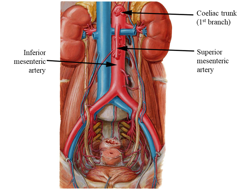
- 27Where do the coeliac trunk + superior/inferior mesenteric arteries supply blood?COELIAC TRUNK = supplies the stomach, spleen, liver & the pancreas.
SUPERIOR MESENTERIC ARTERY = supplies the large part of the gut + head of the pancreas.
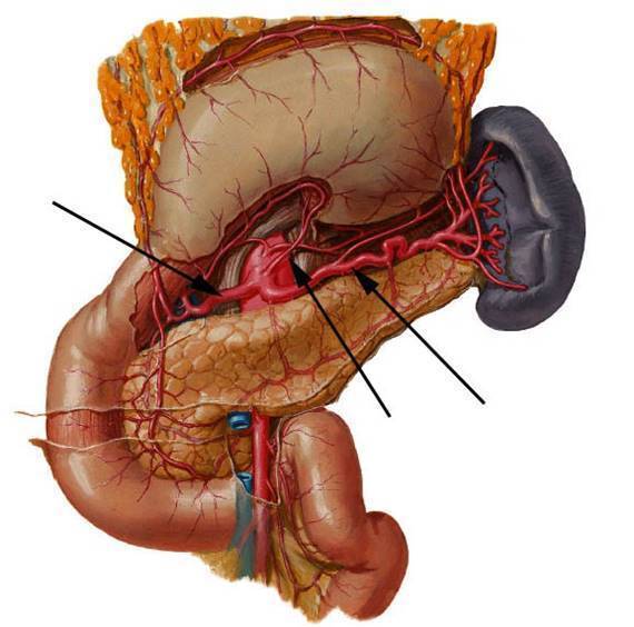
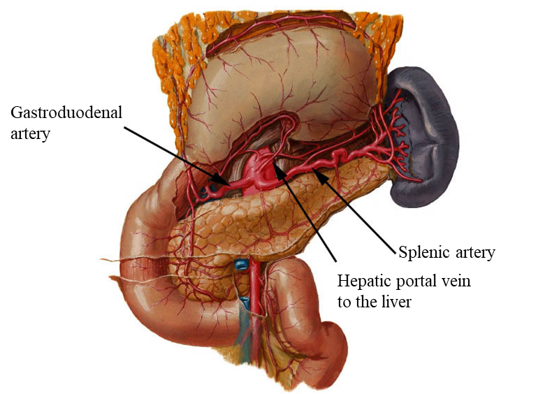
INFERIOR MESENTERIC ARTERY = supplies the gut - 28What is the blood supply of the pancreas?- from the 1st branch of the abdominal aorta - coeliac trunk, via the splenic & gastroduodenal arteries)
- also, the superior mesenteric artery.
- venous drainage to the liver via the hepatic portal vein - 29Label the following: - hepatic portal vein - gastroduodenal artery - splenic artery
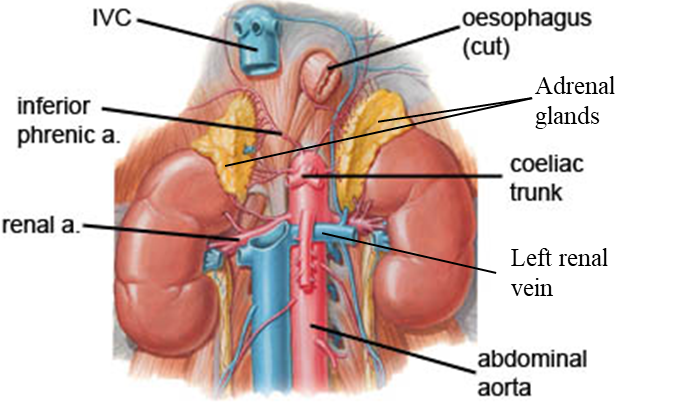
- 30Where are the adrenal (suprarenal) glands located on the body?- located on the superior poles of the kidneys
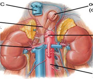
- on 'top' of each kidney - 31What is the blood supply of the adrenal glands?- From the renal artery, inferior phreic artery & abdominal aorta. (3 sources)
ABDOMINAL AORTA + INFERIOR PHRENIC ARTERY = they give rise to the superior & middle adrenal artery.
RENAL ARTERY = gives rise to the inferior adrenal arter.
- Venous drainage is carried out in 2 ways:
* left kidney/side drainage = left renal vein (which merges into the IVC)
* right kidney/side drainage = IVC - 32Label the following: - IVC - inferior phrenic artery - renal artery - abdonimal aorta - left renal vein - coelic trunk - adrenal glands - oesophagus
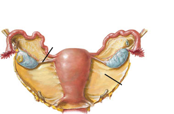
- 33How are the ovaries located in the body?The ovaries are suspended from the posterior of the broad ligament by the mesovarium (part of the broad ligament).
- 34Label the following: - ovarian ligament - ovaries - mesovarium - fimbriae - broad ligament - suspensory ligaments
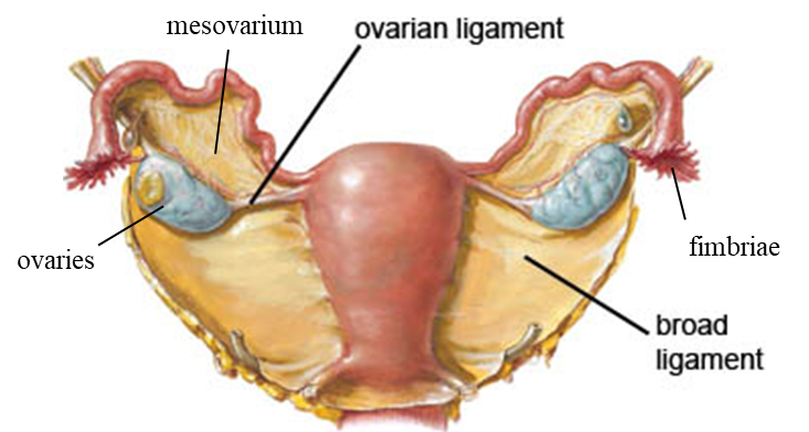
- 35What is the role of Fimbriae?It is located at the end of the fallopian tubes through which the eggs move from the ovaries to the uterus.
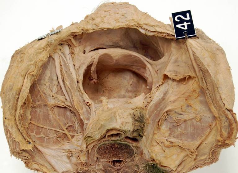
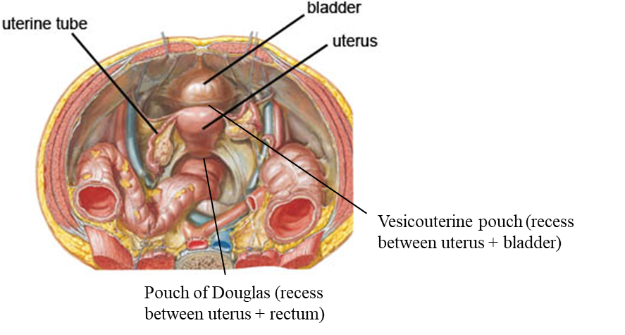
- 36Label the following: - uterine tube - bladder - uterus - rectum - vesicouterine - pouch of Douglas
- 37How are the ovaries supplied with blood?The ovarian arteries supply the blood to the ovaries, which originate from the abdominal aorta.
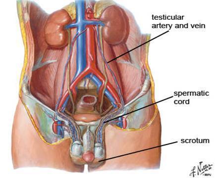
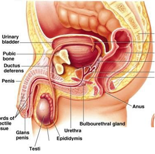
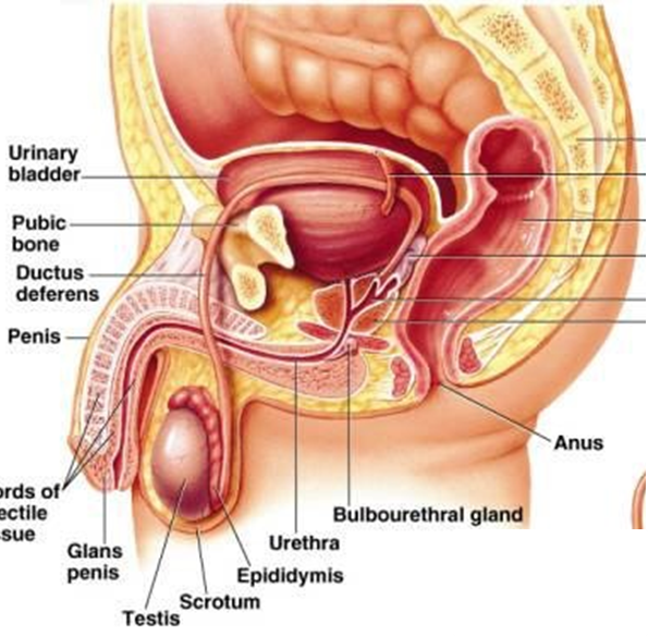
Venous drainage occurs via the ovaries veins. The right side drains into the ovary vein & then into the IVC & the left side drains into the renal vein. - 38Where are the testes located in the body?- suspended (hanged) in the scrotum by the spermatic cord.
- it produces testosterone - 39What does the spermatic cord consist of?* ductus deferens
* testicular artery
* testicular veins - 40What is the wall of the scrotum made out of?the wall of the scrotum contains dartos muscle, contracts/relaxes to regulate the temperature of the testes.
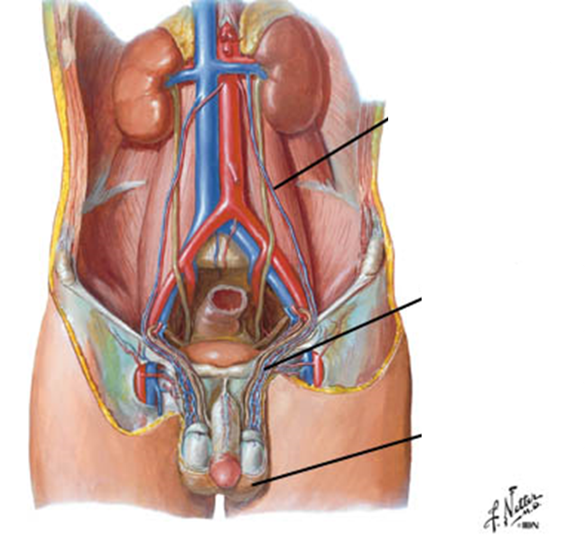
- 41Label the following: - testicular artery & vein - scrotum - spermatic cords
- 42What is venous drainage of the testes?A network around the outside of the spermatic cord, called PAMPINIFORM PLEXUS. This cools the incoming arterial blood.
- The venous drainage in the IVC, from the right testicular vein & in the renal vein, from the left testicular vein. - 43Describe the thymus.- stimulates the maturation of T-cells
- it consists of 2 lobes situated in the upper part of the chest behind the sternum.
- enlarges during childhood, & shrinks to fat after puberty. - 44Describe the pineal gland.- secretes melatonin, which maintains the 'biological clock'.
- large during childhood but shrinks at puberty
- plays a role in sexual development, seasonal breeding & hibernation in animals
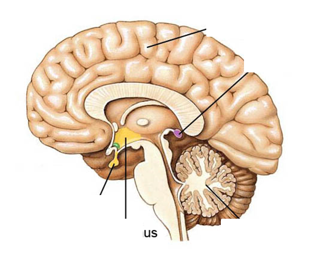
- pineal tumours have been lnked to PRECOCIOUS PUBERTY. - 45Label the following: - cerebral cortex - pituitary gland - hypothalamus - cerebellum
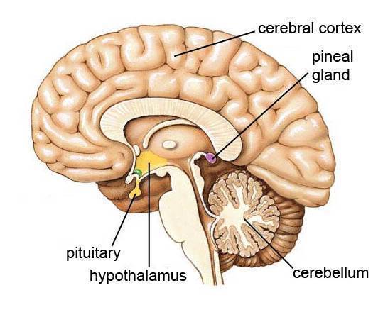
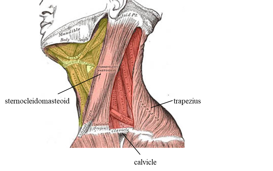
- 46What is in the anterior triangle?- strap muscles
- hyoid
- submandibular salivary gland
- larynx/trachea
- carotid artery
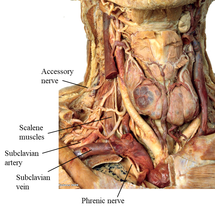
- internal jugular vein - 47What is in the posterior triangle?- accessory nerve
- scalene muscles
- brachial plexus = gives rise to nerves for upper limbs.
- scalenus anterior
- phrenic nerve
- subclavian artery/vein - 48Label the following: - trapezius - clavicle - sternocleidomasteoid
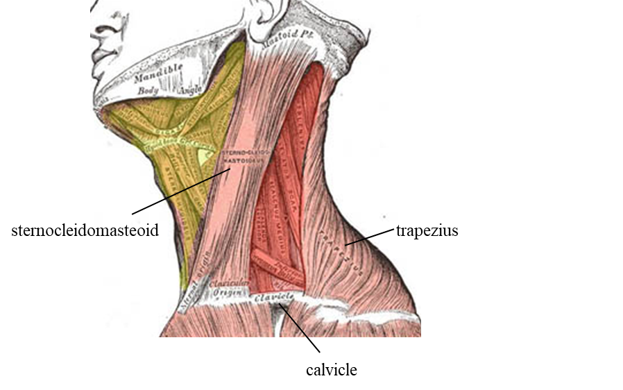
- 49Label the following: - clavicle - trapezius - sternocleidomasteoid
- 50Label the following: - accessory nerve - brachial plexus
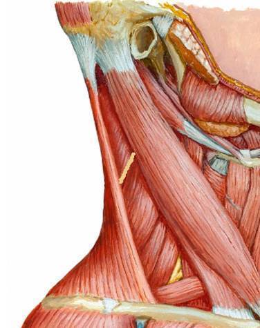
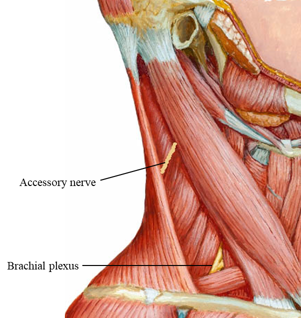
- 51Label the following: - accessory nerve - scalene muscles - subclavian artery - subclavian vein - phrenic nerve
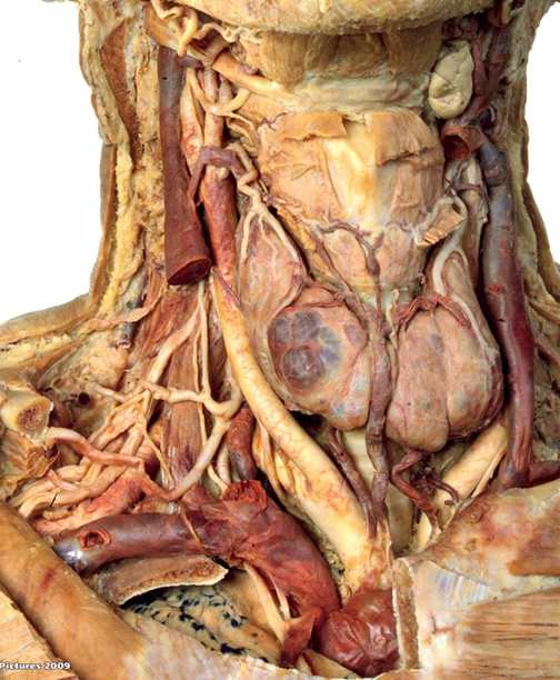
- 52Define chiasma.This is where the 2 optic nerves cross over each other.
- 53What are the 4 major structures in the carotid sheath?1. common carotid artery
2. internal carotid artery
3. internal jugular vein
4. vagus nerve - 54What does retroperitoneal mean?It is a word to describe the placement of the kidneys.
It means that the kidneys are situated behind the peritoneum (membrane that causes the lining of the abdominal cavity). - 55Where does the hilum of kidney lie at?L1 level
- 56What is the transpyloric plane?an imaginary horizontal line, located halfway between the kidneys (very simplified)
- 57At what levels do the right & left kidneys lie at?LEFT = T12 to L3
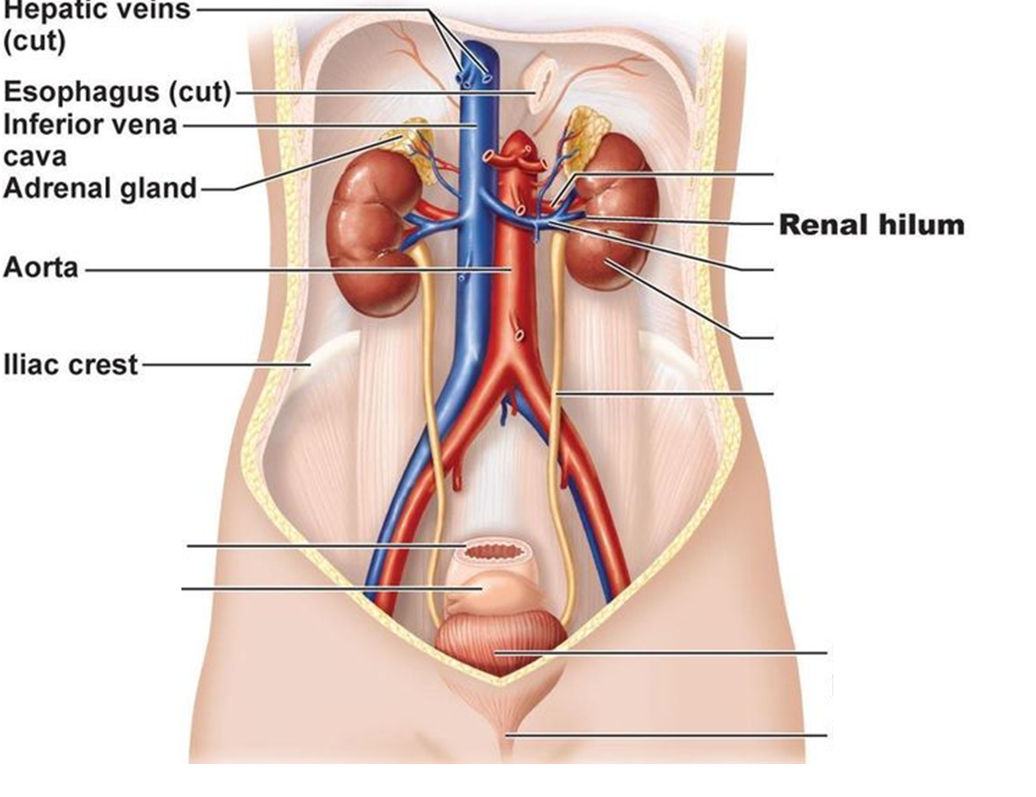
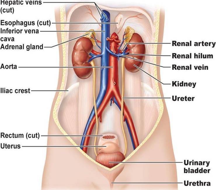
RIGHT = slightly lower, sits just below the diaphragm & posterior to the liver. - 58Label the following: - rectum - uterus - renal artery - renal vein - urinary bladder - urethra
- 59Define splenomegaly.Abnormal enlargement of the spleen.
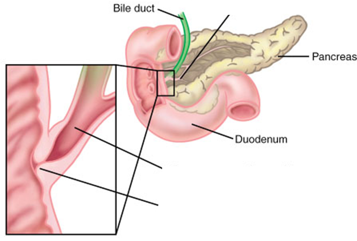
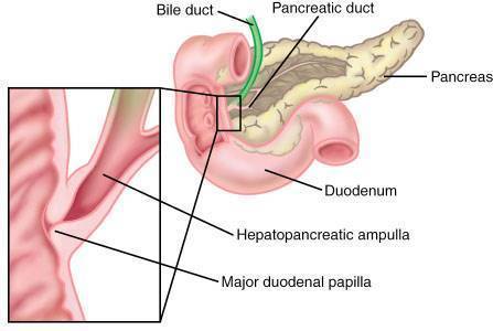
- 60Where does the pancreas secrete its pancreatic digestive enzymes? (exocrine)Via the pancreatic duct at the hepatopancreatic ampulla, along with blie from the gall bladder.
- 61What is the major duodenal papilla?This is where the digestive secretions enter the GI tract.
It is located in the mucosa of the descending part of the duodenum. - 62Label the following: - pancreatic duct -hepatopancreatic ampulla - major duodenal papilla
- 63Define anteverted & anteflexed.ANTEVERTED = inclines forward like the uterus.
ANTEFLEXED = the top of the uterus is forward like a 'C'. - 64What is inguinal canal & what does it contain?This is where the testes descend from to reach the scrotum.
It contains the spermatic cords. - 65What are the 5 layers of scrotum/spermatic cord? (from outer to inner)1. Skin
2. Dartos muscle
3. External spermatic fasica
4. Cremastic muscle + fascia
5. Internal spearmatic fasica - 66What is a varicocele?An abnormal enlargement of the pampiniform venous plexus in the scrotum.
It contains varicose veins in the spermatic cord. - 67What is the function of the dartos muscle?It is a layer of smooth muscle, that contracts/relaxes depending on the external temperature.
This is responsible for the wrinkled apperance of the scrotum. - 68Label the following: - vas / ductus deferens - epididymis
- 69What happens VIA the superficial inguinal ring & at the deep inguinal ring?SUPERFICIAL = this is where epididymis enters the inguinal canal.
DEEP = then the epididymis enters the body cavity via the deep ring. - 70Why is the inguinal canal a common site for hernias to develop?because it is a site of potential weakness.
Information
- Description
- Anatomy Flashcards on The Endocrine System, created by Krupali Parikh on 13/04/2018.
- Created by
- Krupali Parikh
- Views
- 1
- Rating