1532582
Description
Flashcards by Susannah Mackenz, updated more than 1 year ago
|
|
Created by Susannah Mackenz
over 9 years ago
|
|
| Question | Answer |
|
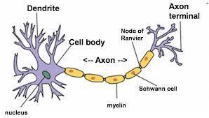
What does this image represent?
Image:
download__3_ (image/jpg)
|
Neuron |
| What is a neuron? | A neuron is the basic building block of the nervous system . |
|
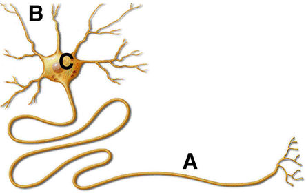
What does C represent?
Image:
Neuron (image/jpg)
|
The cell body (soma) |
|
What does B represent?
Image:
Neuron (image/jpg)
|
The dendrites. |
|
What does A represent?
Image:
Neuron (image/jpg)
|
The axon. |
| The branchlike fibres that are like antennas that collect messages from neighbouring neurons and send them to the cell body are called? | Dendrites |
| Here neighbouring information from other neurons is collected and processed. | The cell body |
| This conducts electrical impulses away from the cell body and to other neurons, muscles, or glands. | Axon |
| -Surround the neurons and hold them in place - Manufacture nutrient chemicals that the neurons need - Form the myelin sheath around some axons - Absorb toxins and waste materials that might damage neurons. | Glial cells |
| Surround neurons and hold them in place | Glial cells |
| Manufacture nutrient chemicals neurons need | Glial cells |
| Form myelin sheath around some axons | Glial cells |
| Absorb toxins and waste materials | Glial cells |
| Fatty insulating layer some neurons have | Myelin sheath |
| Gaps in the myelin sheath that allows impulses to jump, and speeds up transmission | Nodes of Ranvier |
| Protect(s) the brain from toxins | -Blood-brain barrier -Glial cells |
| Nerve activation has how many basic steps? | 3 |
| What are the steps for basic nerve activation? | 1) The neuron is at resting potential 2) Stimulation' resting turns into action potential; nerve impulse generated 3) The neuron is again at rest. |
| What is depolarization and when does it occur? | -The shift from negative to positive voltage -Occurs during the action potential |
| Reversal in the membrane's voltage, from -70mV to +40mV (inside). | Action potential |
| The change in polarization in a neuron membrane that leads to a neural impulse | Action potential |
| The interior of the cell is negative. The exterior of the cell is positive. There is a high concentration of sodium ions outside the cell (in the liquid) and chloride ions (outside cell in the liquid). Negatively charged protein ions (A-) and + potassium ions are inside the neuron. | Resting potential. |
| The inside of the cell is positive; outside, negative. Sodium channels open up; flood into the axon, creating depolarization | Action potential |
| Most sodium outside cell; chloride outside cell Potassium inside cell; negative anion proteins inside cell Gates closed | Resting potential |
| Sodium channels open. Sodium floods into axon. K+ channels still closed | Action potential generated |
| Sodium channels close; potassium channels open | Restoration of resting potential |
| What is the voltage of the refractory period? | Smaller than -70 mV |
| Membrane cannot generate another action potential. | Absolute refractory period |
| All-or-none law | Action potentials occur at a maximum intensity or they do not occur at all. |
| -50 mV | Action potential threshold |
| Graded potentials | Occur when the absolute threshold is not reached but an action potential occurs as a result of many neurons added up. |
| Action potential travels down the axon like a burning fuse | unmyelinated axons |
| High conduction speeds generated from jumping of action potential from node to node | Myelinated axons |
| High conduction speeds generated from jumping of action potential from node to node | Myelinated axons |
| Satiago Ramon Y cajal | Proposed the idea of synapse. |
| Otto Loewi | Proposed the idea of neurotransmitters |
| Gap from the axon terminal of one neuron to the dendrites of another | Synaptic cleft |
| True or false: electrical impulses through a wire are faster than impulses through an axon | True |
| Damage to the myelin sheath can cause | MS Multiple Scorosis |
| When the immune system attacks the Myelin Sheath; impulses are slowed down | MS |
| Chemicals that carry messages across the synapse | Neurotransmitters |
| That excite other neurons OR inhibit their firing | Neurotransmitters |
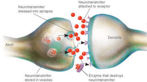
| ______Molecules stored within these _______chambers, within the axon terminal | Neurotransmitters are stored within synaptic vesicles within the axon terminals. |
| When the action potential reaches the end of the axon, the axon terminal releases ________ into the place between the end of the sending neuron( _______) and the membrane of the receiving neuron (______). The space is called _______. | When an action potential reaches the end of the axon, vesicles bind to the membrane and release neurotransmitters into the synaptic space between the pre-synaptic (sending) neuron and the post-synaptic (receiving) neuron. |
| When neurotransmitters cross the synaptic space, they bind to ______ which are? | When neurotransmitters cross the synaptic space they bind to receptor sites, which are large protein molecules embedded in the membrane. |
| Space between the pre-synapse and post-synapse? | Synaptic cleft. |
| Neurotransmitter activity moves from synthesis to deactivation. What is this process? | 1) Synthesis of neurotransmitter 2) Storage in synaptic vesicles 3) Release into synaptic space 4) Binding to receptor sites 5) Deactivation through reuptake or breakdown |
| _________ of neurotransmitter --> storage in ______--> Release into ______--> _________to receptor sites --> Deactivation through _______ or breakdown | Synthesis of neurotransmitter --> Storage in synaptic vesicles--> Release into synaptic space--> Binding to receptor sites--> Deactivation through re-uptake or breakdown |
|
Image:
synapse (image/jpg)
|
Synapse |
| What do A, B, C REPRESENT? | A-Pre-synaptic neuron B- Vesicle C- Synaptic cleft |
| What do D, E, F represent? | D- Dendrite (Post-synaptic neuron) E- Receptor (Collection of which: receptor site) F--Neurotransmitter |
| Neurotransmitters that create depolarization | Excitatory transmitters |
| TRasnmitters that create hyperpolarization | Inhibitory |
| Excitatory--> Depolarizes membrane--> _____likelihood of action potential | Increases |
| Inhibitory--> Hyperpolarizes membrane--> _______likelihood of action potential | Decreases |
| Transmitters are reabsorbed into the pre-synapse | Re-uptake |
| Depression: undersupply Stress and panic disorders: oversupply Inhibitory and excitatory at various sites Involved in learning, memory, wakefulness and eating | Norepinephrine |
| Glutamate | Has powerful excitatory effect |
| GABA | Inhibitory; motor control and control of anxiety; many drugs that help with anxiety disorders target______ |
| ACh | _____; involved in muscle activity and memory; under[production: alzeimer's disease Excitatory Drugs that block _____ can result in muscular paralysis. |
| Dopamine | Can be inhibitory OR excitatory Involved in pleasure, motivation, reward, voluntary movement control, through processes. Parkinsons: not enough _____ LDOPA tries to increase ____production. |
| Serotonin | Often inhibitory; influences mood, eating, sleep, and sexual behaviour -Low levels cause depression -Prozac prevents the reuptake of _____ |
| Endorphins | Reduce pain and increase feelings of well-being; bind to same receptors as morphine and opium, therefore produce similar effects |
|
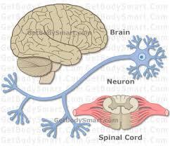
What does this represent?
Image:
images__6_ (image/jpg)
|
The main units of the nervous system |
|
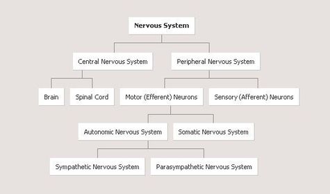
What does this chart visualize?
Image:
5416079_orig (image/jpg)
|
Components of the nervous system |
|
The brain and spinal cord comprise what component of the nervous system?
Image:
Nervous-system (image/jpg)
|
CNS: Central nervous system |
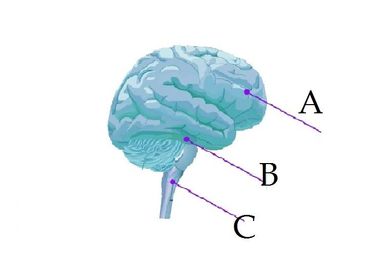
| Label the following components of the brain | A-Forebrain B-midbrain C-hindbrain |
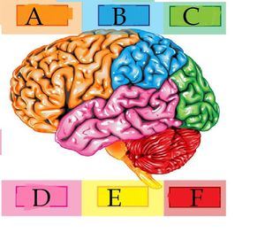
| Label the following | A- Frontal lobe B- Parietal lobe C- Occipital lobe D- tEMPORAL LOBE E-Brain stem F-Cerebellum |
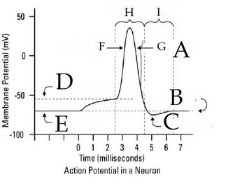
| Label the graph | A--Repolarization B--Resting potential C--Hyperpolarization D--Threshold potential E--Resting potential F--Depolarization G--Repolarization H-- Action potential period I--Refractory period |
| Two major branches of the nervous system | Central and peripheral |
| The hindbrain is composed of the ______ and ______ | Cerebellum and brainstem |
| The nervous system is composed of the _____ and the ______. | Central NS and peripheral NS. |
| The peripheral NS is composed of the ______ and the _______. | Somatic system and autonomic system. |
| The somatic system is a part of the _______nervous system. Its function is? | The somatic system is part of the peripheral system. Its function is that it controls voluntary muscle activation. |
| The autonomic system is part of ______. Its functions are? | The autonomic system is part of the nervous system. Its functions are basically involuntary. It is involved with smooth muscles, cardiac muscle, and glands. |
| The two branches of the autonomic system are? | Sympathetic and parasympathetic. |
| The somatic system | Consist of sensory and motor neurons. Sensory neurons transmit messages from eyes. ears, and other sensory receptors; motor neurons send signals from the brain and spinal cord to the muscles. VOLUNTARY PATHWAY. |
| The autonomic system is involved in? | Respiration, circulation, digestion, motivation, emotional behavior, and stress response. Involuntary Sympathetic and parasympathetic |
| Compare sympathetic and parasympathetic. | Both are part of the peripheral--autonomic--nervous system. Sympathetic: activation or arousal function Parasympathetic: inhibitory, slowing down function |
| Function of the sympathetic system? | -Activation/'arousal - "Fight or flight" - Speeds heart rate -Dilation of pupils; slows digestive system so blood is transferred to muscles; increases rate of respiration |
| Parasympathetic system | Lowers heart rate and respiration rate; constricts pupils; dilates blood vessels; stimulates digestive activity |
| ________ and _______ work together to ensure our body is at a state of equilibrium (homeostasis); coordinated sequence of these activities | Sympathetic "excite" and parasympathetic "relax". |
| Connects spinal cord and brain | Central Nervous System |
| Most nerves enter and leave the CNS through the ______ _______. | Spinal cord |
| ______ _______ are fast responses that involve the senses and the spinal cord (not the brain). | Spinal reflexes |
| Grey matter is surrounded by white (myelinated) matter; this structure cross-section forms an "H". the neurons are protected by back bone vertebrae. | Spinal Cord. |
| The most active energy consumer of all your organs | Your brain |
| Rate of energy metabolism relatively constant throughout the day/night and even increases slightly as you sleep. 2% of body weight; 20 % oxygen consumption at rest. | Your Brain |
| _________tests measure verbal and non-verbal behaviours affected by areas of brain damage. Used in clinical evaluations of people who may have suffered BD through disease or accidents. | Neuropsychological |
| Specific nervous tissue is destroyed by chemicals, electricity, cold or heat; this produces brain damage (lesions). Or can surgically remove areas and study the consequences. Or can be stimulated--producing opposite effects--through electricity or chemicals. This can be constant if an electrode is implanted. | How scientists can test brain damage using sections of the brain and causation |
| PARTS AND FUNCTIONS OF THE BRAIN | STUDY |
| LOBES AND FUNCTIONS OF BRAIN STUDY | STUDY |
| LEFT AND RIGHT HEMISPHERES OF BRAIN | STUDY |
| BRAIN SCANS | STUDY |
| NEUROTRANSMITTERS AND FUNCTIONS | STUDY |
Want to create your own Flashcards for free with GoConqr? Learn more.