15507176
Description

Flashcards by Med Student , updated more than 1 year ago
|
|
Created by Med Student
over 5 years ago
|
|
| Question | Answer |
|
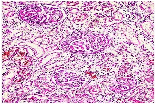
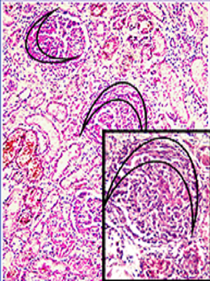
Ans: Rapidly Progressing Glomerulonephritis
1) Pathognomic crescents are seen on the inside of Bowman's capsule. Crescents are formed by proliferation of parietal epithelial cells
2) Glomerular tufts frequently contain fibrin thrombi
3) Tubular epithelial cells may show hyaline droplets and tubular lamina may contain casts, red blood cells and fibrin
4) The interstitium is oedematous and may show early fibrosis
5) Arteries and arterioles may show associated changes of hypertension
Image:
Rpgn (binary/octet-stream)
|
|
|
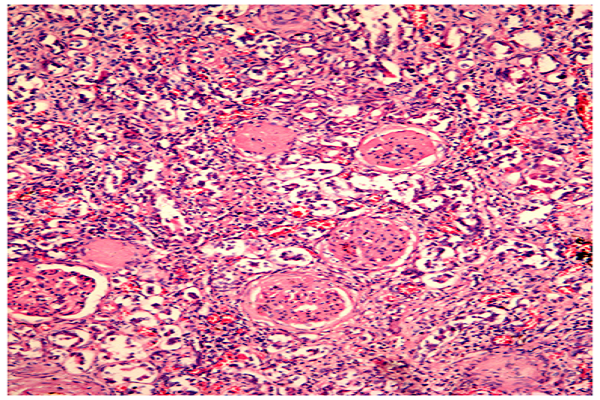
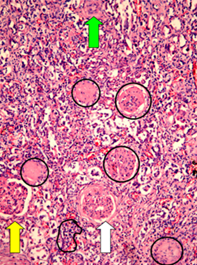
Ans: Sclerotic Glomerulonephritis
The histological finding is mosaic:
1) Almost all glomeruli are hyalinized: homogenously pink, acellular and small size.
2) Lymphocytes form reactive infiltrates around them.
3) Damaged of ‘dead’ nephrones are replaced by connective tissue.
4) Arteriolar walls are thicken their lumens are narrow because of secondary hypertension changes.
5) Only in single glomeruli can show the signs of the initial glomerulonephritis.
6) Single glomeruli and their tubules are preserved and even hypertrophic. They maintain the minimal kidney function in the final stage of the disease.
Image:
Slc Gn (binary/octet-stream)
|
|
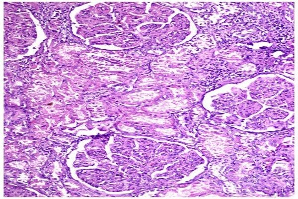
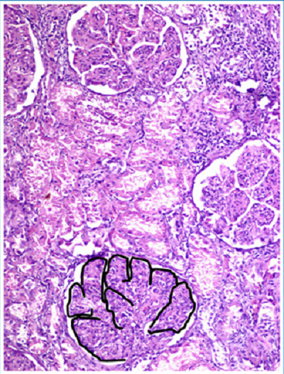
| Ans: Mesangocapillary Glomerulonephritis 1) The glomeruli are enlarged, markedly lobulated and ‘rich’ in cells due to proliferation of mesangial cells (more than 2 cells in a plane section) and accumulation of basement membrane-like material. 2) The capillary walls are thick, their lumens– narrow due to the penetration of mesangium in between the endothelium and basement membrane (so called ‘mesangial interposition’). 3) With silver stain, this phenomenon is seen as a duplication of basement membrane (‘tram rails’ - metaphor). | |
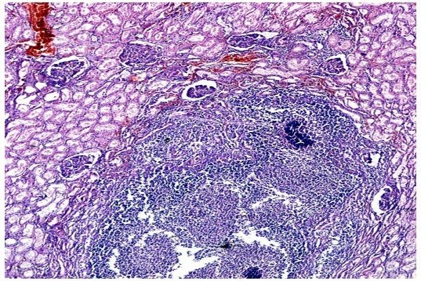
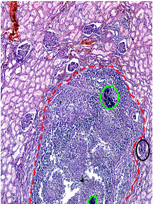
| Ans: Acute Ascending Pyelonephritis 1)Renal cortex is focally destroyed by segmented leukocytic infiltration among which can be found bacterial colonies. 2) Glomeruli even in close proximity to the abscesses, stay long time unaffected. 3) In tubules are formed leukocytic casts (‘cylinders’). | |
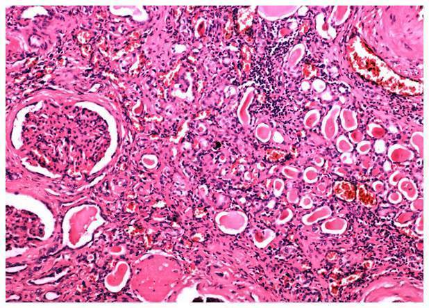
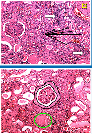
| Ans: Chronic Pyelonephritis Histological triad: 1) In interstitium mostly on the border of cortex/medulla, are found focal infiltrations of lymphocytes and plasma cells. 2) Renal tubules are dilated and filled with pink protein casts resembling the follicular structures of thyroid gland – called ‘strumisation’ of parenchyma 3) Pericapsular glomerular sclerosis. Some kidney bodies are fully sclerosed. In interstitium grows connective tissue, arterioles are with thickened walls and narrow lumens due to secondary hypertension. | |
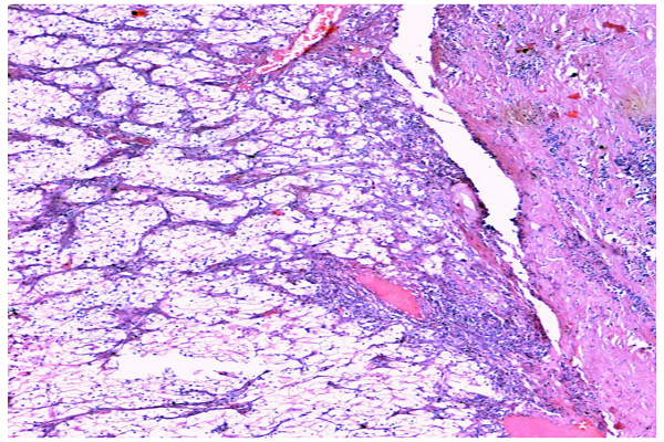
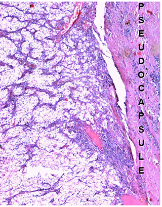
| Ans: Renal Cell Carcinoma 1) Tumor cells with polygonal shape are arranged in nests, tubules and papillae. 2) They have bright cytoplasm and well presented cell membrane. The light cytoplasm is due to extraction of lipids and glycogen from their cytoplasm. 3) Tumor cells infiltrate walls of blood vessels. 4) Among tumor are found necroses and hemorrhages. 5) Stroma is scant, with thin-wall blood vessels and scant lymphoid infiltrations. 6) Tumor is well distinguished with a pseudocapsule (PsC): thick fibrous connective tissue, which at some places appear to have neoplastic infiltration. | |
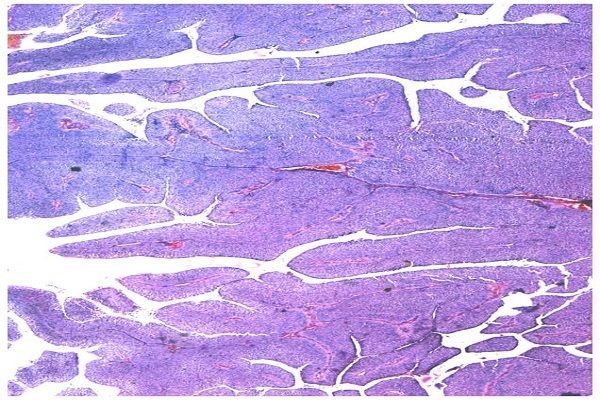
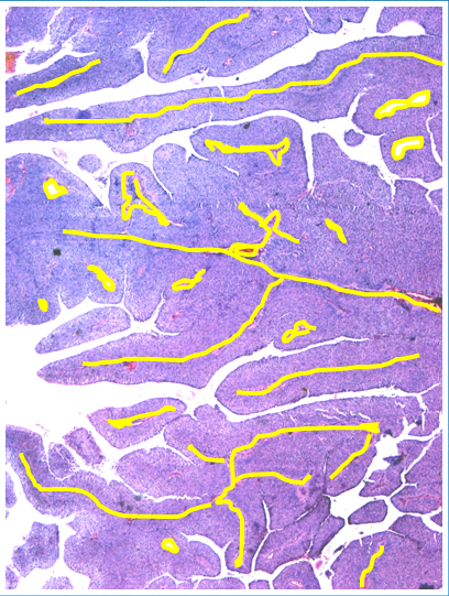
| Ans: Transitional cell carcinoma of urinary bladder 1) Tumor occurs from the mucosa of the urinary bladder. 2) It is composed of a large number of branched papillae, covered with multilayered epithelium, often with anastomoses. 3) They are built up of tender fibro vascular ‘core’, covered with atypical urinary epithelium, which thickness exceeds several times the normal one. 4) On the surface are found so called ‘umbrella’ cells. 5) More differentiated forms have soft papillae and faint cell atypia. 6) In tumor progression are found abnormal mitoses and infiltration in the bladder’s wall. Papillae are short, thick and at some places are fused together. |
Want to create your own Flashcards for free with GoConqr? Learn more.