16055847
Description
Flashcards by Juliette Carroll, updated more than 1 year ago
|
|
Created by Juliette Carroll
about 7 years ago
|
|
| Question | Answer |
| What are the external nares? | Nostrils |
| What is the purpose of the cartilage found in nostrils? | To support their structure during inspiration so that they don't collapse |
| What type of cartilage is found in the nostrils? | Alar cartilage which is very elastic |
| What is the nasal plate? What is its purpose? | External cartilaginous plate in the nostrils to widen nostrils during inspiration |
| Which species' is the philtrum found in? What is its structure? | Sheep and dogs: median groove which extends down from the middle of the nasal plate |
| Why is the nasal plate referred to as the nasolabial plate in cows? | Extends down to include the lips |
| Is the cartilage in the nostrils of horses internal or external? | Internal |
| What is an obligate nasal breather? | A species which can only breathe throught its nostrils and not through the mouth |
| Describe the structure of the false nostril in horses | Blind ending "pouch" in the dorsal region of the nostril in horses |
| What care must be taken when inserting a stomach tube into a horse? | That you insert it into the real nostrils and not the falsie |
| What is the os rostrale in pigs? | Splanchnic bone found in the nose for support |
| What separates the nostrils? | Cartilaginous septum |
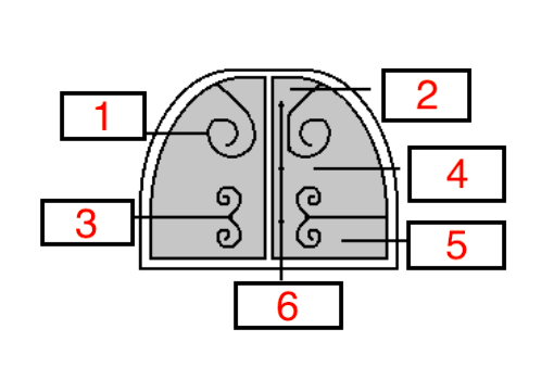
| Name structures 1-6 (transverse section of a nose shown) | 1) Dorsal concha 2) Dorsal meatus 3) Ventral concha 4) Middle meatus 5) Ventral meatus 6) Common meatus |
| In horses, through which structure is a stomach tube passed? | Ventral meatus |
| What are conchae? | Scrolls of bone extending off the skull into the nostrils |
| What are conchae covered in? | Ciliated mucus epithelium |
| What structure within the nasal cavity is responsible for olfaction? | Specialised conchae close to the brain w/sensory nerve endings |
| What does meatus mean? | Passage |
| What structure does the dorsal meatus lead to in some species'? | Frontal sinus |
| Where does the middle meatus lead to? | The ethmoconchae and maxillary sinuses |
| What is the name for the conchae which detect smell? | Ethmoconchae |
| Which meatus leads to the nasopharynx? | Ventral meatus |
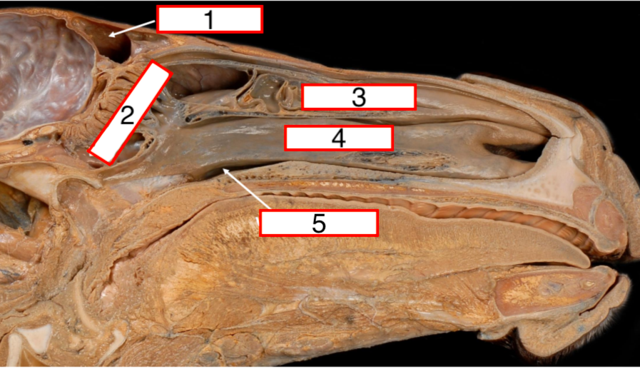
| Name structures 1-5 | 1) Frontal sinus 2) Ethmoconchae 3) Dorsal concha 4) Ventral concha 5) Ventral meatus |
| What are the paranasal sinuses? | Air-filled spaces within the bones of the skull |
| Which are the 2 most important paranasal sinuses? | Frontal and maxillary |
| Where do the paranasal sinuses drain into? | Different parts of the nasal cavity |
| In which species is the maxillary sinus poorly developed? What is it called instead of a sinus? | ~In the dog ~Recess |
| How does the maxillary sinus drain into the nasal cavity? | Via the middle meatus |
| In which species are the sinuses extensive? | Horse |
| What is the maxillary sinus divided into in the horse? | Rostral sinus and caudal sinus |
| How do the frontal and maxillary sinuses drain into the nasal cavity in the horse? | Via the naso-maxillary opening by way of middle meatus |
| How is the structure of the sinuses of the horse clinically important? | Closely related to the cheek teeth and upper jaw so when there is an abscess in a tooth, the pus tends to drain into the sinuses and must be removed by drilling into the skull |
| What is the laryngopharynx? | Bits at the back of the soft palate which borders with the larynx |
| Which structure connects the nasopharynx and auditory tube? | Slight-like opening in lateral nasopharynx connecting it with the auditory tube |
| What is the Hyoid apparatus? | Series of small bones suspending larynx and tongue from skull |
| What is the mastoid proceess? | Point at which hyoid apparatus joins skull (?) |
| Which structure in the hyoid apparatus connects to the skull? | Tympanohyoid cartilage |
| Which is the only bone in the hyoid apparatus which is unpaired? | Basihyoid bone |
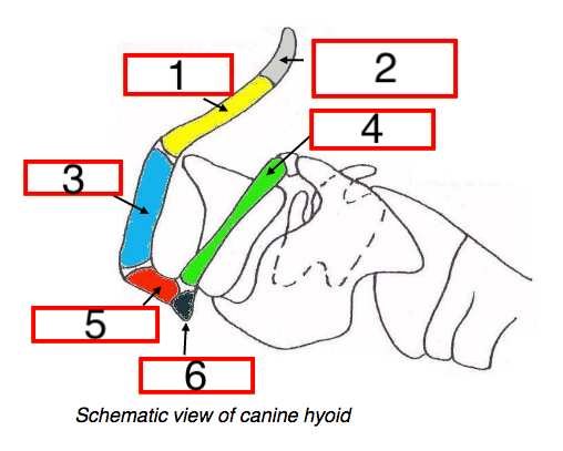
| Name structures 1-6 | 1) Stylohyoid 2) Tympanohyoid cartilage 3) Epihyoid 4) Thyrohyoid 5) Ceratohyoid 6) Basihyoid |
| What is found in the hyoid apparatus of large animals? | Lingual process extending from basihyoid bone |
| What kind of cartilage comprises the epiglottis? | Elastic cartilage |
| What type of cartilage comprises the other sections of cartilage in the larynx besides the epiglottis? | Hyaline |
| What are the functions of the larynx? | ~Regulation of air into trachea ~Vocalisation ~Epiglottis prevents entry of food into trachea during swallowing |
| Name the 4 laryngeal cartilages | 1) Epiglottis 2) Thryoid 3) Arytenoid 4) Cricoid |
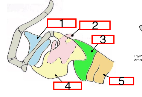
| Name structures 1-5 | 1) Epiglottis 2) Arytenoid 3) Cricoid 4) Thyroid 5) Trachea |
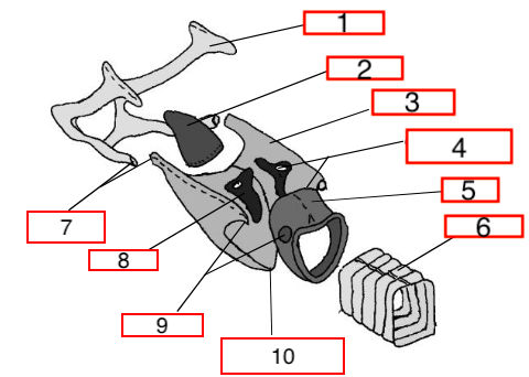
| Name structures 1-10 | 1) Hyoid bone 2) Epiglottis 3) Thyroid lamina 4) Crico-arytenoid articulation 5) Cricoid 6) Trachea 7) Thryo-hyoid articulation 8) Arytenoid 9) Crico-thyroid articulation 10) Thyroid body (junction of laminae) |
| Name the most rostral piece of the larynx | Epiglottis |
| Which piece of cartilage does the epiglottis connect to? | Thyroid cartilage |
| Describe the structure of the thyroid cartilage | U-shaped tube consisting of 2 sheet-like joined together ventrally |
| What 2 structures does the thyroid cartilage connect to? | Thyrohyoid bone dorsocranially Cricoid cartilage caudally |
| Where are the arytenoid cartilages found? | 2 of them within the thyroid cartilage |
| Which cartilage do the arytenoid cartilages articulate with? | Cricoid |
| What structure does the cricoid cartilage join with caudally? | Trachea |
| What 2 structures does the cricoid cartilage join with cranially? | Thyroid cartilage Arytenoid cartilages |
| What is the glottis? | Diameter of the airway determined by distance between vocal ligaments |
| Describe the structure of the vocal cords | ~Vocal ligaments covered in mucosa ~Originate in ventral process of arytenoid cartilage and run down to join floor of thyroid |
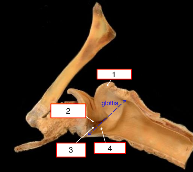
| Name structures 1-4 (equine larynx shown) | 1) Arytenoid 2) Ventricular fold 3) Laryngeal ventricle 4) Vocal fold |
| What is the difference between the vocal ligament and the vocal fold? | Vocal fold is vocal ligament covered in mucosa |
| Where does the dorsal cricoarytenoideus muscle lie? What type of muscle is it? | ~Between the cricoid and arytenoid cartilages positioned dorsally ~Intrinsic muscle of the larynx |
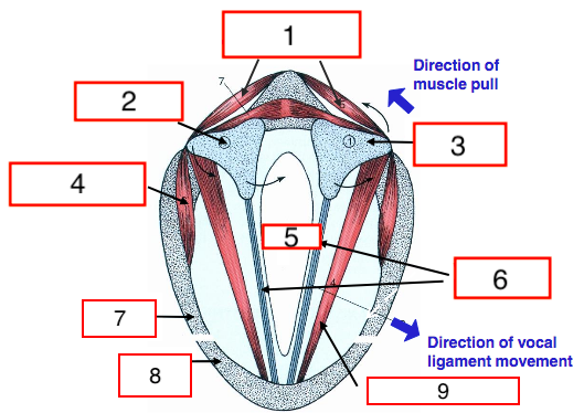
| Name structures 1-4 (transverse section of canine larynx) | 1) Dorsal cricoarytendoideus 2) Crico-aytenoid articulation 3) Arytenoid cartilage 4) Lateral cricoarytenoideus |
| Name structures 5-9 (transverse section of canine larynx) | 5) Glottis 6) Vocal ligaments 7) Cricoid cartilage 8) Thyroid cartilage 9) Thyroarytenoideus |
| What does contraction of the dorsal cricoarytenoideus muscle do to the size of the glottis? | Increases it by causing the arytenoid cartilages to rotate |
| What happens to the vocal ligaments when the dorsal cricoarytenoideus muscle contracts? | They are abducted |
| Name the nerve which innervates the dorsal cricoarytenoideus | Recurrent laryngeal nerve |
| What does contraction of the lateral cricoarytenoideus muscles do? | Causes adduction of vocal ligaments |
| What is the function of the thyroarytenoideus muscle? | Adjust tension in the vocal ligaments |
| What is laryngeal hemiplegia? | Damage to the recurrent laryngeal nerve causing paralysis of the dorsal cricoarytenoid muscle |
| Which side is laryngeal hemiplegia most likely to occur on? | Left |
| An inability to do what makes it difficult for animals w/laryngeal hemiplegia to enlarge their glottis? | Unable to abduct the ipsilateral vocal fold and therefore enlarge the glottis |
| What is roaring? | Noise produced by air moving over loose vocal cord which cannot be abducted when the animal is moving quickly (in horses and sometimes in dogs) |
| What is done to correct laryngeal hemiplegia? | Hobday or "tie back" operation |
| Where does the trachea run from and to? | Cricoid cartilage to bifurcation at base of heart |
| Name the ligament which attaches the cricoid cartilage to the trachea | Cricotracheal ligament |
| Name the structure which connects the cartilaginous rings of the trachea to one another | Annular ligaments consisting of connective tissue |
| What type of muscle is the trachealis muscle made of? | Smooth |
| Where does most of the control of the diameter of the airway occur? | In the bronchi/bronchioles |
| Where does the trachealis muscle lie in a) carnivores? b) herbivores? | a) Outside cartilaginoues rings b) Inside cartilaginoues rings |
| What kind of epithelium lines the trachea? | Pseudostratified columnar ciliated epithelium |
| What is found in the mucosa of the trachea? | Mucous glands |
| Where specifically is the guttoral pouch located? | Between the nasopharynx and the middle ear |
| What does it mean that the epiglottis is retrovelar in horses? | Sits behind the soft palate |
| How does the epiglottis being retrovelar in horses mean that it is an obligate nasal breather? | Air simply hits a "brick wall" when is passes the soft palate in the mouth because the epiglottis extends behind it |
| How is the guttoral pouch divided into left and right sides? | Thin membrane |
| What separates each of the compartments of the guttoral pouch into medial and lateral sides? | Stylohyoid bone |
| Which kinds of infection are common in the guttoral pouch? | Fungal |
| Which is the only species (vets focus on) which has a guttoral pouch? | Horses |
| What is the function of the guttoral pouch? | Unknown |
| What is the pharyngotubal opening? | Opening which connects the guttoral pouch with the nasopharynx and has flaps |
| What is the anatomical name for the guttoral pouch? | Diverticulum of the Auditory Tube (DAT) |
| Describe the structure of the guttoral pouch? | An air filled pouch with a capacity of 300-500ml |
| Which important structure pass through the guttoral pouch? | Blood vessels and nerves |
| How can fungal infections in the guttoral pouch be dangerous? | Can eat away at walls of BVs found there |
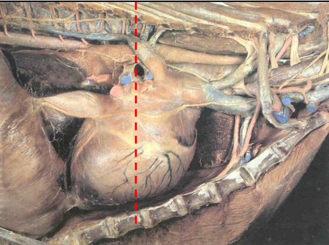
| Roughly where on this image does the trachea bifurcate to the left and right bronchi? What is this region called? | Base of heart |
| Where does the tracha lie in relation to the oesophagus a) cranially? b) at the point of the heart and more caudally? | a) On the right b) Ventral |
| What do segmental bronchi aerate? | Broncho-pulmonary segments (cone-shaped) |
| At which point does the cartilaginous support in the resp. tract stop? | At the bronchioles |
| Which feature of the resp. tract can be found in pigs and ruminants? | Tracheal bronchus |
| Where does the tracheal bronchus come off of the main trachea in respect to bifurcation of the trachea? | Comes off at 3rd rib and trachea bifurcates at 5th |
| Which structure does the tracheal bronchus aerate? | Cranial lobe of the right lung |
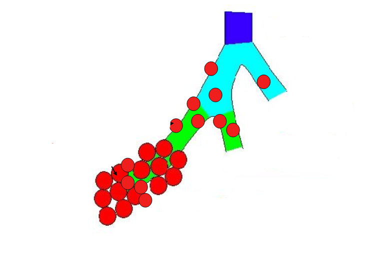
| Which is the alveolus and which is the alveolar saccule on the image shown? | Alveolus is single red sphere Aveolar saccule is the bunch of grapes |
| Which structures attach to the lungs at the hilus? | Primary bronchus Pleurae (?) Arteries/veins Nerves Lymphatics |
| What does the colour of the lungs depend on? | Amount of blood in them |
| What are the 2 types of blood supply that the lungs receive? | Pulmonary Systemic |
| What is the purpose of the systemic circulation to the lungs? | To provide them w/ox. blood so that they can function |
| Name the vessels in the pulmonary circulation which supply the lungs | Pulmonary artery Pulmonary vein |
| Name the vessels in the systemic circulation which supply the lungs | Bronchial arteries Bronchial veins |
| What structure can be seen at the cardiac notch? | Heart |
| Name strutures 1-5 | 1) Caudal lobe 2) Diaphragm 3) Cranial lobe 4) Middle lobe 5) Heart |
| Indicate the cardiac notch on the image | |
| Which is the costal surface of the lungs? | That which touches the ribs |
| What is the main difference between the structure of the lungs of a dog and that of other mammals eg. horse? | They have deep fissures between each lobe |
| What is the cranial lobe of a dog divided into? | Cranial and caudal parts |
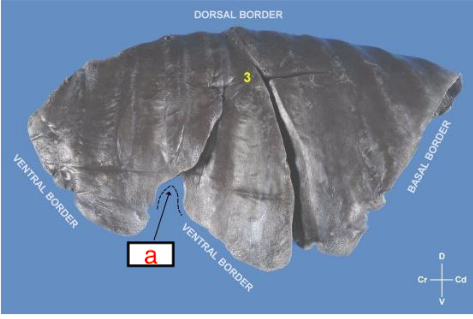
| Which species does this lung belong to? What is arrow a pointing at? | Dog. A is pointing at the cardiac notch |
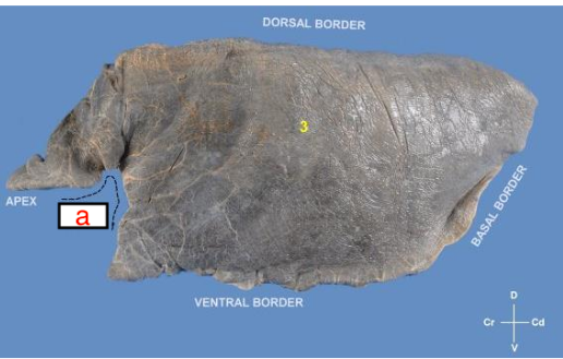
| Which species does this lung belong to? What is indicated by a? | Horse. A shows cardiac notch |
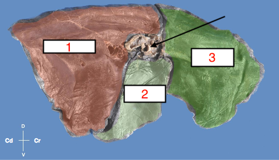
| Name structures 1-3. What is the arrow pointing at? | 1) Caudal lobe 2) Caudal part of cranial lobe 3) Cranial part of cranial lobe Arrow is pointing at hilus |
| Name structures 1 and 2 | 1) Caudal lobe 2) Cranial lobe |
| How is the cranial lobe of dogs different from other animals? | Subdivided into cranial and caudal parts |
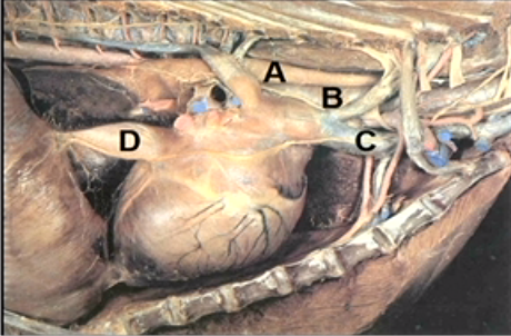
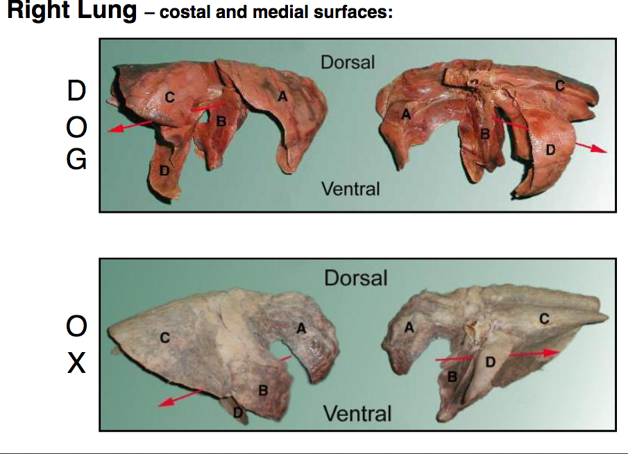
| What is lobe D known as? What does the red arrow represent? | D: Accessory lobe Red arrow shows passage of vena cava |
| How many lobes are present in the right lung in dogs and cows? | 4 |
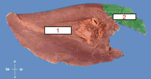
| How many lobes are present in the right lung in horses? | 3 |
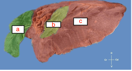
| Which of a, b and c represents the accessory lobe? Which species does this lung belong to? | b: belongs to horse |
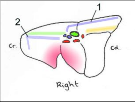
| Is this a left or a right lung? | Right |
| Which structure entering the lungs at the hilus is the most dorsal? | Bronchus |
| What are lobulations? | Sections of lung tissue (alveoli?) surrounded by connective tissue |
| Which structure entering the lungs at the hilus is the most cranial? | P. artery |
| Why is a tracheal impression (in fixed specimens) not seen on the left lung? | Because it lies to the right of the oesophagus |
| Why is a tracheal impression (in fixed specimens) not seen on the caudal lobe of the lung? | It bifurcates into left and right bronchi at the 5th rib so doesn't pass through caudal lobe |
| Where is the impression (in fixed specimens) of the oesophagus seen? Why is this? | Caudal lobe of right lung Both lobes of left lung Lies to the left of the trachea and therefore only seen on caudal lobe of right lung once trachea has bifurcated but seen on both lobes of left lung |
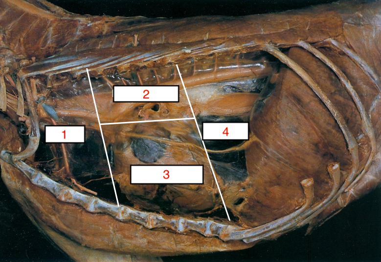
| What are 1 and 2 pointing at? | 1) Azygous vein 2) Cranial vena cava and internal thoracic vein (?) |
| How are pleura often named (besides parietal and visceral)? | After the structure they cover eg. costal pleura is parietal pleura which covers the ribs |
| What are the functions of the pleurae? | To reduce friction Ventilate lungs |
| Which type of pleura is included in mediastinal pleura? | Pericardial pleura |
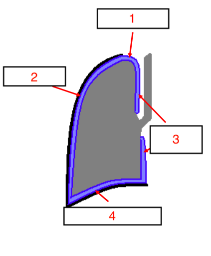
| Name structures 1-4 | 1) Cupula pleurae 2) Costal pleura 3) Mediastinal pleurae 4) Diaphragmatic pleura |
| Which 3 types of pleura comprise the parietal? | ~Costal ~Mediastinal ~Diaphragmatic |
| Which type of pleura is particularly vulnerable to injury? | Cupula |
| Why is the cupula pleura vulnerable to injury? | Extends beyond the thoracic cavity into neck and is therefore not protected by ribs |
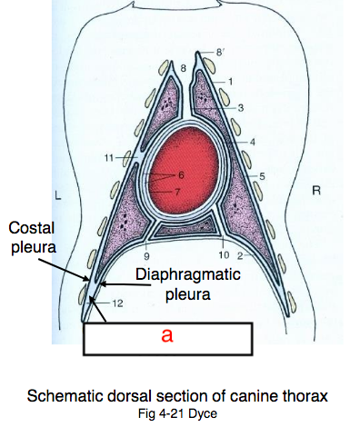
| Which area in the thoracic cavity can be used to sample pleural fluid? | Costodiaphragmatic recess |
| Name the 2 pleural recesses | Costodiaphragmatic Costomediastinal |
| Why are there recesses present within the pleura? | Lungs do not completely fill the pleural cavity |
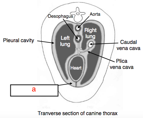
| What is the plica vena cava? | Fold in mediastinal pleura which covers vena cava |
| What is a pointing at? | Costodiaphragmatic recess |
| What is a pointing at? | Costomediastinal recess |
| What is the mediastinum? | Central compartment between the right and left pleural cavities |
| What structures can be found in the mediastinum? | Heart Oesophagus Blood vessels Nerves Lymphatic Thymus gland (in young animals) |
| What surrounds the mediastinum? What is significant about this strucutre? | ~Mediastinal pleura ~Very deliate |
| Why does an infection in one pleural cavity in the horse spread easily to the other? | Fenestrations in mediastinal pleura |
| In which regions do the 2 mediastinal pleura come in contact with each other? | Cranial and caudal to heart |
| Which species has a delicate but complete mediastinal pleura? | Dog |
| Why is it sometimes necessary to ventilate both lungs when only one collapses in a dog? | Pressure is so great that it causes rupture of mediastinal pleura |
| Why does infection/collapsed lung not spread from one pleural cavity to the other in cows? | Thick mediastinal pleura |
| Is an infection likely to pass from one pleural cavity in dogs to the other? | No (possible?) |
| Name divisions 1-4 of the mediastinum (left side shown) | 1) Cranial 2) Middle dorsal 3) Middle ventral 4) Caudal |
| What is the NAV term for muzzle? | Rostrum |
Want to create your own Flashcards for free with GoConqr? Learn more.