1976889
| Question | Answer |
| Fristch and Hitzig | 1870s; What happens if you apply electricity to the brain? - took dogs, anesthesia, removed top of skull, stimulate brain * Reactions to stimulation of the brain were specific to the area stimulated and also repeatable suggesting that the brain controls muscles and thus behavior. |
| Bartholow | What happens if you stimulate the human brain? - patient Mary Rafferty with hole in head --> stimulate * Once again, stimulation of the brain was selective (only producing a response when touching a specific area) and reliable. |
| John Flynn | What happens if you stimulate deeper regions in a controlled manner? - used an electrode to stimulate deeper regions - stimulated hypothalamus and other areas 1) quiet biting attack - make self smaller 2) affective attack - emotional * The stimulation resulted in something that was more than just a bunch of twitches. It looked like real behavior. |
| Wilder Penfield | 1950s How could we help people with epilepsy? - find the focus/locus of the seizure - take off hunk of skulls --> stimulate various portions of the brain STIMULATION AREAS: - middle - muscles twitch - back - saw imagery, heard noises, smelled scents, felt touches (sensory experiences) ; different area - hear songs, hear bells - some areas stimulated memories * The brain is linked with sensation, emotion, and cognition. |
| Deep Brain Stimulation (DBS) | implanting electrical stimulus into the brain; used with Parkinsons and depression |
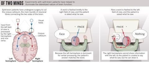
| Gazzaniga | People could usually feel when epilepsy would happen. They felt some sort of "aura." Where is this "aura" location? - tried to keep seizures localized - severed corpus callosum (localized seizures to one hemisphere) |
| Corpus callosum | wide flat bundle of neural fibers that allows the left side of the brain to speak with the right side; located in the middle between two hemispheres |
| 1) Connections in the brain are crossed | Left side of brain --> right side of body Right side of brain --> left side of body |
| 2) There is some laterality in the brain. | Some things are on one side and not the other. Right handed people: - language, details on left side - facial recognition, patterns on right side |
| Experiments on people with severed corpus callosums | Screen in front; can't see but can feel 1) right hand feels --> left side brain --> speaks (says what the object is) 2) left hand feels --> right side brain --> can't speak (because language is on the left side) Eventually left hand --> right brain --> facial expression (frowns) --> recognizes it is the wrong object (able to communicate with left brain this way) |
| Experiments on people with severed corpus callosums (facial) | - take pictures of two people (half of one, half of other) - expose in one visual field - right visual field --> left hemisphere --> sees contours, lines - left visual field --> right hemisphere --> sees faces |
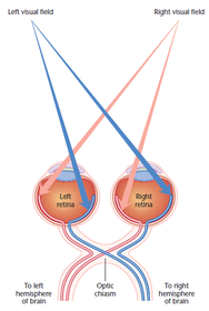
| Hemispheres & visual fields | right visual field --> left half of each retina --> left hemisphere left visual field --> right half of each retina --> right hemisphere |
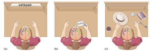
| Effects of damage to the corpus callosum | Right visual field (band) --> left retinas --> left hemisphere (language) --> writes out band using right hand Left visual field (hat) --> right retinas --> right hemisphere (visual) --> left hand points to hat |
| Nissl | - worked with stains; discovered the Nissl stain - could see a lot of internal anatomy in the brain (areas with tons of cells vs little, big cells vs small) - saw two diff types of cells (big neurons vs small glia) - stains most of the cells in the brain |
| Types of Glia | 1) astrocyte 2) oligodendrocytes 3) microglia 4) radial glia |
| Astrocytes | - line all the spaces between the brain & the outside world - helps synchronize activity of neurons - remove waste material when neurons die - control amount of blood flow |
| Oligodendrocytes | - have myelin on the outside - form myelin in the central nervous system Schwann cell: a glial cell that makes myelin outside of the brain, in the body |
| Microglia | - scavenger cells - mini Roombas in the brain - function like part of the immune system |
| Radial glia | - guide migration of neurons & axons during development - provide a scaffold for cells to move - only present during development |
| Golgi & Caja | 1873 - Golgi found a way to stain nerves with silver salts - stained some cells without affecting others - Cajal looked at these stains and looked at infant brains * Cajal discovered that the nervous system is composed of separate cells. |
| Golgi stain | stains between 1-5% of neurons in the brain --> can see individual stains/cells |
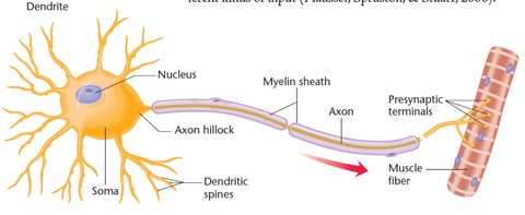
| Structure of a neuron | |
| Dendrites vs axons | DENDRITES: - receiving - acute angle between dendritic branches/spines and dendrite - bumpy, lumpy, big - many AXONS: - transmitting - right angle between axon and axon collaterals (branches) - smooth - usually only one - axon terminal (swell at tip where axon releases chemicals) |
| Afferent, efferent, and intrinsic (interneuron) | Afferent - admission - bringing information into a structure Efferent - exit - carrying information away from the structure Intrinsic/interneuron - cell's dendrite & axons are all located within a single structure |
| blood-brain barrier | unbroken wall of cells that surrounds the blood vessels of the brain and spinal cord - Uncharged particles (CO2, O2) and fat soluble molecules can cross passively. - Active transport pumps glucose, amino acids, iron, hormones, etc. into the brain & spinal cord. * Most adult neurons rely on glucose (and thiamine / vitamin B1 to use it). |
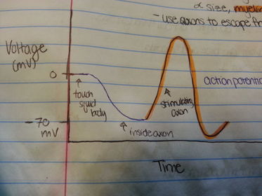
| Hodgkins and Huxley | Nobel prize 1963 - wanted to measure electricity (the diff in voltage between point A and point B) - microelectrode, amplifier, oscilloscope (cathode ray tube) - used a giant squid axon (~ 1 mm across) --> stimulate cell with electrode --> look at oscilloscope |
| Why use a giant squid axon? | conduction velocity - speed of signal along the axon proportional to size and amount of myelinatin |
| Hodgkins and Huxley Results | |
| Resting Potential | -70 mV; due to chemical (concentration) and electrical gradient involving the cell; interior of cell is slightly negative relative to exterior |
| sodium-potassium (Na-K) pump | |
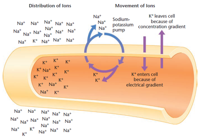
| The sodium-potassium pump creates ______? | chemical and electrical gradients CHEMICAL: - More Na+ outside - More K+ inside - potassium channels slightly open, potassium can leak out (low conc --> high conc) --> ELECTRICAL: - less positive charges on inside - also negatively charged proteins & chloride ions - potassium thus wants to enter the cell (more positive area --> more negative area to even out charge) |
| potassium flow in/out of the neuron | |
| Nernst equation | V = ± 60 log [K_out] / [K_in] KNOW THAT: K+ naturally wants to be outside cell Na+ naturally wants to be in the cell Cl- naturally wants to be outside the cell |
| Action potential graph | |
| What happens during an action potential? | - Membranes have channels (proteins that are like pores) - These channels are gated (can be opened or closed). DEPOLARIZATION: 1) voltage-gated sodium channels open up 2) Na+ goes through pore and starts to flood inside of cell (since inside is relatively negative and has lower concentration of Na+) 3) peak of action potential --> Na+ gates close HYPERPOLARIZATION: 1) voltage-gated potassium channels open 2) potassium flows out (no longer negative charge in cell, much more concentrated on inside) 3) chloride ions flow in (more concentrated outside membrane, cell is positive) BACK TO RESTING POTENTIAL: 1) sodium-potassium pump works to restore normal concentrations |
| refractory period | - period of time where the neuron resists the production of further action potentials - sets an upper boundary on the # of action potentials that can occur at one point in time absolute: membrane cannot produce an action potential, regardless of the stimulation relative: a stronger than usual stimulus is necessary to initiate an action potential |
| Summary graph of refractory period | |
| All or None Laws | For a given neuron, all action potentials are approximately the same amplitude (size), velocity, and shape. |
| Propogation of the action potential | transmission of the action potential down an axon |
| Properties of a graded potential | 1) passive - doesn't cost energy 2) graded - put in more current, go a longer distance 3) fast |
| Properties of an action potential | 1) active - cell is expending energy 2) all or none 3) relatively slow |
| Why does an impulse not travel backwards? | Because of the refractory period of the axons that already had an action potential |
| Myelin | insulating material made up of fats and proteins; enhances speed of conduction - no myelin --> action potential - myelinated sections --> graded potential * myelinated sections of the axon have no sodium channels underneath |
| saltatory conduction | same as myelinated conduction; the jumping of action potentials from node of Ranvier to other node |
| T. R. Elliott | applied adrenaline directly to the surface of heart, stomach, pupils --> produces same effects as those of the sympathetic nervous system Elliott suggested that sympathetic nerves stimulate muscles by releasing a chemical. |
| Otto Loewi | stimulated vagus nerve of frog thereby decreasing heart rate --> took liquid from heart --> put liquid into second frog's heart --> second frog heart decreased rate of beating |
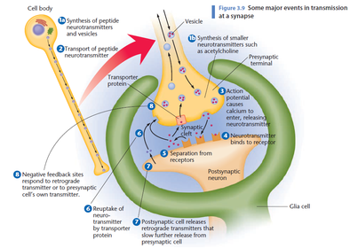
| Steps at the synapse | |
| Sequence of events at the synapse | 1) Synthesis of neurotransmitters (axon terminals) and neuropeptides (cell body) 2) Transport of neuropeptides to axon terminal/dendrite 3) Action potential travels down axon --> voltage gated calcium channel opens --> Ca^2+ enters the axon terminal --> neurotransmitters released in vesicles into the synaptic cleft 4) Released molecules attach specifically to receptor molecules and change activity of postsynaptic neuron 5) Neurotransmitter molecules separate from receptors 6) Neurotransmitter molecules taken back into presynaptic neuron for recycling or diffuse away 7) Some post synaptic cells send reverse messages to control further release of neurotransmitters by presynaptic cells. |
| Synapse | mechanism by which information is transferred in the nervous system gap is called synaptic cleft point where conduction is slow but information processing is complicated |
| ionotropic synapse | neurotransmitter binds to receptor --> opens channels for some type of ion - instantaneous changes that go away quickly - effects localized to one point on the membrane - depend mostly on GABA (inhibatory) or glutamate (excitatory) |
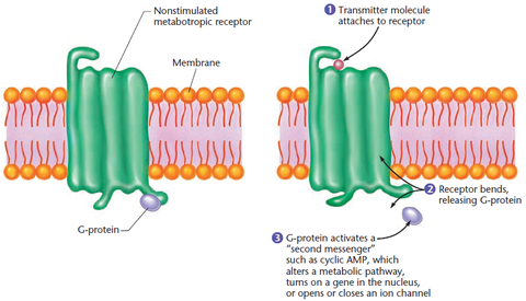
| metabotropic synapse | neurotransmitters released --> bind to receptor --> causes second messenger concentration to rise --> second messenger diffuse around cell causing changes - effects last for longer than ionotropic, influence activity over most all of the cell (not just one part of the membrane) |
| Types of neurotransmitters | - amino acids - neuropeptides (chains of amino acids) - acetylcholine - monoamines - purines - gases (e.g. nitric oxide, released by stimulated neurons, dilate nearby blood vessels) |
| Acetylcholine | found at the neuromuscular junction; cholinergic cells are cells that use acetylcholine |
| Monoamine | - serotonin (5HT) - catecholamines (e.g. dopamine [DA], epinephrine, norepinephrine [NE]) |
| Amino acids | - glutamate (excitatory) - GABA (inhibatory) |
| Ligand gated channel | proteins in the membrane that are formed like channels and can be opened when a specific ligand (molecule) binds to them |
| Neurotransmitters vs neuropeptides | NEUROTRANSMITTERS: - released mainly at axon terminal - single action potential can make them get released - released immediately adjacent to receptors NEUROPEPTIDES: - released by cell bodies, dendrites, and sides of axons - requires repeated stimulation to get released - not released often but a substantial amount is released when they are - diffuse widely throughout the brain via metabotropic receptors |
| Metabotropic synapse diagram | |
| glutamate | amino acid that depolarizes (excites) a cell via ionotropic pathways |
| GABA | amino acid that hyperpolarizes (inhibits) the cell via ionotropic pathways |
| Dale's Law | Originally stated: Any neuron will release one and only one neurotransmitter at all its synapses. Changed over time: Any neuron will release the same cocktail of neurotransmitters (possibly both ionotropic and metabotropic) |
| Quantic release | Voltage only changes in integer units. You don't ever release half a vesicle. |
| How do you clean out the synaptic cleft? | a) enzymatic breakdown (e.g. acetylcholinesterase, monoamine oxidase (MAO), catechol-O-methyltransferase (COMT) ) b) reuptake (picked up by the cleft and used again) |
| Sherrington | conducted experiments on reflexes FOUND: a) reflexes are slower than conduction along an axon b) several weak stimuli presented at different times or slightly different locations produce a stronger reflex than a single stimulus c) one muscle becomes excited; another group becomes relaxed |
| Implications of Sherrington's experiment | - Sherrington measured the total distance that the impulse traveled and calculated the speed needed to travel in the specific amount of time measured FOUND: - speed was slower than previously measured action potential velocities * Concluded that some process must slow conduction down --> probably occurs where neurons communicate with each other --> established existence of synapses |
| Results of Sherrington's experiment presented visually | |
| Temporal summation | repeated stimuli within a close period of time add together and may reach threshhold --> stimulate an action potential |
| Spatial summation | synaptic inputs from separate locations combine their effects on a neuron; e.g. pinching two places at once sums together |
| Excitatory post-synaptic potential (EPSP) | graded depolarization caused by opening of ligand-gated channels letting in positively charged ions - function is to determine whether the axon will send an action potential or not |
| Inhibitory post-synaptic potential (IPSP) | temporary hyperpolarization of the membrane (moving away from threshhold) caused when K+ gates are selectively opened (and K+ leaves the cell) or when Cl- ions enter the cell - function is to determine whether the axon will send an action potential or not |
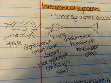
| Hiearchical synapses | - some synapses are louder than others - closer the synapse is to the axon hillock, the more potent it is (faster it reaches somewhere) |
| Effects of drugs on dopamine | |
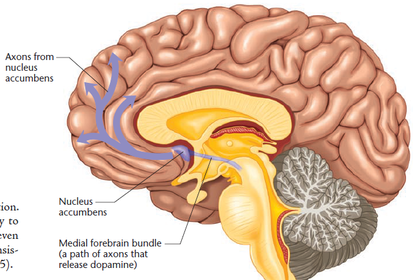
| Nucleus accumbens | location in the brain that nearly all drugs affect; where more dopamine is released |
| Myasthenia gravis | - 14/100,000 people - autoimmune disease - protein part of post synaptic receptor for acetylcholine begins to get eaten away |
| Parkinson's Disease characteristics | - associated with aging - movement poor - muscle rigidity - resting tremor (shake when they're at rest) |
| Parkinson's Disease possible causes | basal ganglia (base of forebrain) - helps you decide on movement --> substantia nigra (sends dopamine-releasing axons to the caudate nucleus and putamen) striatum is no longer innervated by dopamine - dopaminergic neurons progressively die off * increased inhibition of the thalamus, decreased excitation of the cerebral cortex |
| L-DOPA | precursor to dopamine |
| Parkinson's treatment | - take L-DOPA as a daily pill, cross blood-brain barrier, neurons convert into dopamine - doesn't work forever or - don't want to clean out the synapse - break down MAO (monoamineoxidase) so it can't break down neurotransmitters or - antioxidant drugs - brain tissue transplants - drugs that prevent apoptosis of further cells |
| Hungtinton's Chorea | - genetic disorder - doesn't show up until you're 30-40 - causes toxic gain of function (neurons in striatum --> gabanergic interneurons eventually die off) - huntington protein (increases neurotransmitter release, impairs mitochondria, |
| C-A-G repeats | The higher the number of C-A-G repeats, the earlier the probable onset of the disease. |
| Antagonist | a drug that blocks the effects of a neurotransmitter |
| Agonist | a drug that mimics or increases the effects of a neurotransmitter |
| Affinity | how well a drug fits into a receptor; like a key into a lock |
| Efficacy | a drug's tendency to activate the receptor |
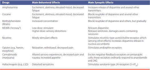
| Stimulant drugs | - increase excitement, alertness, and activity while elevating mood and decreasing fatigue - impair attention and learning - correlated with "impulsiveness" 1) amphetamine 2) cocaine 3) heroin 4) morphine 5) methylphenidate (Ritalin) 6) methylenedioxymethamphetamine (MDMA) |
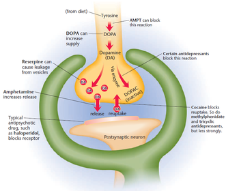
| Amphetamine | - increase the amount of dopamine in presynaptic terminals - nonspecific ORDINARILY: - presynaptic terminal reabsorbs dopamine with protein called dopamine transporter WITH AMPHETAMINE: - transporter is reversed causing cell to release dopamine instead of reabsorbing it AFTERWARD: - dopamine washes out of the synapse faster than the presynaptic cell can replace it --> user crashes |
| Cocaine | - blocks the reuptake of dopamine, norepinephrine, and serotonin --> prolongs effects AFTERWARD: - dopamine washes out of the synapse faster than the presynaptic cell can replace it --> user crashes |
| Methylphenidate (Ritalin) | - block reuptake of dopamine at brain receptors - prolonged use may lead to fearfulness - use of ritalin when young does not necessarily lead to stimulant drug use later in life |
| Methylenedioxymethamphetamine (MDMA) | - stimulant at low doses - increases release of dopamine - higher doses --> increases serotonin release (causing perception changes) - large amount of MDMA damage neurons that contain serotonin WHY? - high body temperature - excess serotonin is broken down and one of the products is h2o2 |
| Nicotine | - stimulates the nicotonic receptor (type of acetylcholine receptor) in CNS and nerve-muscle junction of skeletal muscles - increases dopamine release in nucleus accumbens (similar to cocaine) * excites acetylcholine receptors on neurons that release dopamine |
| Opiates | drugs derived from the opium poppy 1) morphine 2) heroin (enters brain faster than morphine, more addictive) 3) methadone - relax people, decrease sensitivity to pain - brain produces endorphines (endogenous morphines) --> indicated opiates relieve pain by acting on receptors in the brain INDIRECTLY ACTIVATE DOPAMINE RELEASE: - stimulate endorphin synapses --> - inhibit ventral tegmental neurons that release GABA (neurotransmitter that inhibits firing of dopamine neurons) --> net effect is to increase dopamine release |
| Marijuana (Δ9-THC) - Δ9-tetrahydrocannabinol | - reported significant impairments of memory and cognition - cannaboid receptors among most abundant receptors in mammalian except in medulla (area that controls breathing and heartbeat) - anandamide and 2-AG bind to cannaboid receptors - located on presynaptic neuron --> release anadamide or 2-AG as retrograde transmitters --> travel back and inhibit further release - cannabinoids inhibit GABA --> decrease inhibition (increase activity) of neurons that release dopamine in the nucleus accumbens h - relieve nausea by inhibiting serotonin type 3 synapses (5-HT3) |
| Hallucinogenic drugs | - drugs that distort perception - LSD chemically resembles serotonin and stimulates serotonin type 2A (5-HT2A) receptors |
| Alcohol | - combines with GABA to produce longer effects than GABA would by itself - increases stimulation at both dopamine and opiate receptors |
| Summary of drugs & their effects | |
| Tranquilizers | benzodiazepine --> stimulate gabanergic neurons by turning off neurons that are inhibiting gabanergic neurons |
| Ways to affect the synapse (antagonistic) | 1) Block formation of the neurotransmitter 2) Block release of neurotransmittion (turn down synapse) 3) Block receptors (receptors bound with some other compound) |
| Ways to affect the synapse (agonistic) | 4) Promote formation of neurotransmitter 5) Promote release of neurotransmitters 6) Stimulate receptors (muscarin stimulates metabotropic synapses vs nicotine stimulates ionotropic) 7) Block breakdown of neurotransmitters (by Catechol-O-methyl transferase [COMT] or monoaminoxide [MAO]) 8) Block reuptake of neurotransmitters |
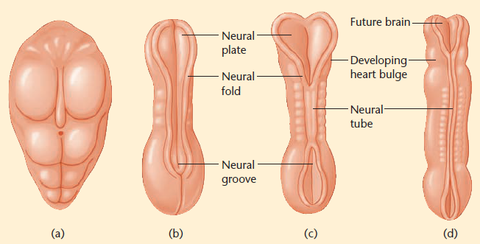
| Early development of vertebrate brain | |
| Proliferation | the production of new cells |
| Migration | the movement of cells after cells have differentiated as neurons or glia guided by chemicals known as immunoglobins and chemokines |
| Differentiation | when the neuron forms its axons & dendrites the axon grows first; dendrites form after migrating neuron reaches its destination |
| Myelination | the process by which the glia produce the insulating fatty sheaths that accelerate transmission in vertebrate axons spinal cord --> hindbrain --> midbrain --> forebrain |
| Synaptogenesis | the formation of synapses |
| New neurons grow in the adult brain? | Yes. Some in the olfactory receptors and the hippocampus |
| Development of the brain | neural plate --> folds in --> neural groove --> closes up at top --> neural tube |
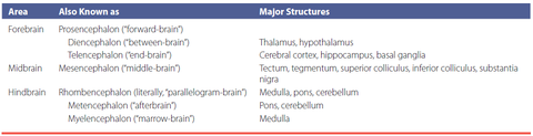
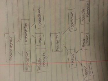
| Three different pouches of the brain | Top --> bottom prosencephalon (forebrain) - telencephalon (front) - diencephalon (back) mesencephalon (midbrain) rhombencephalon (hindbrain) - metencephalon - myelincephalon |
| Endocast | cast of the inside of the skull cavity |
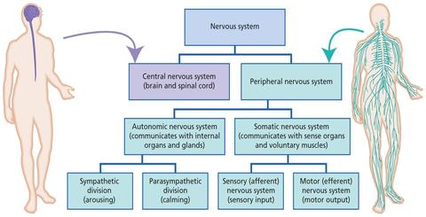
| Organization of the nervous system | |
| Central nervous system (CNS) | brain & spinal cord |
| Peripheral nervous system | - connection from the central nervous system to the rest of the body - communication |
| Somatic nervous system | - part of peripheral nervous system - muscles, skin information - split into: SENSORY MOTOR |
| Autonomic | - guts, digestive tract, smooth muscle - SPLIT INTO: SYMPATHETIC PARASYMPATHETIC |
| Sensory nervous system | afferent (going into the body) under somatic nervous system |
| Motor nervous system | Efferent under somatic nervous system |
| Sympathetic nervous system | arousal responses (heart beat increase, tense muscles) under autonomic nervous system postganglionic release norepinephrine; sweat glands (acetylcholine) |
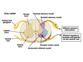
| Spinal cord | - starts at the base of the brain - goes down through the vertebrae - can bend, axons project out from holes in the vertebrae - nerves start out perpendicular at the top --> spinal cord stretches --> gravity pushes spinal cord down --> nerves start pointing downward |
| Nerve | collection of axons outside the nervous system |
| Tract | collection of axons inside nervous system |
| Spinal cord nerves | 31 pairs 8 - cervical 12 - thoracic 5 - lumbar 5 - sacral 1 - coccygeal |
| Directions in the brain | front --> back rostral --> caudal midline --> outside medial --> lateral back --> tummy dorsal --> ventral |
| Cut of spinal cord | |
| Path of information in the spinal cord | IN: sensory information --> dorsal root --> dorsal horn OUT: motor information --> ventral horn --> ventral root |
| Structure of brain | |
| Lamina | a row or layer of cell bodies separated from other cell bodies by a layer of axons and dendrites |
| Column | a set of cells perpendicular to the surface of the cortex, with similar properties |
| Tract | A set of axons within the CNS, known as a projection |
| Nerve | A set of axons in the periphery either from CNS to a muscle/gland or from a sensory organ to the CNS |
| Nucleus | a cluster of neuron cell bodies within the CNS |
| Ganglion | A cluster of neuron cell bodies, usually outside the CNS (as in the sympathetic nervous system) |
| Gyrus | a protuberance on the surface of the brain |
| Sulcus | A fold or groove that separates one gyrus from another |
| Fissure | a long, deep sulcus |
| Bell-Magendie law | the principle that entering dorsal roots (axon bundles) carry sensory information and exiting ventral roots carry motor information |
| Grey matter | cell bodies, dendrites, densely packed in the middle of the spinal cord |
| White matter | myelinated axons (outside of spinal cord) |
| Parasympathetic nervous system | relaxing responses (e.g. lower heart rate, start digesting) aka craniosacral system (consists of cranial nerves and nerves from the sacral spinal cord) - postganglionic axons release acetylcholine |
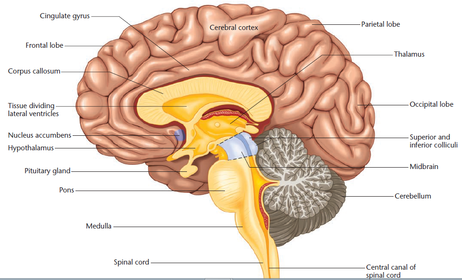
| Medulla | aka medulla oblongata; just above spinal cord - controls vital reflexes including breathing, heart rate, vomiting, salivation, coughing, etc through the cranial nerves |
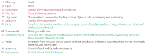
| Cranial nerves | nerves that control sensation from the head, muscle movements in the head, and much of the parasympathetic output to the organs |
| Major divisions of the vertebrate brain | |
| Cranial Nerves | |
| Pons | anterior (behind) and ventral (to the underside) to the medulla many axons in the pons cross from one side of the brain to the other |
| Reticular formation | descending - controls motor areas of the spinal cord ascending - sends output to much of cerebral cortex, selectively increasing arousal and attention in one area or another |
| Raphe system | sends axons to much of the forebrain, modifying the brain's readiness to respond to stimuli |
| Cerebellum | large hindbrain structure with many folds needed for balance & coordination, shifting attention, sensory timing, etc contains 50% of cells in the brain, bigger in animals that navigate 3D space |
| Ventricles | remnant of the fluid filled tube in the middle of the brain; filled with cerebrospinal fluid |
| Meninges | membrane around the brain; there are three |
| tectum | top of mesencephalon divided into front & back superior colliculus & inferior colliculus |
| superior colliculus | front of tectum; helps you process visual info, put things together in sensory space |
| inferior colliculus | back of tectum; helps in processing auditory informamtion |
| tegmentum | bottom of mesencephalon; contains part of the reticular formation goes all throught the brain & spinal cord related to states of consciousness |
| substantia nigra | located in the mesencephalon; gives rise to the dopamine-containing pathway that facilitates readiness for movement |
| Thalamus | pair of structures (left and right) in the center of the forebrain most sensory information goes here first --> processed --> sends output to the cerebral cortex |
| Hypothalamus | small area of brain ventral to thalamus involved in feeding, drinking, temperature regulation, sexual behavior, fighting, etc. |
| Mnemonics for cranial nerves | On old Olympus' towering top, a Finn and German viewed some hops. Some say marry money, but my brother says bad boys marry money. |
| Parts of the brain | |
| decussation of pyramids | where the spinal cord ends and the brain starts |
| Organization of the brain structures (e.g. medulla, pons, etc.) |
Want to create your own Flashcards for free with GoConqr? Learn more.