2225111
Description
Flashcards by helen.rebecca, updated more than 1 year ago
|
|
Created by helen.rebecca
about 9 years ago
|
|
| Question | Answer |
| What is the maximum magnification and resolution of a light microscope? | Resolution= 200nm Magnification= x1,400 |
| What is the maximum magnification and resolution of a transmission electron microscope? | Resolution= 0.5nm Magnification= x300,000 |
| What is the maximum magnification and resolution of a scanning electron microscope? | Resolution= 0.5nm Magnification= x300,000 |
| What is the difference between magnification and resolution? | Magnification is how many times bigger than the object the image is. Resolution is how detailed the image is. |
| Why do samples need to be stained before they are viewed through a microscope? | In light and TEM microscopes, light passes through the sample. Staining results in an image because the dye is taken up more by certain organelles. |
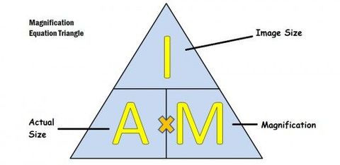
| How is magnification calculated? | Magnification= Image size/ real size |
| What is the function of the plasma (cell) membrane? | It regulates the movement of substances in and out of the cell. Contains receptor molecules that can respond to chemicals such as hormones. |
| What is the function of the cell wall? Where is it found? | It supports plant cells as it's a rigid structure made of cellulose. It's found on the outside of plant cells. |
| What is the function of the nucleus? Where is it found? | Contains nucleolus which makes RNA for ribosomes. It's covered in a nuclear envelope, which has nuclear pores to allows passage of substances between nucleolus and cytoplasm. It's found in all eukaryotic cells. |
| What is the function of a lysosome? What is the structure of a lysosome? | It contains digestive enzymes. They are used to break down worn out cell components and to digest invading cells. They are round and are surrounded by a membrane to keep their enzymes separate from the cell's cytoplasm. |
| Describe the function and structure of a ribosome. | They are very small organelles that can either float freely or be attached to the RER. They are the site at which proteins are produced. |
| Describe the function and structure of the rough endoplasmic reticulum. | It's a system of membranes enclosing a fluid filled space that is encrusted with ribosomes. The RER processes proteins that have been made in the ribosomes. |
| Describe the structure and function of the smooth endoplasmic reticulum. | It's a system of membranes enclosing a fluid filled space, but it DOESN'T have any ribosomes. It synthesises and processes lipids. |
| Describe the structure and function of a vesicle. | It's a small, fluid filled sac found in the cytoplasm that's surrounded by a membrane. It transports substances in and out of the cell (via the plasma membrane). Some are formed by the golgi or the SER & RER, and others are formed at the cell surface, |
| Describe the structure and function of the Golgi apparatus. | It's a group of flat, fluid filled sacs. Vesicles are often found along the edges. It processes and packages proteins and lipids, as well as making lysosomes. |
| Describe the function and structure of a mitochondrion. | It's an oval shaped cell with a double membrane. (the inner membrane is called the cristae, which contains the matrix, where enzymes involved in respiration are found.) It's the site of aerobic respiration where ATP is produced. |
| Describe the structure and function of chloroplast. | It's a small flattened structure (only found in plant cells). It has a double membrane. It's where photosynthesis takes place in plants. |
| Describe the structure and function of a centriole. | They are small hollow cylinders containing a ring of microtubules. They come in pairs at right angles to one another and have a 9+2 arrangement. They create the spindles used in mitosis. |
| Describe the structure and function of cilia. | They are small hair like structures, found in the lungs, Fallopian tubes and trachea. They have a 9+2 arrangement and are made of microtubules. The microtubules allow the cilia to move, so they can 'waft' things along (e.g. mucus in the trachea) |
| Describe the function and structure of a flagellum. | On eukaryotic cells, they're like cilia but longer. They stick out from the cell surface and are also made from microtubules with a 9+2 arrangement. The microtubules contract to make the flagellum move, which can help the cell to move. |
| Name the 5 units of measurement used in microscopy, from smallest to largest. | Picometre (pm) Nanometre (nm) Micrometre (µm) Millimetre (mm) Metre (m) |
| Outline the process of protein production. | 1) Proteins are made at the ribosomes 2) Ribosomes on RER make proteins that are excreted or attached to cell membrane. Free ribosomes make proteins that stay in cytoplasm. 3) New proteins produced at RER are folded & processed in RER. 4) They're transported from RER to golgi in vesicles. 5) Proteins are processed more in golgi. 6) The proteins enter more vesicles to be transported around the cell. |
| Name the 4 functions of the cytoskeleton. | 1) Microtubules support the cells organelles, keeping them in position 2) They also help to strengthen the cell & maintain its shape 3) They help to transport materials within the cell (e.g. the movement of chromosones.) 4) They also help the cell to move (e.g. the movement of the cilia and flagella) |
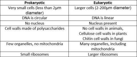
| Compare the structure of prokaryotic and eukaryotic cells. |
Want to create your own Flashcards for free with GoConqr? Learn more.