6863925
Description

Flashcards by RadTech Fairy, updated more than 1 year ago
|
|
Created by RadTech Fairy
about 9 years ago
|
|
| Question | Answer |
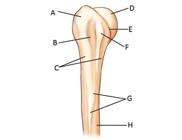
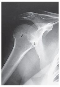
| A. greater tubercle (tuberosity) B. intertubercular groove (bicipital) C. surgical neck D. head E. anatomic neck F. lesser tubercle (tuberosity) G. deltoid tuberosity H. humeral body (shaft) | |
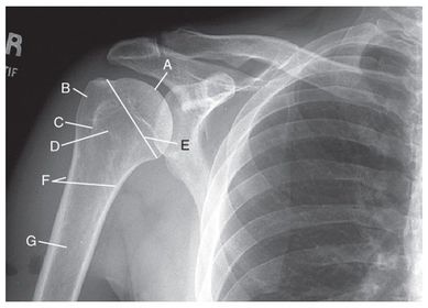
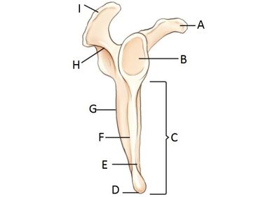
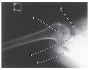
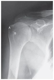
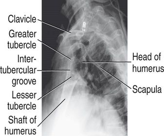
| A. humeral head B. greater tubercle C. intertubercular groove (bicipital) D. lesser tubercle E. anatomic neck F. surgical neck G. humeral body | |
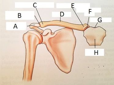
| A. acromion B. acromioclavicular joint (AC) C. acromial extremity D. clavicular body E. sternal extremity F. sternoclavicular joint (SC) G. jugular notch H. manubrium of sternum | |
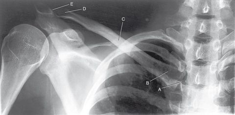
| A. sternoclavicular joint (SC) B. sternal extremity C. body D. acromial extremity E. acromioclavicular joint (AC) | |
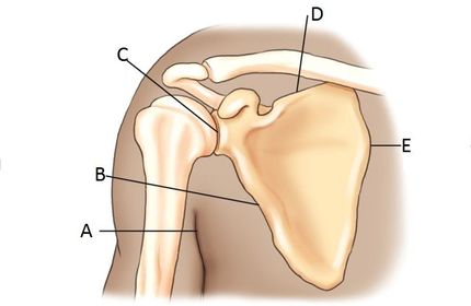
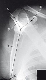
| A. axilla B. lateral (axillary) border C. scapulohumeral joint (glenohumeral) D. superior border E. medial border | |
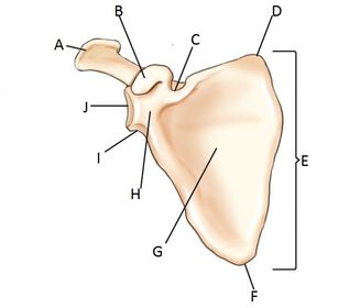
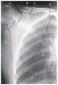
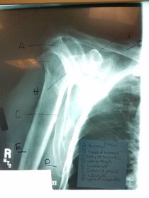
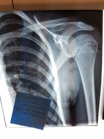
| A. acromion B. coracoid process C. scapular notch D. superior angle E. body (blade, wing, ala...) F. inferior angle G. costal surface (anterior) subscapular fossa H. neck I. lateral angle J. glenoid cavity (fossa) | |
| A. supraspinous fossa B. crest of spine C. acromion D. dorsal surface (posterior) E. infraspinous process | |
| A. coracoid process B. glenoid cavity (fossa) C. body D. inferior angle E. ventral (costal) surface F. lateral (axillary) border G. dorsal surface H. spine of scapula I. acromion | |
| A. acromion B. neck of scapula C. scapular notch D. superior angle E. medial (vertebral) border F. inferior angle G. lateral (axillary) border H. glenoid cavity OR scapulohumeral joint | |
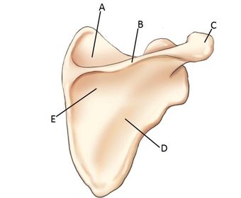
| A. acromion B. coracoid process C. inferior angle D. spine of scapula E. body of scapula | |
| A. coracoid process B. glenoid cavity OR scapulohumeral joint C. spine of the scapula D. acromion | |
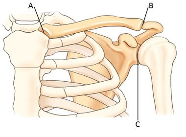
| A. sternoclavicular joint (SC) B. acromioclavicular joint (AC) C. scapulohumeral joint (glenoid cavity) | |
| Which type of joint is the glenoid cavity? | spheroidal ball and socket |
| Which type of joints are the sternoclavicular and acromioclavicular joints? | plane gliding |
| AC Dislocation | the distal clavicle is displaced superiorly |
| AC Joint Separation | trauma to the upper shoulder region that causes a partial or complete tear of the AC ligament |
| An injury of the anteroinferior aspect of the glenoid labrum is called a _____ _____ . | Bankart Lesion |
| A compression Fx of the articular surface of the posterolateral aspect of the humeral head is called ___-___ ____ ; it's usually associated with dislocations of the humeral head | Hill-Sachs Defect |
| Idiopathic Chronic Adhesive Capsulitis "frozen shoulder" | disability of the shoulder joint caused by chronic inflammation in and around the joint |
| Impingement Syndrome | impingement of the greater tuberosity and soft tissues during abduction of the arm |
| Rotator Cuff Injuries | refer to injury to the rotator cuff muscles: teres minor supraspinatus infraspinatus subscapularis |
| Shoulder Dislocation | traumatic removal of the humeral head from the glenoid cavity |
| Tendonitis | inflammatory condition of the tendon that usually results from a strain |
| External Rotation shows: | -epicondyles parallel to IR greater tubercle |
| Internal Rotation shows: | - epicondyles perpendicular to IR lesser tubercle |
| Neutral Rotation shows: | - epicondyles 45 degrees angled to IR - palm of hand facing inward |
| What are the routine exams for a HUMERUS (trauma and nontrauma) | AP Lateral |
| AP Humerus *routine | 40 SID 70-85 kVp 14x17 IR grid CR @ midhumerus must see entire humerus |
| Rotational Lateral Humerus *routine lateromedial or mediolateral projections | 40 SID 70-85 kVp 14x17 IR grid CR @ midhumerus LATEROMEDIAL- body obliqued with affected side against bucky, internally rotate arm so epicondyles are perpendicular to IR MEDIOLATERAL- oblique pt. 20-30 degrees from PA, keep elbow flexed 90 degrees |
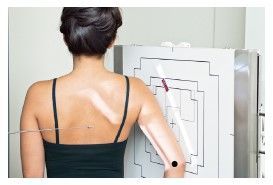
| Horizontal Beam Lateral Humerus *routine OR trauma lateromedial projection | 40 SID 65-85 kVp 10x12 IR nongrid CR @ midpoint of distal 2/3 of humerus |
| Why would you perform a transthoracic lateral projection of the humerus? | when there's trauma to the humerus and you cannot rotate the arm |
| Transthoracic Lateral Humerus *trauma only | 40 SID 70-85 kVp 14x17 IR grid CR @ mid-diaphysis through the thorax expose during orthostatic respiration (gentle, short, shallow breaths) *if pt. in too much pain to drop shoulder and elevate the opposite shoulder over the head, angle CR 10-15 degrees cephalad |
| What are the routine exams for the SHOULDER (nontrauma)? | AP external rotation AP internal rotation Grashey Y-View Axial |
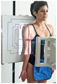
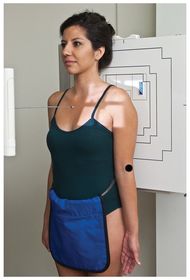
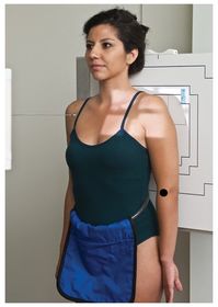
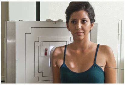
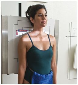
| AP External Rotation Shoulder (proximal humerus) *nontrauma | 40 SID 70-85 kVp 10x12 IR grid CR @ 1 in inferior to coracoid process expose on expiration *pt palm of hand supinated *epicondyles parallel to IR *greater tubercle in profile |
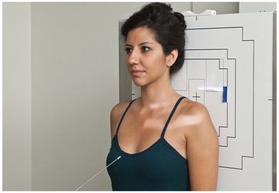
| AP Internal Rotation Shoulder (proximal humerus) *nontrauma | 40 SID 70-85 kVp 10x12 IR grid CR @ 1 in inferior to coracoid process *pt palm of hand pronated *epicondyles perpendicular to IR *Lesser tuberosity in profile |
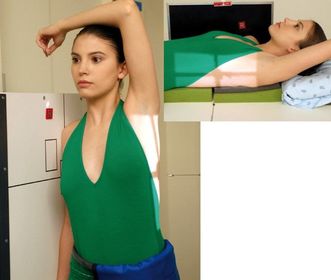
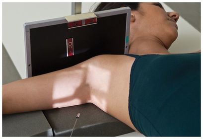
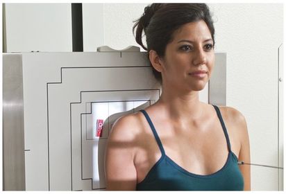
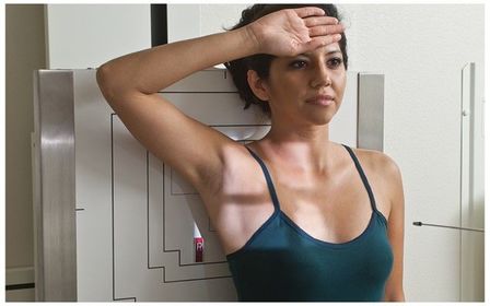
| Inferosuperior Axial Shoulder *nontrauma *SPECIAL* "Lawrence Method" | 40 SID 70-80 kVp 10x12 IR grid CR directed medially 25-30 degrees cephalad, centered horizontally into axilla and humeral head. *keep arm flexed @ 90 degrees, if less than, decrease CR angle to 15-20 degrees *palm up *lesser tuberosity in profile |
| What are the routine exams for a SHOULDER (trauma series) | AP neutral rotation Transthoracic lateral (lawrence) Scapular Y lateral |
| AP Neutral Rotation Shoulder *trauma | 40 SID 70-85 kVp 10x12 IR grid CR @ midscapulohumeral joint - 3/4 in. inferior and slightly lateral to corocoid process *expose on expiration |
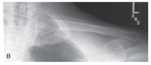
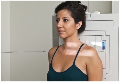
| Transthoracic Lateral Shoulder *trauma | 40 SID 70-85 kVp 10x12 IR grid CR directed through thorax to the level of the affected surgical neck *expose during orthostatic breathing |
| Scapular Y Lateral Shoulder *trauma | 40 SID 70-85 kVp 10x12 IR grid CR perpendicular to IR, directed @ scapulohumeral joint Expose on suspended breath *pt obliqued 45-60 degrees, and abduct arm slightly |
| What are some special projections of the shoulder that can be used for non trauma patients? | -Tangential Bicipital Groove Shoulder -Grashey Method |
| Intertubercular (Bicipital) Groove Shoulder *nontrauma *SPECIAL | 40 SID 55-75 kVp 8x10 IR nongrid CR perpendicular to IR, directed to groove area at midanterior margin of humeral head *expose on suspended breath *pt can be erect or supine |
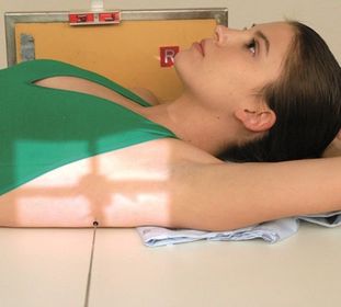
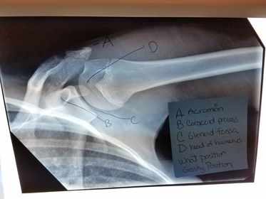
| Glenoid Cavity Posterior Oblique Shoulder *nontrauma "Grashey Method" *SPECIAL | 40 SID 70-85 kVp 10x12 IR grid CR perpendicular to IR, centered to scaphohumeral joint - 2 in inferior and medial from the superolateral border of shoulder *expose on suspended breath *pt obliqued 35-45 degrees toward the affected side - opens up glenoid cavity |
| Which projections of the shoulder require you to expose on orthostatic respiration? | Any transthoracic projection |
| What are the routine exams for the CLAVICLE? | AP AP Axial |
| AP Axial Clavicle | 40 SID 65-85 kVp 10x12 IR grid CR angled 15-30 degrees cephalad to midclavicle *expose during suspended breathing *asthenic pts require 10-15 degrees more angle than hypersthenic pts |
| AP Clavicle | 40 SID 65-85 kVp 10x12 IR grid CR perpendicular to IR, centered at midclavicle *must have both AC and SC joints on image |
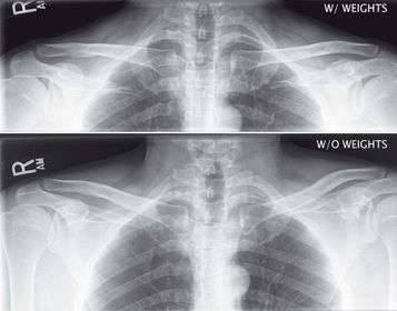
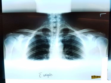
| AP AC and SC Joints weight bearing and non weight bearing | 72 SID 65-80 kVp 14x17 IR crosswise CR @ manubrium expose during suspended respiration *take first exposure without weights *8-10lb weights should be attached to wrists so the arms and clavicles will be relaxed for the 2nd projection |
| What are the routine exams of the SCAPULA? | AP Lateral |
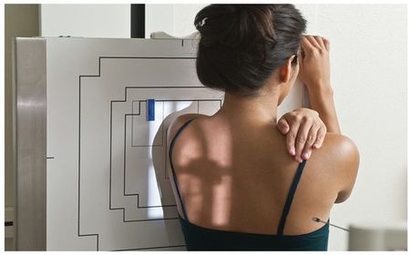
| AP Scapula Erect | 40 SID 70-85 kVp 10x12 IR grid minimum of 3 sec exposure time with optional breathing technique (3-4) seconds preferred CR perpendicular to midscapula, 2 in inferior to coracoid process, or to level of axilla and 2 in medial |
| 45 degree LAO Scapula | 40 SID 75-85 kVp no AEC - manual only 10x12 IR grid CR @ midvertebral border of scapula *this view best demonstrates the body of the scapula |
| 60 degree LAO Scapula | 40 SID 70-85 kVp no AEC - manual only 10x12 IR grid CR @ midvertebral border of scapula *best demonstrates the acromion and coracoid processes |
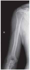
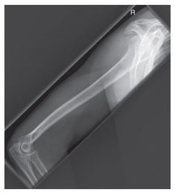
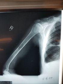
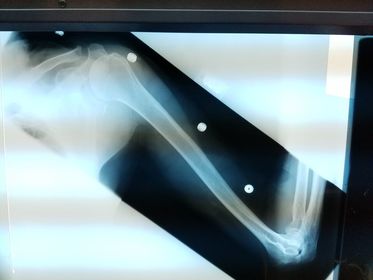
| Which projection/position is this? | PA Lateral Humerus |
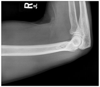
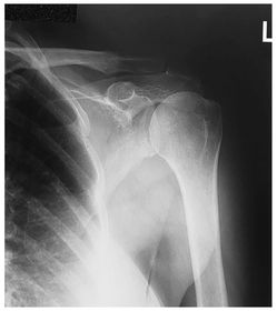
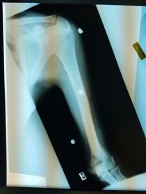
| Which projection/position is this? | AP External Humerus ________________________ Epicondyles are parallel to IR |
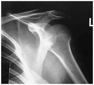
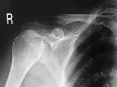
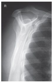
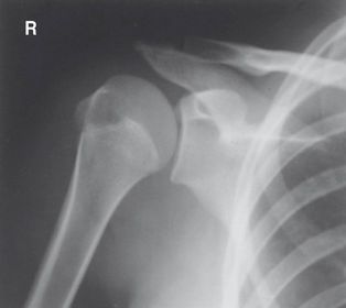
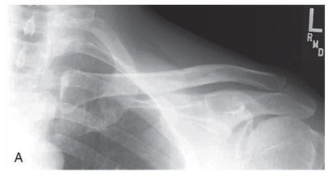
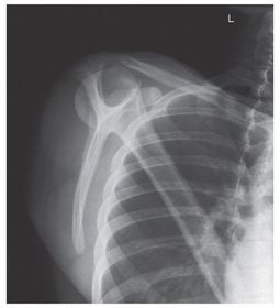
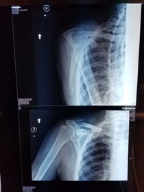
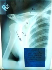
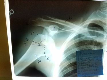
| Which projections/positions are these? (top->bottom) | TOP: Scapular Y-View _________________________ BOTTOM: AP External Rotation (greater tubercle in profile) |
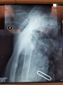
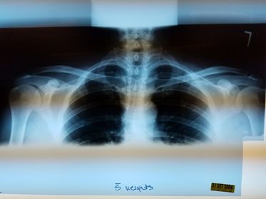
| Which projection/position is this? | Transthoracic Shoulder ___________________ ribs are blurred out from orthostatic breathing |
| Bilateral AC Joints w/ weights | |
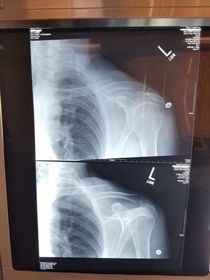
| which projection/position are these? | TOP: AP Clavicle __________________ BOTTOM: AP Axial Clavicle |
| Projection/position? | Lateral Humerus |
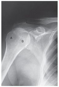
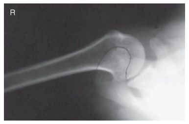
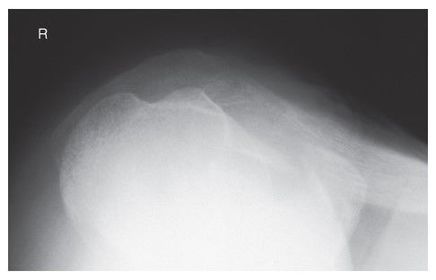
| On the inferosuperior axillary x-ray, what anatomical feature is in profile? | LESSER TUBEROSITY |
| When the proximal humerus is in anatomical position, which anatomical landmark is most lateral? | GREATER TUBEROSITY |
| For an inferosuperior axillary projection, where does the CR exit the body? | AC Joint |
| Where is the CR centered for a transthoracic humerus? | at the level of the surgical neck through the thorax |
| For an AP internal rotation projection of the shoulder, the proximal humerus is in _____ position | LATERAL |
| For an AP scapula projection, the arm should be abducted at least ___ degrees | 90 |
| The shoulders are aligned in the same transverse plane using a 14x17 IR for which projection? | AC joints |
| A 10x12 IR is placed crosswise above the shoulders for which projection? | AP clavicle |
Want to create your own Flashcards for free with GoConqr? Learn more.