7061764
Description

Flashcards by RadTech Fairy, updated more than 1 year ago
|
|
Created by RadTech Fairy
over 7 years ago
|
|
| Question | Answer |
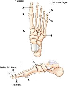
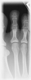
| A. distal phalanx B. middle phalanx C. proximal phalanx D. head E. body F. base G. tuberosity 5th metatarsal | |
| A. Interphalangeal (IP) joint B. Metatarsophalangeal (MTP) joint C. CR location @ 3rd MTP joint D. distal interphalangeal joint (DIP) E. proximal interphalangeal joint (PIP) F. tarsometatarsal joint (TMT) G. DIP H. PIP I. MTP J. TMT K. IP - 1st digit L. sesamoid | |
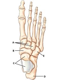
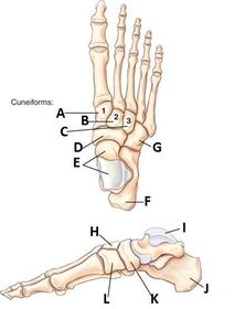
| A. 3 cuneforms (right->left: medial, inter, lateral) B. navicular C. talus D. calcaneus E. cuboid | |
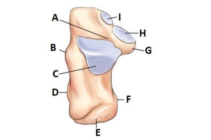
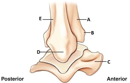
| A. calcaneal sulcus B. peroneal trochlea (trochlear process) C. posterior articular process D. lateral process E. tuberosity F. medial process G. sustentaculum tali H. middle articular surface I. anterior articular surface | |
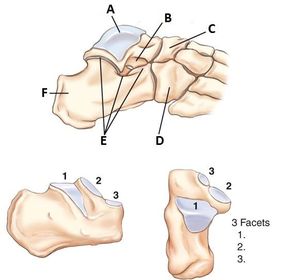
| A. talus B. sinus tarsi C. navicular D. cuboid E. talocalcaneal joint F. calcaneus 1. posterior facet 2. middle facet 3. anterior facet | |
| A. medial cuneiform (1st) B. intermediate cuneiform (2nd) C. lateral cuneiform (3rd) D. navicular E. talus F. calcaneus G. cuboid H. intermediate cuneiform I. talus J. calcaneus K. navicular L. medial cuneiform | |
| A. 1st metatarsal B. 1st cuneiform C. navicular D. talus E. calcaneus F. calcaneus G. talus H. cuboid I. cuboid J. 3rd cuneiform K. 2nd cuneiform L. 1st cuneiform | |
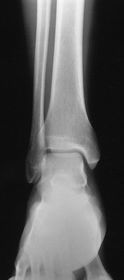
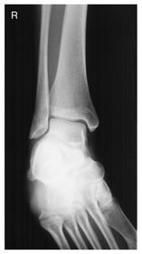
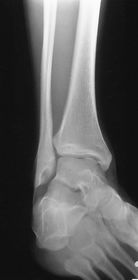
| ANKLE FRONTAL VIEW: A. fibula B. anterior tubercle C. lateral malleolus D. talus E. medial malleolus F. tibial plafond G. tibia | |
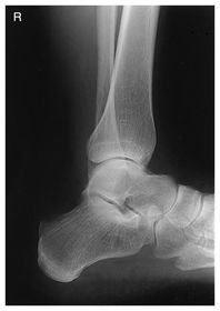
| ANKLE LATERAL VIEW: A. tibia B. anterior tubercle C. talus D. lateral malleolus E. fibula | |
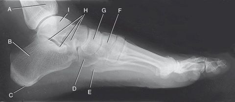
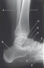
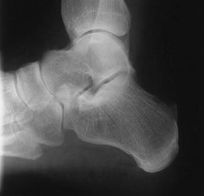
| A. tibia B. calcaneus C. tuberosity of calcaneus D. cuboid E. 5th metatarsal tuberosity F. superimposed cuneiforms G. navicular H. subtalar joint I. talus | |
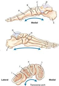
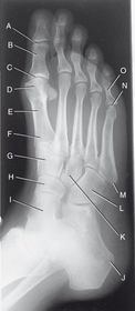
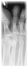
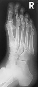
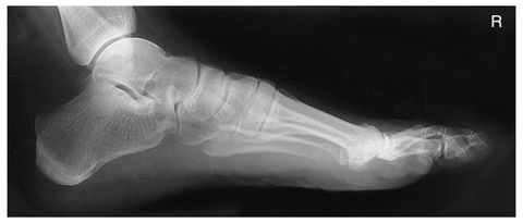
| A. IP joint first toe B. proximal phalanx of first toe C. MTP joint of first toe D. head of first metatarsal E. body of first metatarsal F. base of first metatarsal G. 2nd or intermediate cuneiform H. navicular I. talus J. tuberosity of calcaneus K. 3rd or lateral cuneiform L. cuboid M. tuberosity of 5th metatarsal base N. 5th MTP joint O. proximal phalanx 5th digit | |
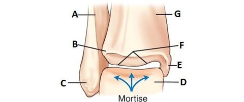
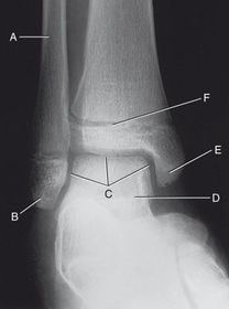
| A. fibula B. lateral malleolus C. "open" mortise joint of ankle D. talus E. medial malleolus F. tibial epiphyseal plate | |
| A. fibula B. calcaneus C. cuboid D. 5th metatarsal tuberosity E. navicular F. talus G. sinus tarsi H. anterior tubercle I. tibia | |
| Which metatarsal Fx is most common? | base of 5th metatarsal tuberosity |
| The 1st cuneiform is between the _____ and the ____ | 1st metatarsal and navicular |
| the cuboid is on the ____ side of the foot | lateral |
| which tarsal bone is located directly anterior to talus? | navicular |
| Which tarsal bone is on the medial side between the talus and 3 cuneiforms? | Navicular |
| What is the name of the small space between the talus and calcaneus? | Sinus Tarsi |
| What bone is superior to the calcaneus? | Talus |
| Which cuneiform will articulate with the 3rd metatarsal? | 3rd cuneiform |
| Which bones make up the ankle joint? | distal tibia distal fibula talus |
| How can you tell if an x-ray of a lateral ankle is truly lateral? | the distal fibula must superimpose half of the distal tibia |
| For a standard oblique of the ankle, the ankle is in ___ degree rotation For the mortise the ankle is in ___ degree obliquity | 45 15-20 |
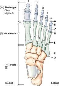
| How many bones are found in the foot? | 26 |
| What is the strongest and largest of the tarsal bones? | Calcaneus |
| The sustentaculum tali is found on the ____ | calcaneus |
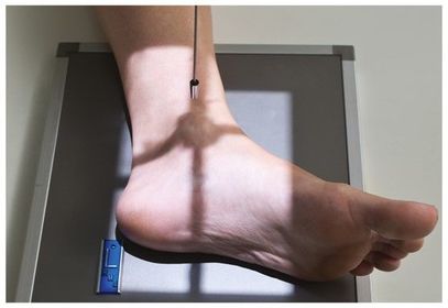
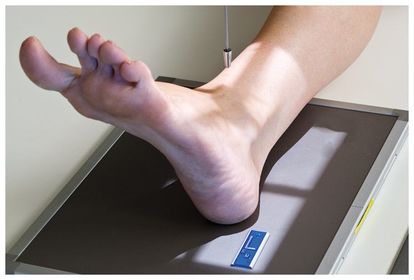
| What are some technical considerations for toes, feet, and ankle projections? | -40 SID -small focal spot -table top - nongrid -50-70 kVp -10x12 or smaller IR -place marker on lateral side of appendage |
| What are the routine studies for the toes? | AP Oblique Lateral |
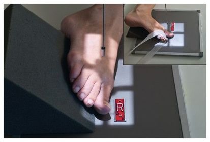
| AP Toes | - CR < 10-15 degrees toward calcaneus @ MTP joint - can use a 15 degree wedge w/ CR perpendicular as an alternative |
| Lateral Oblique Toes | - use lateral oblique for 4th and 5th digits to reduce OID - CR perpendicular to IR @ affected digit's MTP joint |
| Medial Oblique Toes | - use medial oblique for 1st - 3rd digits to reduce OID - CR perpendicular to IR @ affected digit's MTP joint |
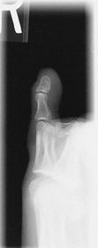
| Lateral Toe | - CR perpendicular to IR - directed to IP joint for 1st digit, and PIP for 2nd-5th digits |
| Why are the lateral and oblique projections usually done mediolateral? | It's easier for the patient to position their foot into a mediolateral projection - you get true laterals and better obliques when the patient can cooperate. |
| What makes a digit x-ray repeatable? | too much/too little rotation exposure factors MUST SEE up to half of the metatarsal DO NOT COLLIMATE OFF DIGIT! |
| What are the routine studies for a foot? | AP Oblique Lateral |
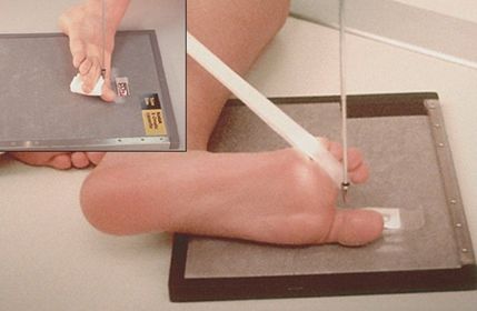
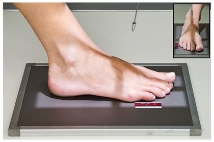
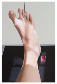
| AP Foot | - CR < posteriorly toward the heel 10 degrees - CR centered @ base of 3rd metatarsal |
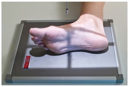
| AP Oblique Foot | - location of foreign bodies - CR perpendicular @ base of 3rd metatarsal - should be at least 30 degrees obliqued |
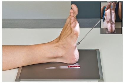
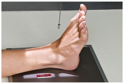
| Lateral (mediolateral) Foot | - location of foreign bodies - CR perpendicular @ medial cuneiform - MUST HAVE at least 1 in of tib/fib shown with the open joint |
| What makes a foot x-ray repeatable? | coning off anatomy rotation exposure factors motion |
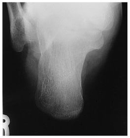
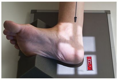
| What are the routine views of the calcaneus? | Plantodorsal Axial Lateral |
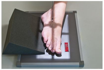
| Plantodorsal Axial | - foot should be flexed 90 degrees - CR < 40 degrees cephalad from long axis of foot (down into the heel) - center @ base of 3rd metatarsal to emerge at a level just distal to lateral malleolus |
| Lateral (mediolateral) Calcaneus | - CR perpendicular @ 1 in distal/inferior to medial malleolus - angling the foot up slightly will open up the joint spaces for a true lateral projection |
| What makes a calcaneus x-ray repeatable? | coning off anatomy rotation exposure factors motion |
| What are the routine views of the ankle? | AP Mortise Lateral |
| AP Ankle | - CR perpendicular @ point midway between the malleoli - make sure the lower leg is not rotated! |
| AP Mortise | - Internally rotate entire leg and foot 15-20 degrees until intermalleolar line is parallel to IR - CR perpendicular @ midpoint between malleoli - should have space all around fibula |
| Lateral (mediolateral) Ankle | - use sponges to keep the foot and leg in true lateral position - CR perpendicular @ medial malleolus |
| *special* AP Oblique Medial Rotation | - rotate foot and leg medially 45 degrees - CR perpendicular @ midpoint between malleoli |
| What makes an ankle x-ray repeatable? | coning off anatomy rotation exposure factors motion closed joint spaces on Mortise |
| Which tarsal forms an aspect of the ankle joint? | Talus |
| Which is the smallest of the cuneiforms? | Intermediate Cuneiform |
| Which tarsal is found on the medial side of the foot between the talus and cuneiforms? | Navicular |
| Which is the largest of the cuneiforms? | Medial Cuneiform |
| Which tarsal articulates with the 2nd, 3rd, and 4th metatarsals? | Lateral Cuneiform |
| Which is the most superior tarsal bone? | Talus |
| Which bone articulates with the first metatarsal? | Medial Cuneiform |
| Which tarsal is a common site for bone spurs? | Calcaneus |
| Which is a tarsal found anterior to the calcaneus and lateral to the lateral cuneiform? | Cuboid |
| Which is the 2nd largest tarsal bone? | Talus |
| Which projection has the CR angled 10-15 degrees posteriorly into the heel? | AP Toes |
| Which projection has the CR angled 10 degrees posteriorly into the heel? | AP Foot |
| Which projection has the CR angled 40 degrees cephalad into the heel? | Inferosuperior Axial Calcaneus |
| Which projection has the foot rotated inward 15-20 degrees? | AP Mortise Ankle |
| Which projection has the foot rotated inward 45 degrees? | AP Medial Oblique Ankle |
| Which projection best demonstrates the lateral mortise joint? | AP Mortise |
| Which projection best demonstrates the distal tibulofibular joint articulation? | AP Medial Oblique Ankle |
| Which projection best demonstrates the talus bone and medial joint? | AP Ankle |
| Ossification | Bone Development |
| Os Calcis | Calcaneus |
| Which projection(s) have the patella perpendicular to the table/IR? | Lateral Foot Lateral Ankle Lateral Toe |
| Which projection best demonstrates the sinus tarsi, calcaneus, and talus? | Oblique Foot |
| For which projections should you make sure the foot is dorsiflexed to get open joint spaces? | AP Mortise Ankle AP Oblique Ankle Lateral Ankle Lateral Foot |
| If you need more exposure time but your mAs and kVp need to stay the same, what should you do? | Decrease mA Increase time _____________________ mA sec mAs 2 1 3 1 2 3 |
Want to create your own Flashcards for free with GoConqr? Learn more.