7145171
Description

Flashcards by RadTech Fairy, updated more than 1 year ago
|
|
Created by RadTech Fairy
about 9 years ago
|
|
| Question | Answer |
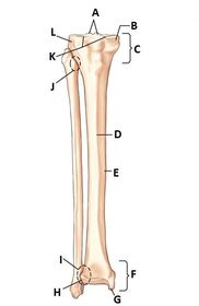
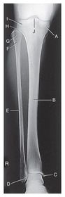
| A. intercondyloid eminence B. medial condyle C. proximal extremity D. anterior crest E. body of tibia F. distal extremity G. medial malleolus H. fibular notch (on tibia) I. distal tibiofibular joint J. proximal tibiofibular joint K. articular facets L. lateral condyle | |
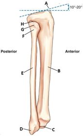
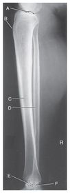
| A. tibial plateau B. body of tibia C. medial malleolus D. lateral malleolus E. fibular body F. neck G. head H. apex | |
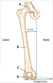
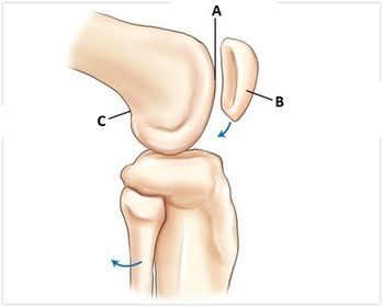
| A. hip joint B. femoral body C. patella D. trochlear groove | |
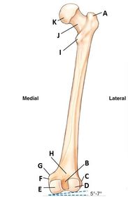
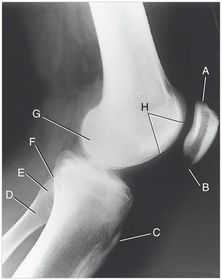
| A. greater trochanter B. intercondylar fossa (notch) C. lateral epicondyle D. lateral condyle E. medial condyle F. medial epicondyle G. adductor tubercle H. popliteal surface I. lesser trochanter J. neck K. head | |
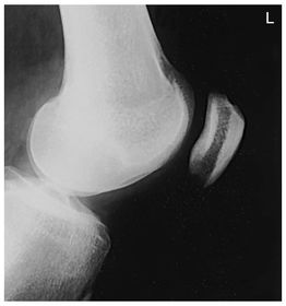
| A. patellar surface B. patella C. popliteal surface | |
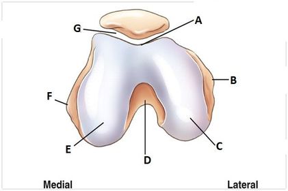
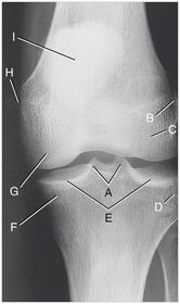
| A. trochlear groove B. lateral epicondyle C. lateral condyle D. intercondylar fossa E. medial condyle F. medial epicondyle G. patellofemoral joint | |
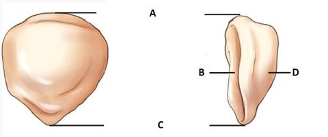
| A. superior border (base) B. posterior surface C. apex (inferior surface) D. anterior surface | |
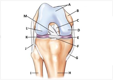
| A. patellar surface B. femur C. PCL D. medial condyle E. medial meniscus F. transverse ligament G. tibial medial collateral ligament (MCL) H. tibia I. fibula J. fibular lateral collateral ligament (LCL) K. lateral meniscus L. lateral condyle M. ACL | |
| A. medial condyle (tibia) B. tibia body C. medial malleolus D. lateral malleolus E. fibula body F. neck fibula G. head fibula H. apex fibula I. lateral condyle tibia J. intercondylar eminence | |
| A. intercondylar eminence B. tibial tuberosity C. body tibia D. body fibula E. medial malleolus F. lateral malleolus | |
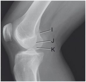
| I. adductor tubercle J. lateral condyle K. medial condyle | |
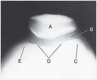
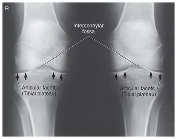
| A. medial and lateral intercondylar tubercles B. lateral epicondyle femur C. lateral condyle femur D. lateral condyle tibia E. articular facets tibia (tibial plateau) F. medial condyle tibia G. medial condyle femur H. medial epicondyle femur I. patella | |
| A. base of patella B. apex of patella C. tibial tuberosity D. neck of fibula E. head of fibula F. apex of head of fibula G. superimposed lateral and medial condyles H. patellar surface | |
| A. patella B. femoropatellar joint C. lateral condyle D. patellar surface E. medial condyle | |
| The articular facets making up the tibial plateau slopes posteriorly at _____ | 10-20 degrees |
| Another term for the patellar surface of the distal femur is __________ | Intercondylar Sulcus Trochlear Groove |
| Which bone is the longest and strongest bone in the body? | Femur |
| The adductor tubercle is viewed in only one projection, which is it? | Lateral Knee *under rotation of knee* |
| if you add a grid when you normally don't use one, how will you adjust your technique? | increase kVp |
| If you are x-raying through dense anatomy, how will you change your technique? | Increase mAs |
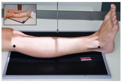
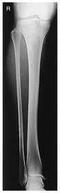
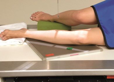
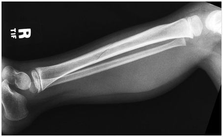
| AP Tib/Fib | 40 SID 14x17 IR tabletop - pt supine w/ leg rotated inward 5 degrees w/ foot dorsiflexed for TRUE AP - CR perpendicular @ midleg *must see ankle and knee joints on image - you can overlap 2 images if 1 doesn't fit both joints* |
| Lateral TIb/Fib Mediolateral | 40 SID 14x17 IR tabletop can angle IR diagonally to fit entire tib/fib on - CR perpendicular @ midleg *must see ankle and knee joints on image - you can overlap 2 images if 1 doesn't fit both joints* |
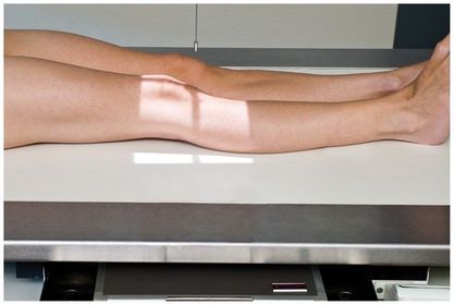
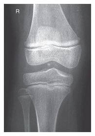
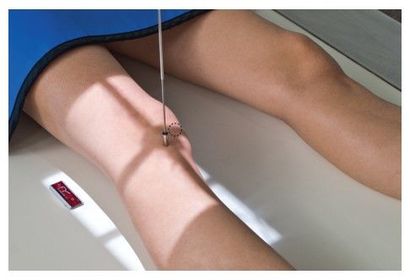
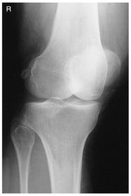
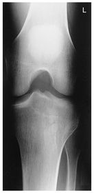
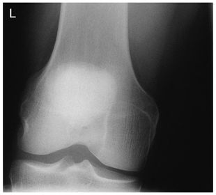
| AP Knee | pt supine w/ leg rotated internally 5 degrees to get knee in TRUE AP - CR parallel to tibial plateau @ 1/2 in distal to apex of patella *angle CR 3-5 degrees CAUDAL for ASTHENIC pts* *angle CR 3-5 degrees CEPHALIC for HYPERSTHENIC pts* leave CR angled 0 degrees for average sized pts. |
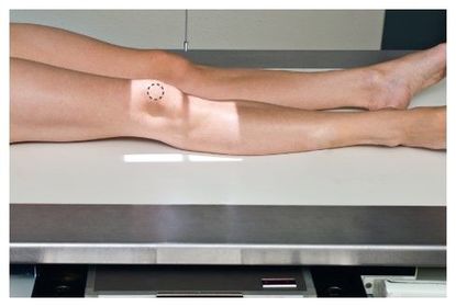
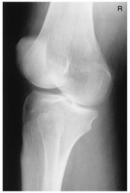
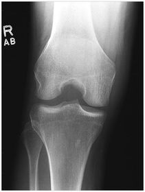
| AP Medial Oblique Knee | pt supine w/ knee rotated medially 45 degrees - CR parallel to tibial plateau @ 1/2 in distal to apex of patella *will see LATERAL condyles best along with the APEX OF FIBULA* |
| AP Lateral Oblique Knee | pt supine w/ knee rotated externally 45 degrees - CR parallel to tibial plateau @ 1/2 in distal to apex of patella * will see MEDIAL condyles best along with 1/2 of patella free of superimposition of the condyles* |
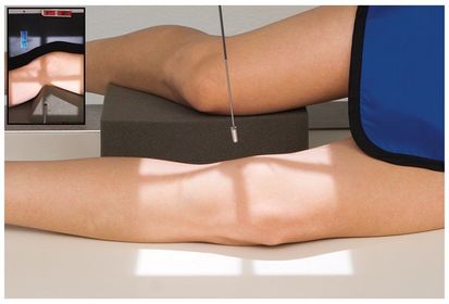
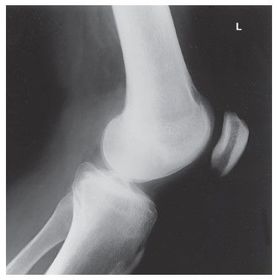
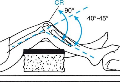
| Lateral Knee Mediolateral | 40 SID 10x12 IR in table BUCKY - pt lateral recumbent w/ knee flexed 20-30 degrees so patella is perpendicular to IR - CR angled 5-7 degrees cephalic @ 1 in distal to medial epicondyle (Always angle for lateral) *seeing the adductor tubercle indicates the pt is underrotated* *seeing the distal borders of femoral condyles NOT superimposed indicates an incorrect cephalic CR angle* |
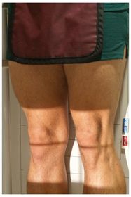
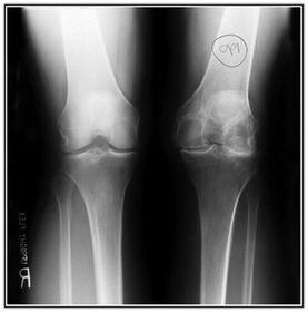
| AP Bilateral Weight Bearing Knees | 40 SID 14x17IR in wall BUCKY MANUAL TECHNIQUE for bilateral PHOTOTIME for unilateral - CR perpendicular to IR @ 1/2 in below apex of patella for average sized pts - CR angled 5-10 degrees cephalic for hypersthenic pts |
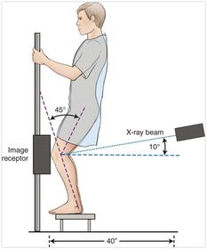
| Rosenberg Method PA Bilateral Knees | 40 SID 14x17IR in wall BUCKY - pt erect squatted back so knees are flexed 45 degrees - CR angled 10 degrees caudal @ popliteal crease |
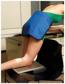
| Camp Coventry Intercondylar Fossa PA Axial Projection | 40 SID 14x17 IR in BUCKY pt prone/knee flexed 40-50 on sponge CR perpendicular to lower leg/ angled 40-50 degrees @ popliteal crease *looking @ ICF* |
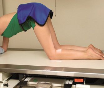
| Holmblad Method Intercondylar Fossa *alternative to camp coventry* PA Axial Alternative | pt on all fours w/ knees flexed 20-30 degrees CR perpendicular to lower leg @ popliteal crease |
| Holmblad Method Intercondylar Fossa Variations *alternatives to Camp Coventry* PA Axial Alternatives | pt positions vary over table, wheelchair, or chair - CR perpendicular to lower leg w/ leg flexed 20-30 degrees |
| Beclare AP Axial Intercondylar Fossa | - 40 SID - 8x10 IR tabletop built up on sponge - pt supine/knee flexed 40-45 degrees - CR perpendicular to lower leg angled 40-45 cephalic @ 1/2 in distal to apex of patella |
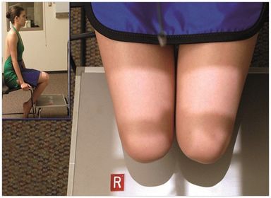
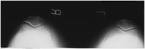
| Merchant Bilateral Method Patella | pt seated with knees flexed 40 degrees on merchant board - CR angled 30 degrees caudal @ midway between the patellae *over flexion of knees will pull the patellae into the trochlear groove* |
| PA Patella | pt prone CR perpendicular @ mid femoropatellar joint |
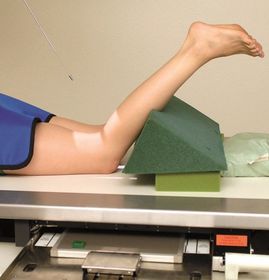
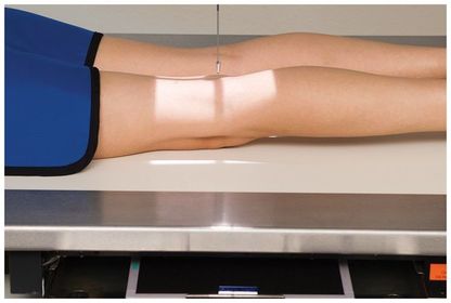
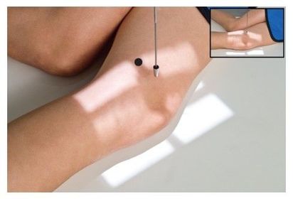
| Lateral Patella Mediolateral | pt lateral recumbent, knee flexed 5-10 degrees ONLY - CR perpendicular to mid femoropatellar joint *adductor tubercle reveals underrotation of knee* *distal borders of femoral condyles must be superimposed - if not it reveals incorrect CR angulation* |
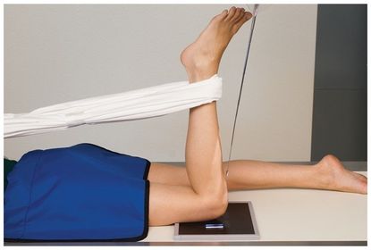
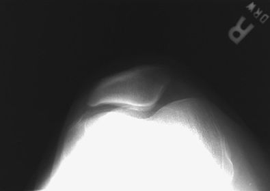
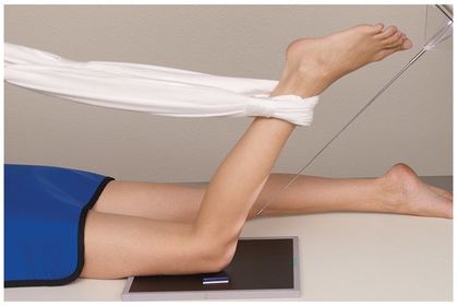
| Settegast Method Patella | pt prone knee flexed 90 degrees - CR angled 10-20 degrees to lower leg @ femoropatellar joint |
| Houghston Method Patella | pt prone knee flexed 55 degrees - CR angled 15-20 degrees to lower leg @ femoropatellar joint |
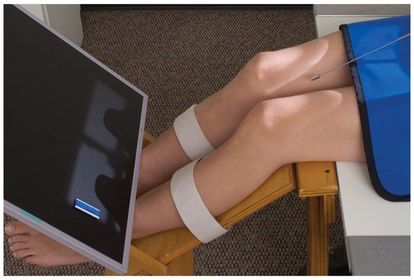
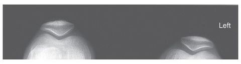
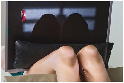
| Inferosuperior Bilateral Patellae Sunrise | 40-45 degree flexion of knees - CR angled 10-15 degrees cephalic from perpendicular @ femoropatellar joint |
| Hobbs Modification Patella Alternative to Inferosupeior Bilateral Patellae | pt seated knees flexed slightly - IR placed on foot stool to reduce OID - 48-50 SID minimum - CR perpendicular to IR @ mid femoropatellar joint |
| A palpable landmark used to position the knee is the ______ | epicondyle |
| Which structures serve as shock absorbers within the knee joint? | Menisci |
| For a mediolateral projection of the lower leg, the knee is flexed _______ degrees | 20-30 degrees |
| For a mediolateral projection of the knee, the CR is angled ______ centered at ___ inch distal to the apex of the patella | 5-7 degrees cephalic 1/2 in. |
| For the PA axial projection - Camp-Coventry Method - for intercondyloid fossa, the knee is flexed ____ | 40-50 degrees |
| What does the annode-heel effect determine about projections of the lower leg? | The heavier portion of the leg (knee->hip) should be on the cathode side of the table |
| Which projections have the CR perpendicular to, and centered at mid-leg? | AP leg Lateral leg |
| Which projection of the patella has the patient prone with knee flexed 55 degrees? | Hughston Method Patella |
| How much are the knees flexed for the inferosuperior axial projection (sunrise) of the patella? How much is the CR angled? | 40-45 degrees 10-15 degrees cephalic @ mid femoropatellar joint |
| Which projection of the patella has the patient seated with knees flexed 40 degrees on a board or over the table? How much is the CR angled and in which direction? | Merchant Bilateral Tangential Patellae 30 degrees caudal @ midway between patellae |
| How much is the knee flexed for the lateral-mediolateral projection of the patella? | 5-10 degrees |
| Why is PA preferred over AP for the patella? | Less OID with PA |
| With the PA patella projection AND the AP knee projection, how much should the leg be rotated to get TRUE AP and PA? | 5 degrees internally |
| Which projection of the Intercondylar Fossa has the patient supine with the IR built up on a kleenex box under the knee? How much is the CR angled? | Beclere - AP Axial CR perpendicular to lower leg : if leg is flexed 45 degrees, CR is angled 45 degrees |
| How is the CR angled for the Camp-Coventry - PA Axial projection of Intercondylar Fossa? | CR is perpendicular to lower leg leg flexed 40-45 degrees CR angled 40-50 degrees caudal |
| Which projection of the knees has the patient erect on a stool squatted back with knees flexed 45 degrees | Rosenberg - PA Axial Bilateral Weight Bearing Knees |
| Which projection of the knees has the CR perpendicular to the IR in the wall bucky? | AP Bilateral Weight Bearing Knees |
| How should the feet be positioned for the AP Bilateral Weight Bearing projection of the knees? | rotated 5 degrees inward to get True AP |
| What is seen on a projection of the lateral knee when it is under rotated? | Adductor Tubercle |
| What is seen on a projection of the lateral knee when the CR is not angled adequately? | The distal borders of the femoral condyles will NOT be superimposed |
| Which projection of the knee best visualizes the fibular apex? | AP Oblique with Medial Rotation |
| Which projection of the knee best visualizes 1/2 of the patella free of superimposition from the condyles of the femur? | AP Oblique with Lateral Rotation |
| How much will you angle the CR for knee projections on a patient that is hypersthenic? (greater than 24cm diameter) | 3-5 degrees cephalic |
| How much will you angle the CR for knee projections on patients that are asthenic? (less than 19cm diameter) | 3-5 degrees caudal |
Want to create your own Flashcards for free with GoConqr? Learn more.