
7430784
Description

| Question | Answer |
| Acceptable PA CXR | |
| Acceptable Lateral CXR | |
| What makes a CXR repeatable? | - poor inspiration - clipping anatomy - artifacts - excessive rotation |
| How will a pneumothorax or a pneumonectomy appear on a radiograph? | lung will not have any markings, or appear blacked out |
| what could excessive lung markings on a radiograph indicate? | conditions like pulmonary edema, fibrosis, or compression of lung tissue |
| The air/fluid separation line on a CXR acts similarly to water in a cup, how will it appear on a radiograph? | A defined line of brightness against a darkened area of air, it will tilt as the patient tilts |
| Where will the tip of an endotracheal (air) tube be positioned in the lungs? | 1-2 in. superior to the carina |
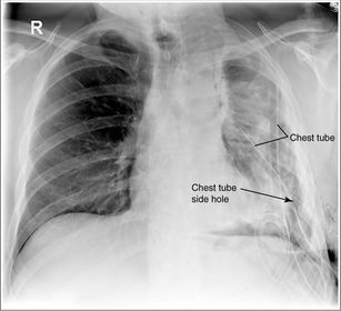
| Where will the tip of a pleural drainage (fluid) tube be placed? | between 5th and 6th intercostal spaces |
| How is the diaphragm positioned on a hypersthenic patient? | high |
| How is the diaphragm positioned on an asthenic patient? | low |
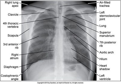
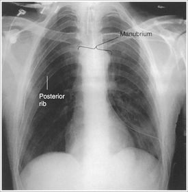
| How will the midcoronal plane be positioned against the IR to prevent rotation and keep the lung fields symmetric on a PA CXR? | plane is parallel to the IR |
| How can you determine rotation on a PA CXR? | SC joints should be level and symmetric |
| If a patient is rotated to the right for a PA CXR, how will the SC joints appear on the radiograph? | the RIGHT SC joint will be superimposed within the spine the LEFT SC joint will be free of superimposition the LEFT lungs will be elongated because they are farther away from the IR - they will be magnified |
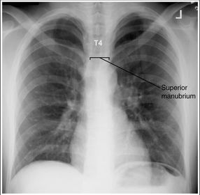
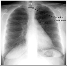
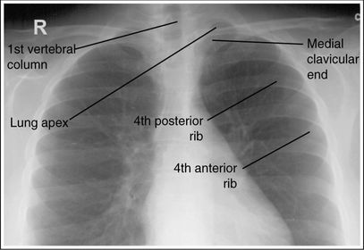
| How can you tell if the patient's shoulders were elevated on a PA CXR? | the clavicles are level and the manubrium is at T2-3 with only about an inch of apex seen above clavicle ______________________________________ When shoulders are elevated, the lateral sides of the clavicles are elevated |
| If the superior portion of a patient's midcoronal plane is tilted forward towards the IR, how will the radiograph appear? | manubrium situated @ T5 or lower more than 1 in. of apex will be seen above the clavicles |
| If the superior portion of a patient's midcoronal plane is tilted backward away from the IR, how will the radiograph appear? | manubrium situated @ T1-2 or higher less than 1 in. of apex will be seen above the clavicles |
| Which directions will the lungs expand? | Transversely Anteroposteriorly Vertically |
| Which direction of lung expansion is the greatest? | Vertical |
| Which pathology would require a PA CXR with expiration? | pneumothorax foreign body localization |
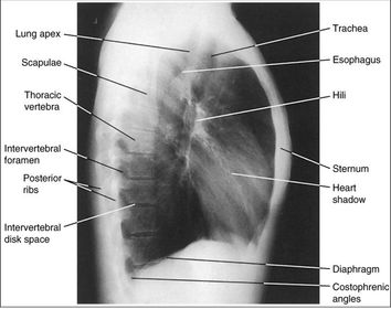
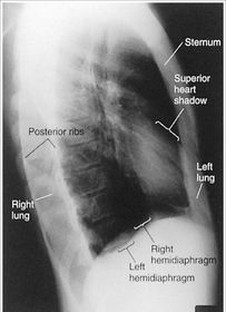
| How will the midcoronal plane be positioned with the IR to prevent rotation on a LAT CXR? | midcoronal plane is perpendicular to IR |
| What's the best way to determine rotation on a LAT CXR? | if there's more than 0.5 in. of space between the posterior ribs and spine |
| How can you tell if the patient was rotated to the LEFT for a LAT CXR? | The heart shadow will extend anteriorly past the sternum |
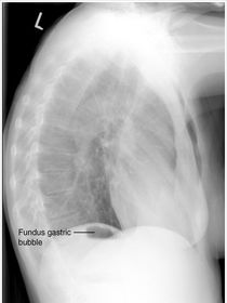
| How can you tell if the patient was rotated to the RIGHT for a LAT CXR? | The gastric bubble will appear under the right hemidiaphragm, and the right lung will extend anteriorly beyond the sternum |
| On a well inspired LAT CXR, where will the hemidiaphragm be seen? | below T11 |
| Which way is the patient rotated? | Right side is more anterior |
| Which way is the patient rotated? | Left side is more anterior |
| Which way is the superior midcoronal plane tilted? | FORWARD |
| Which way is the superior midcoronal plane tilted? | BACKWARD |
| Which way is the patient rotated? | RIGHT SIDE ANTERIORLY |
| Which way is the patient rotated? | LEFT SIDE ANTERIORLY |
| Inaccurate CR angulation will _____ or _____ the heart and lungs on an AP CXR | elongate or foreshorten |
| If you have to use a 40-48 SID for an AP CXR, how will the image appear? | heart will be magnified |
| If your CR is angled too caudal on an AP CXR, how will the image look? | manubrium will be inferior to T4 more than 1 in. of apex will be seen above clavicle posterior ribs will be more vertical |
| If your CR is angled too cephalic on an AP CXR, how will the image look? | manubrium will be superior to T4 less than 1 in. of apex will be seen above the clavicle posterior ribs will be more horizontal |
| What's wrong with this AP CXR? | the CR was angled too caudal |
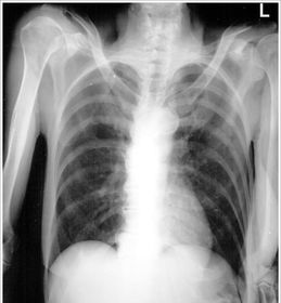
| What's wrong with this AP CXR? | pt. leaned to the left, shoulders elevated |
| If there's fluid in right lung, which decubitus CXR would you perform? | Right lateral decubitus fluid falls |
| If there's fluid in the left lung, which decubitus CXR would you perform? | Left lateral decub fluid falls |
| If there's air in the right lung, which decubitus CXR would you perform? | left lateral decubitus air rises |
| If there's air in the left lung, which decubitus CXR would you perform? | right lateral decubitus air rises |
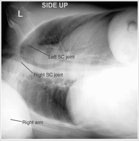
| How can you determine if the patient was rotated for a decubitus CXR? | Whichever side is closer to the IR will have elongated posterior ribs, and the SC joint will be least superimposed by the vertebral column |
| Which side of the patient is rotated closer to the IR? | LEFT |
| How can you determine if the patient was properly positioned for an AP Lordotic CXR? | The clavicles should be level completely out of the lung field |
| What's the positioning error on this AP Lordotic CXR? | Patient's midcoronal plane is NOT at a 45 degree angle with the IR (not leaned back enough) |
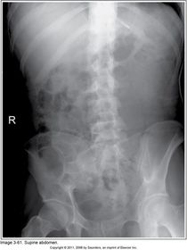
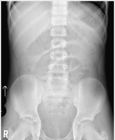
| How is rotation determined on an ABD xray? | The iliac wing that's longer is the side that's rotated closer to the IR |
| Which way is the pt rotated? | RIGHT |
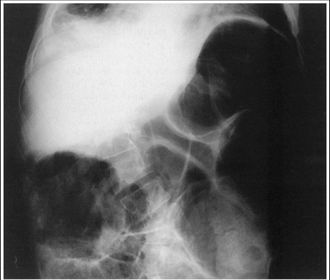
| What's wrong with this ABD XR? | overexposed patient off centered motion |
| What's wrong with this ABD XR? | underexposed clipped diaphragm |
| Which side should be down on a lateral decubitus ABD and why? | Left Side Down because the gastric bubble is on the left side, so we don't want to confuse it with possible free air in the ABD |
| How can you determine rotation on a Decubitus ABD XR? | Iliac Wings Spinous Processes |
| Which side of the patient is closer to the IR? | Left side |
| What's wrong with this decub ABD XR? | left side cut off bra artifact |
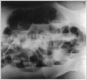
| What's the repeatable error in this decub ABD XR? | Diaphragm cut off |
| TECHNIQUE INCREASE OR DECREASE? Atelectasis | partial or total collapse of lung Increase |
| TECHNIQUE INCREASE OR DECREASE? Cardiomegaly | enlarged heart increase |
| TECHNIQUE INCREASE OR DECREASE? CHF | Congestive Heart Failure increase |
| TECHNIQUE INCREASE OR DECREASE? Pleural Effusion | free fluid in the pleural cavity increase |
| TECHNIQUE INCREASE OR DECREASE? Pneumonia | Excessive fluid built up in the alveoli Increase |
| TECHNIQUE INCREASE OR DECREASE? Pneumonectomy | partial or total removal of lung increase, but don't use AEC over affected area |
| TECHNIQUE INCREASE OR DECREASE? Pulmonary Edema | swelling in the lungs increase |
| TECHNIQUE INCREASE OR DECREASE? Emphysema | deteriorated lung tissue density decrease |
| TECHNIQUE INCREASE OR DECREASE? Pneumothorax | air in the pleural cavity decrease |
| TECHNIQUE INCREASE OR DECREASE? Ascites | free fluid in the peritoneum increase |
| TECHNIQUE INCREASE OR DECREASE? Calcified Stones | gallstones increase |
| TECHNIQUE INCREASE OR DECREASE? Bowel Obstruction | blockage decrease |
| TECHNIQUE INCREASE OR DECREASE? Barium Studies | increase in atomic # increase |
| TECHNIQUE INCREASE OR DECREASE? Iodine Studies | water soluble keep technique the same |
0 comments
Want to create your own Flashcards for free with GoConqr? Learn more.