7493166
Description

Flashcards by RadTech Fairy, updated more than 1 year ago
|
|
Created by RadTech Fairy
almost 9 years ago
|
|
| Question | Answer |
| mAs influences | density brightness shades of black |
| kVp influences | contrast shades of gray |
| higher kVp will produce | more contrast and shades of gray |
| short-scale contrast | high contrast few shades of gray low kVp _____________ finger, toe, femur, humerus, elbow, etc... |
| long-scale contrast | low contrast many shades of gray high kVp _________________ abdomen, chest, fluoroscopy, etc.... |
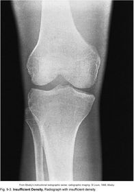
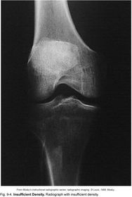
| Density | the quantity of photons reaching the IR |
| Insufficient Density image appears too bright (too many x-ray photons) | |
| Excessive Density image appears too dark (too few x-ray photons) | |
| Spatial Resolution | Detail Sharpness |
| Which factors contribute to SIZE distortion? | Far SID Short OID |
| Which factors contribute to SHAPE distortion? | tube angle anatomical centering |
| What are 2 forms of distortion? | distortion of size (magnification, foreshortening, elongation, etc.) distortion of shape (centering, tube angle, tilted IR, etc.) |
| Window Level | changes brightness |
| if an image is too dark, you can increase brightness by ________ window level | increasing |
| if an image is too light, you can decrease brightness by _________ window level | decreasing |
| Contrast | Combined result of the quality of the x-ray beam, quality of radiation, quality of the IR, and computer processing and display |
| TRUE OR FALSE? elements of high anatomic numbers absorb radiation dose | TRUE: Ca+ in bones has a high atomic number, therefore it absorbs radiation and appears white on a radiograph |
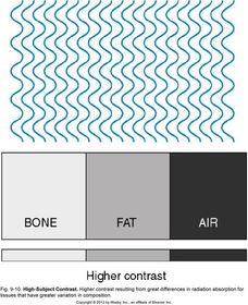
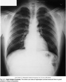
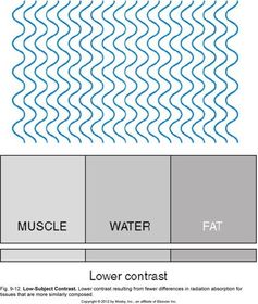
| Subject Contrast | radiation will be absorbed differently throughout the body for regions of high atomic numbers vs. regions of low atomic numbers |
| A radiographic image that has a greater resolution minimizes the amount of __________ of the anatomic structural lines | unsharpness |
| Good diagnostic quality is achieved by _______ _________ and by _______ _____ . | maximizing resolution minimize distortion |
| TRUE OR FALSE? increasing the bit depth of digital pixels, will increase the shades of gray within each pixel | TRUE |
| TRUE OR FALSE? increasing the number of pixels in a matrix will decrease the image quality | FALSE having more pixels increases potential for better contrast |
| TRUE OR FALSE? With DR, you can't overexpose the patient and have a good radiograph. | FALSE. Even if your image comes out overexposed you can postprocess the image by changing your window level and still create an acceptable radiograph without re-exposing the patient. |
| The exposure indicator provides a numeric value indicating the level of radiation exposure to the __________ | digital image receptor. |
| Window level adjusts ________ | brightness |
| Window width adjusts ________ | contrast |
| wide window width will produce _________ and a narrow window width will produce ________ | low contrast high contrast |
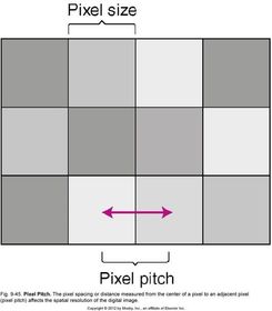
| Pixel Pitch | distance between two pixels where they come together in the middle |
| Visibility of Recorded Details includes: | -brightness -density -contrast |
| Sharpness of Recorded Details includes: | - spatial resolution - distortion |
| Brightness | amount of luminance |
| Density | amount of blackness - controlled by mAs |
| Contrast | quality of radiation absorption within the body - controlled by kVp |
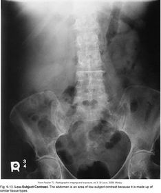
| Low-Subject Contrast | -similar tissue types - similar shades of gray *ABDOMEN* |
| High-Subject Contrast | - varying tissue types - varying shades of black -> gray -> white *CHEST* |
| High contrast, with low kVp and few shades of gray describes _____ _____ contrast | Short - Scale |
| Low contrast, with high kVp and many shades of gray describes _____ ______ contrast | Long - Scale |
| Size Distortion is characterized by ______ and _______ | Magnification OID/SID |
| Shape Distortion is characterized by _______ _______ and ______ | elongation foreshortening tube angle patient positioning |
| Using a grid will ________ contrast and ________ density. Therefore, you should _______ _____ when using a grid | increase decrease increase mAs |
| What are 3 examples of decreasing exposure time? | 1. decreasing SID increases speed 2. increase mA, decrease time 3. increase kVp |
| The area of the anode surface which receives the beam of electroms from the cathode is known as _____ _____ | FOCAL SPOT |
| As focal spot size decreases, _________ decreases, and ________ increases | unsharpness recorded detail |
| Bit Depth | how the exit radiation is recorded controls the pixel brightness |
| Matrix | rows and columns of the image |
| Pixel | each individual box in a matrix |
Want to create your own Flashcards for free with GoConqr? Learn more.