9791779
Description
Flashcards by Ben Harries, updated more than 1 year ago
|
|
Created by Ben Harries
over 6 years ago
|
|
| Question | Answer |
|
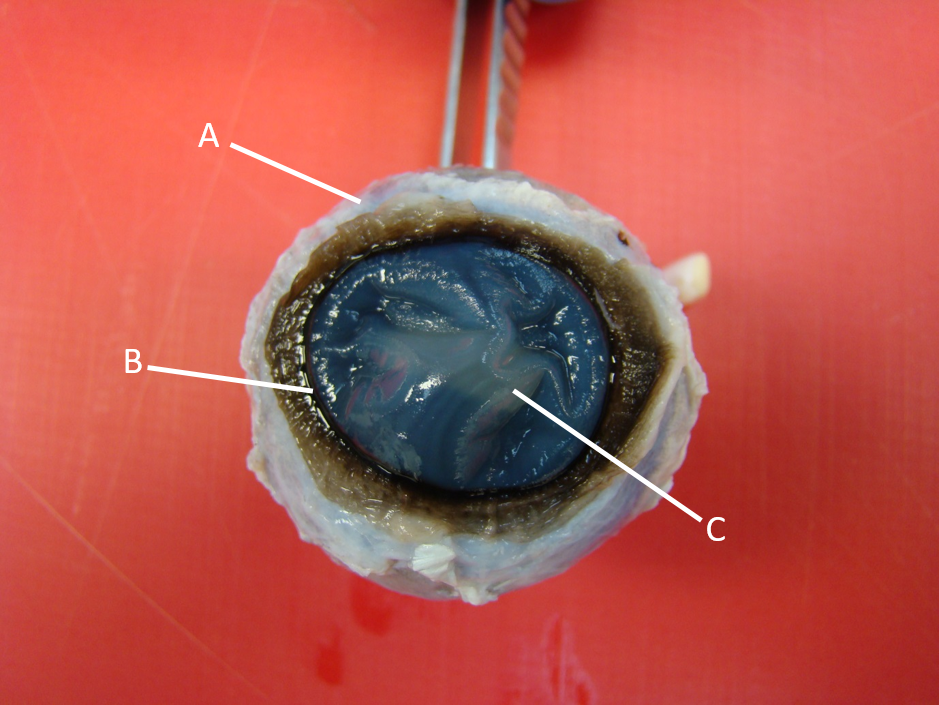
Image:
Eye1 (binary/octet-stream)
|
A - Sclera B - Limbus C - Cornea |
|
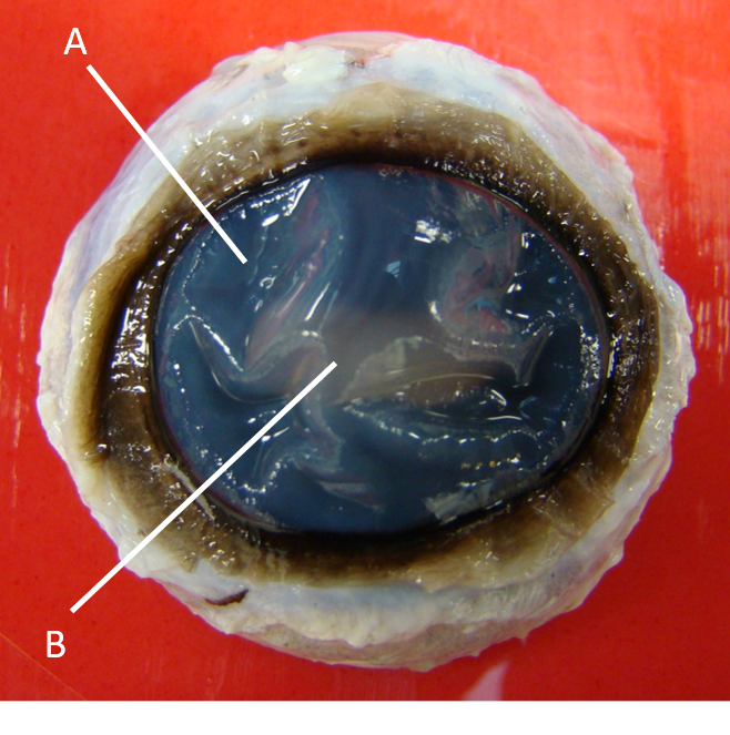
Image:
Eye2 (binary/octet-stream)
|
A - Iris B - Pupil |
|
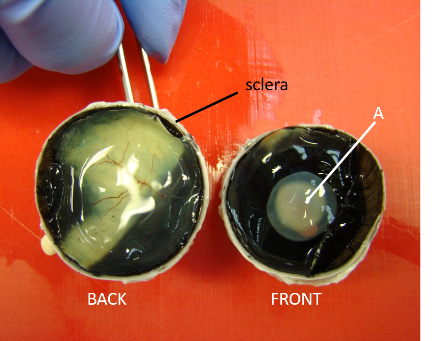
Image:
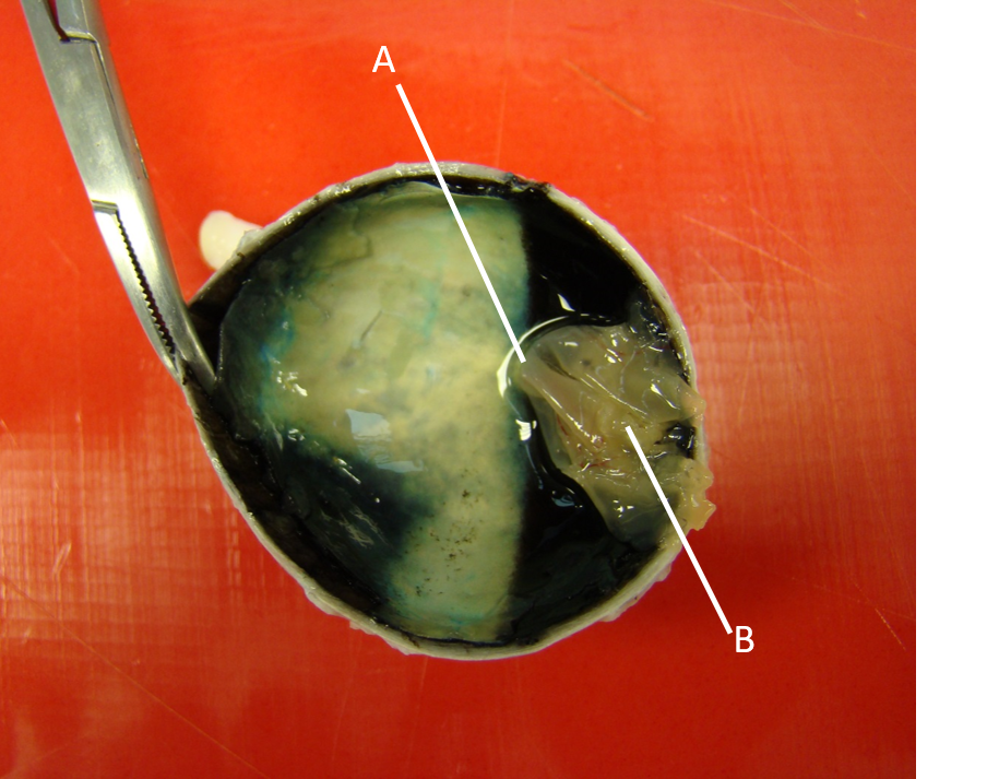
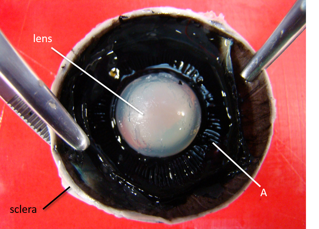
Eye3 (binary/octet-stream)
|
A - Lens |
|
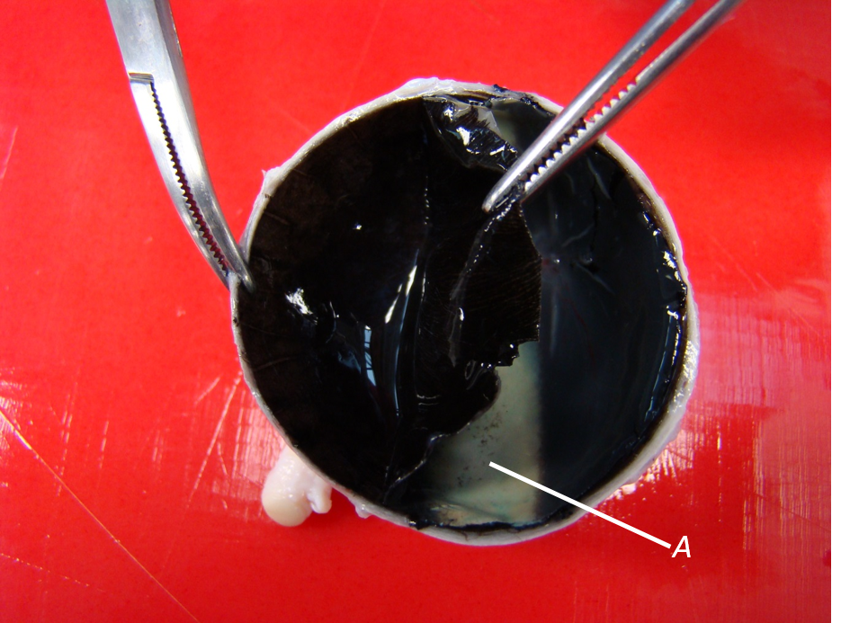
Image:
Eye4 (binary/octet-stream)
|
A - Tapetum Lucidum |
|
Image:
Eye5 (binary/octet-stream)
|
A - retinal point of attachment at optic disc B - Nervous layer of retina |
|
Image:
Eye6 (binary/octet-stream)
|
A - Ciliary Process |
| What structures make up the orbit? What is the adaptation in enclosed orbits? | Frontal, lacrimal, zygomatic, sphenoid & palatine bones; completed by the orbital ligament. Frontal process of zygomatic bone & zygomatic process of frontal bone meet to enclose orbit. |
|
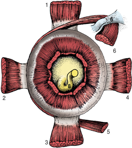
Image:
Eye M (binary/octet-stream)
|
1 - Dorsal rectus m. 2 - Lateral rectus m. 3 - Ventral rectus m. 4 - Medial rectus m. 5 - Ventral oblique m. 6 - Dorsal oblique m. 7 - Retractor bulbi m. 8 - Optic nerve |
| What muscle rotates the eye upwards? | Dorsal rectus |
| What muscle rotates the eye downwards? | Ventral rectus |
| What muscle rotates the globe laterally? | Lateral rectus |
| What muscle rotates the globe medially? | Medial rectus |
| What muscle rotates the dorsal globe medially & ventrally? | Dorsal oblique |
| What muscle rotates the ventral globe medially & dorsally? | Ventral oblique |
| What muscle retracts the globe into the orbit? | Retractor bulbi |
| How is the canthus secured to the bone? How does the dog differ? | Canthus on either side secured to the bone by canthal/palpebral ligaments. In dog the ligament is absent and function replaced by the retractor anguli m. |
| What muscle contracts to close the eyelids? | Orbicularis Oculi m. |
| Name the innervation of the orbicularis oculi m. | Facial n. (CN VII) |
| What muscles lift the medial & lateral portions of the upper lid? | Medial & lateral retractor anguli oculi m. |
| What muscle is the main elevator of the upper eyelid? | Levator palpebrae superioris m. |
| What nerve innervates the medial & lateral retractor anguli oculi m.? | Facial n. (CN VII) |
| What nerve innervates the levator palpebrae superioris m.? | Oculomotor n. (CN II) |
| What nerve supplies sensory innervation to the eyelids? | Trigeminal n. (CN V) |
| What branches of the trigeminal nerve supply what parts of the eyelid? | Ophthalmic branch - innervates most of upper lid & medial part of lower lid Maxillary branch - innervates lower lid & joins with ophthalmic to supply lateral portion of upper lid |
| Name the 3 layers of the tear film, their production site and function | Lipid layer - produced in meibomian glands; reduces evaporation & creates barrier at lid margin Mucin layer - produced by conjunctival goblet cells; stabilises tear film Aqueous layer - produced from lacrimal & 3rd eyelid glands; provides lubrication, protection & nutrition to epithelium |
| Name the 3 layers of the globe and their function | Fibrous layer - supports eyeball shape Uvea - provides nutrition to structures of the eye & alters light transmission Neural layer - the retina |
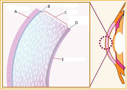
| A - Epithelium B - Bowman's layer C - Stroma D - Descemet's membrane E - Endothelium | |
| What is the function of the cornea? | Transmit & reflect light |
| Describe the structure of the anterior epithelium | Squamous on the outside, becomes columnar at base, rests on basement produced by columnar epithelium cells; epithelium linked to middle stroma by fine fibrils |
| Describe the structure of the stroma | Main body of the cornea; Consists of collagen fibrils with some keratocytes and a ground substance of proteoglycans & GAGs |
| Give the function of the endothelium | Produces Descemet's membrane and maintains corneal clarity by actively pumping water out of the stroma |
| Describe the structure of the uvea | Middle layer of the globe. Iris & ciliary body form anterior uvea; choroid forms the posterior uvea |
| Give the function of the iris | Constricts or dilates to vary amount of light entering posterior chamber of the eye and altering depth of focus |
| Describe the mechanism & innervation of the iris | Constrictor muscle of the iris is a smooth muscle sphincter which sits in stroma of pupillary zone; parasympathetic innervation via oculomotor n. (CN II) |
| Describe the structure & function of the ciliary body | Consists of the ciliary process, ciliary body muscles & forms part of iridocorneal angle. Ciliary body produces aqueous humour, anchors zonular fibres (which suspend the lens) & muscular portion enables lens accomodation |
| What makes up the iridocorneal angle and what is it function? | Where the root of the iris, the anterior ciliary body & corneoscleral junction meet. Main site of drainage of aqueous humour. |
| Describe the layers of the choroid | Suprachoroidea - forms transition between sclera & choroid Large vessel layer - contains vascular plexus mainly consisting of veins which coalesce to form the cortex veins Medium vessel layer - contains vessels & tapteum lucidum (fibrous in herbivores, cellular in carnivores) Choriocacapillaris - innermost layer of vessels, consists of fenestrated capillaries which supply the retina |
| What is the function of aqueous humour? | Provide nutrition to the lens & cornea |
| How is aqueous humour produced? | Diffusion - solutes down conc. gradient into aqueous Ultrafiltration - occurs due to differences in hydrostatic pressure in ciliary body, capillaries & IOP Active secretion - active transport of Na by non-pigmented ciliary epithelium into aqueous which brings water across too |
| Where does the aqueous drain from? | Iridocorneal angle - majority through pectinate ligament & ciliary cleft of ICA and then via aqueous plexus to the scleral venous circulation |
| Where is the vitreous humour found? | Fills posterior segment of globe |
| What is the function of vitreous humour? | Transmits light & lends physical support to the globe; also storage of nutrients & waste products for the retina |
| What is the function of the lens? | Focus light on the retina |
| What structures are visible in the fundus? | Optic disc, retina, RPE & sometimes the sclera |
| Give the differences between cone & rod photoreceptors | Cone - enable colour vision & sharp acuity Rod - enable vision of shape & motion |
Want to create your own Flashcards for free with GoConqr? Learn more.