
Flowchart
Brain and Cranial Nerves BMS 111
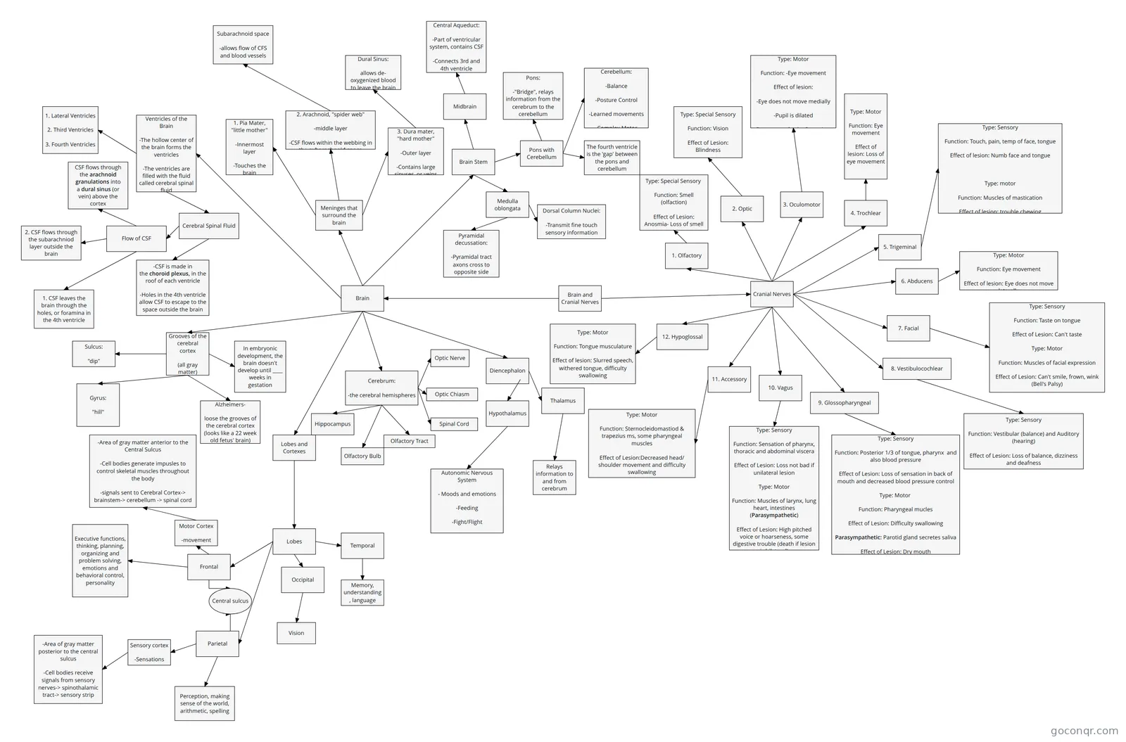
Flowchart: Brain and cranial nerves
Flowchart content
Brain and Cranial Nerves
Brain
Cranial Nerves
Meninges that surround the brain
1. Pia Mater, "little mother"
-Innermost layer
-Touches the brain
2. Arachnoid, "spider web"
-middle layer
-CSF flows within the webbing in the subarachnoid space
Subarachnoid space
-allows flow of CFS and blood vessels
3. Dura mater, "hard mother"
-Outer layer
-Contains large sinuses, or veins
Dural Sinus:
allows de-oxygenized blood to leave the brain
Ventricles of the Brain
-The hollow center of the brain forms the ventricles
-The ventricles are filled with the fluid called cerebral spinal fluid
1. Lateral Ventricles
2. Third Ventricles
3. Fourth Ventricles
Cerebral Spinal Fluid
Flow of CSF
-CSF is made in the choroid plexus, in the roof of each ventricle
-Holes in the 4th ventricle allow CSF to escape to the space outside the brain
2. CSF flows through the subarachniod layer outside the brain
1. CSF leaves the brain through the holes, or foramina in the 4th ventricle
CSF flows through the arachnoid granulations into a dural sinus (or vein) above the cortex
Grooves of the cerebral cortex
(all gray matter)
Sulcus:
"dip"
Gyrus:
"hill"
Lobes and Cortexes
In embryonic development, the brain doesn't develop until ____ weeks in gestation
Alzheimers-
loose the grooves of the cerebral cortex (looks like a 22 week old fetus' brain)
Lobes
Frontal
Parietal
Occipital
Temporal
Central sulcus
Executive functions, thinking, planning, organizing and problem solving, emotions and behavioral control, personality
Perception, making sense of the world, arithmetic, spelling
Motor Cortex
-movement
Sensory cortex
-Sensations
Vision
-Area of gray matter anterior to the Central Sulcus
-Cell bodies generate impusles to control skeletal muscles throughout the body
-signals sent to Cerebral Cortex-> brainstem-> cerebellum -> spinal cord
-Area of gray matter posterior to the central sulcus
-Cell bodies receive signals from sensory nerves-> spinothalamic tract-> sensory strip
Cerebrum:
-the cerebral hemispheres
Memory, understanding, language
Hippocampus
Olfactory Bulb
Olfactory Tract
Optic Nerve
Optic Chiasm
Spinal Cord
Diencephalon
Thalamus
Hypothalamus
Brain Stem
Autonomic Nervous System
- Moods and emotions
-Feeding
-Fight/Flight
-Sex
Relays information to and from cerebrum
Midbrain
Pons with Cerebellum
Medulla oblongata
Central Aqueduct:
-Part of ventricular system, contains CSF
-Connects 3rd and 4th ventricle
Pons:
-"Bridge", relays information from the cerebrum to the cerebellum
Cerebellum:
-Balance
-Posture Control
-Learned movements
-Complex Motor Patterns
The fourth ventricle is the 'gap' between the pons and cerebellum
Pyramidal decussation:
-Pyramidal tract axons cross to opposite side
Dorsal Column Nuclei:
-Transmit fine touch sensory information
1. Olfactory
2. Optic
3. Oculomotor
4. Trochlear
5. Trigeminal
6. Abducens
7. Facial
8. Vestibulocochlear
9. Glossopharyngeal
10. Vagus
11. Accessory
12. Hypoglossal
Type: Special Sensory
Function: Smell (olfaction)
Effect of Lesion: Anosmia- Loss of smell
Type: Special Sensory
Function: Vision
Effect of Lesion: Blindness
Type: Motor
Function: -Eye movement
Effect of lesion:
-Eye does not move medially
-Pupil is dilated
Parasympathetic: Constricts pupil
Type: Motor
Function: Eye movement
Effect of lesion: Loss of eye movement
Type: Sensory
Function: Touch, pain, temp of face, tongue
Effect of lesion: Numb face and tongue
Type: motor
Function: Muscles of mastication
Effect of lesion: trouble chewing
Type: Motor
Function: Eye movement
Effect of lesion: Eye does not move laterally
Type: Sensory
Function: Taste on tongue
Effect of Lesion: Can't taste
Type: Motor
Function: Muscles of facial expression
Effect of Lesion: Can't smile, frown, wink (Bell's Palsy)
Parasympathetic: tears, salivation
Type: Sensory
Function: Vestibular (balance) and Auditory (hearing)
Effect of Lesion: Loss of balance, dizziness and deafness
Type: Sensory
Function: Posterior 1/3 of tongue, pharynx and also blood pressure
Effect of Lesion: Loss of sensation in back of mouth and decreased blood pressure control
Type: Motor
Function: Pharyngeal mucles
Effect of Lesion: Difficulty swallowing
Parasympathetic: Parotid gland secretes saliva
Effect of Lesion: Dry mouth
Type: Sensory
Function: Sensation of pharynx, thoracic and abdominal viscera
Effect of Lesion: Loss not bad if unilateral lesion
Type: Motor
Function: Muscles of larynx, lung heart, intestines (Parasympathetic)
Effect of Lesion: High pitched voice or hoarseness, some digestive trouble (death if lesion is bilateral)
Type: Motor
Function: Sternocleidomastiod & trapezius ms, some pharyngeal muscles
Effect of Lesion:Decreased head/ shoulder movement and difficulty swallowing
Type: Motor
Function: Tongue musculature
Effect of lesion: Slurred speech, withered tongue, difficulty swallowing
Information
- Description
- Undergrad Anatomy Flowchart on Brain and Cranial Nerves BMS 111, created by Kimi Nyquist on 10/10/2017.
- Created by
- Kimi Nyquist
- Views
- 1
- Rating