
Mind map
Spinal Cord - Tracts (p79-89)
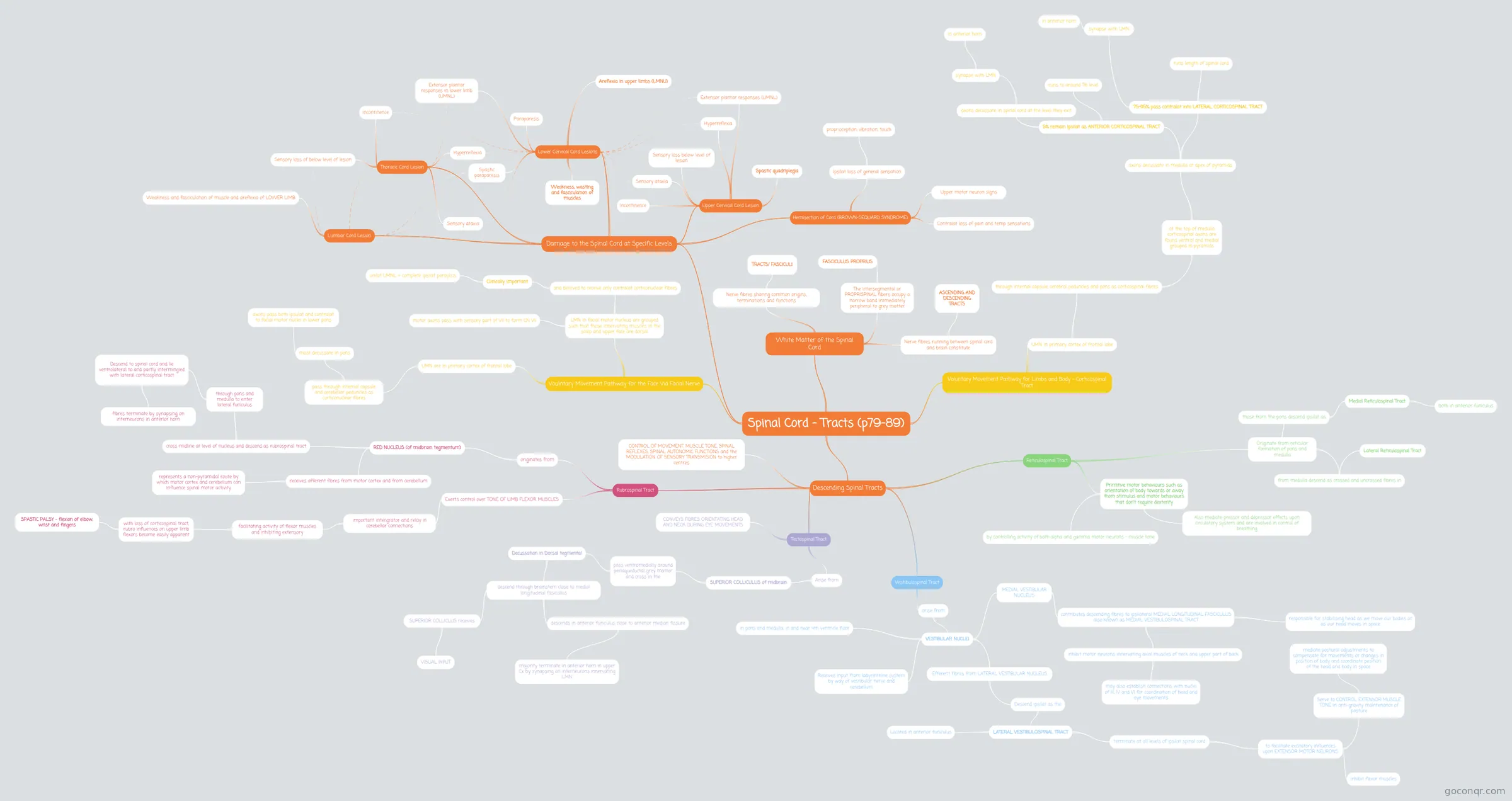
Mind map: Spinal Cord Tracts and Movement Pathways
Map content
Spinal Cord - Tracts (p79-89)
White Matter of the Spinal Cord
- Nerve fibres sharing common origins, terminations and functions
- TRACTS/ FASCICULI
- The intersegmental or PROPRISPINAL fibers occupy a narrow band immediately peripheral to grey matter
- FASCICULUS PROPRIUS
- Nerve fibres running between spinal cord and brain constitute
- ASCENDING AND DESCENDING TRACTS
- Nerve fibres sharing common origins, terminations and functions
Descending Spinal Tracts
- CONTROL OF MOVEMENT, MUSCLE TONE, SPINAL REFLEXES, SPINAL AUTONOMIC FUNCTIONS and the MODULATION OF SENSORY TRANSMISION to higher centres
- Rubrospinal Tract
- originates from
- RED NUCLEUS (of midbrain tegmentum)
- cross midline at level of nucleus and descend as rubrospinal tract
- through pons and medulla to enter lateral funiculus
- Descend to spinal cord and lie ventrolateral to and partly intermingled with lateral corticospinal tract
- fibres terminate by synapsing on interneurons in anterior horn
- Descend to spinal cord and lie ventrolateral to and partly intermingled with lateral corticospinal tract
- through pons and medulla to enter lateral funiculus
- receives afferent fibres from motor cortex and from cerebellum
- represents a non-pyramidal route by which motor cortex and cerebellum can influence spinal motor activity
- cross midline at level of nucleus and descend as rubrospinal tract
- RED NUCLEUS (of midbrain tegmentum)
- Exerts control over TONE OF LIMB FLEXOR MUSCLES
- important intergrator and relay in cerebellar connections
- facilitating activity of flexor muscles and inhibiting extensory
- with loss of corticospinal tract, rubro influences on upper limb flexors become easily apparent
- SPASTIC PALSY - flexion of elbow, wrist and fingers
- with loss of corticospinal tract, rubro influences on upper limb flexors become easily apparent
- facilitating activity of flexor muscles and inhibiting extensory
- important intergrator and relay in cerebellar connections
- originates from
- Tectospinal Tract
- Arise from
- SUPERIOR COLLICULUS of midbrain
- pass ventromedially around periaqueductal grey matter and cross in the
- Decussation in Dorsal tegmental
- descend through brainstem close to medial longitudinal fasicullus
- SUPERIOR COLLICLUS receives
- VISUAL INPUT
- descends in anterior funiculus close to anterior median fissure
- majority terminate in anterior horn in upper Cx by synapsing on interneurons innervating LMN
- SUPERIOR COLLICLUS receives
- descend through brainstem close to medial longitudinal fasicullus
- Decussation in Dorsal tegmental
- pass ventromedially around periaqueductal grey matter and cross in the
- SUPERIOR COLLICULUS of midbrain
- CONVEYS FIBRES ORIENTATING HEAD AND NECK DURING EYE MOVEMENTS
- Arise from
- Vestibulospinal Tract
- arise from
- VESTIBULAR NUCLEI
- in pons and medulla, in and near 4th ventricle floor
- Receives input from labyrinthine system by way of vestibular nerve and cerebellum
- Efferent fibres from LATERAL VESTIBULAR NUCLEUS
- Descend ipsilat as the
- LATERAL VESTIBULOSPINAL TRACT
- Located in anterior funiculus
- terminate at all levels of ipsilat spinal cord
- to facilitate excitatory influences upon EXTENSOR MOTOR NEURONS
- Serve to CONTROL EXTENSOR MUSCLE TONE in anti-gravity maintenance of posture
- mediate postural adjustments to compensate for movements or changes in position of body and coordinate position of the head and body in space
- inhibit flexor muscles
- Serve to CONTROL EXTENSOR MUSCLE TONE in anti-gravity maintenance of posture
- to facilitate excitatory influences upon EXTENSOR MOTOR NEURONS
- LATERAL VESTIBULOSPINAL TRACT
- Descend ipsilat as the
- MEDIAL VESTIBULAR NUCLEUS
- contributes descending fibres to ipsilateral MEDIAL LONGITUDINAL FASCICULUS also known as MEDIAL VESTIBULOSPINAL TRACT
- inhibit motor neurons innervating axial muscles of neck and upper part of back
- may also establish connections with nuclei of III, IV and VI for coordination of head and eye movements
- responsible for stabilising head as we move our bodies or as our head moves in space
- inhibit motor neurons innervating axial muscles of neck and upper part of back
- contributes descending fibres to ipsilateral MEDIAL LONGITUDINAL FASCICULUS also known as MEDIAL VESTIBULOSPINAL TRACT
- VESTIBULAR NUCLEI
- arise from
- Reticulospinal Tract
- Originate from reticular formation of pons and medulla
- those from the pons descend ipsilat as
- Medial Reticulospinal Tract
- both in anterior funiculus
- Medial Reticulospinal Tract
- from medulla descend as crossed and uncrossed fibres in
- Lateral Reticulospinal Tract
- those from the pons descend ipsilat as
- Primitive motor behaviours such as orientation of body towards or away from stimulus and motor behaviours that don't require dexterity
- by controlling activity of both alpha and gamma motor neurons - muscle tone
- Also mediate pressor and depressor effects upon circulatory system and are involved in control of breathing
- Originate from reticular formation of pons and medulla
Damage to the Spinal Cord at Specific Levels
- Upper Cervical Cord Lesion
- Spastic quadriplegia
- Hyperreflexia
- Extensor plantar responses (UMNL)
- Incontinence
- Sensory loss below level of lesion
- Sensory ataxia
- Lower Cervical Cord Lesions
- Weakness, wasting and fasciculation of muscles
- Areflexia in upper limbs (LMNU)
- Spastic paraparesis
- Paraparesis
- Extensor plantar responses in lower limb (UMNL)
- Thoracic Cord Lesion
- Hyperreflexia
- Incontinence
- Sensory loss of below level of lesion
- Sensory ataxia
- Lumbar Cord Lesion
- Weakness and fasciculation of muscle and areflexia of LOWER LIMB
- Hemisection of Cord (BROWN-SEQUARD SYNDROME)
- Ipsilat loss of general sensation
- proprioception, vibration, touch
- Upper motor neuron signs
- Contralat loss of pain and temp sensations
- Ipsilat loss of general sensation
- Upper Cervical Cord Lesion
Voluntary Movement Pathway for Limbs and Body - Corticospinal Tract
- UMN in primary cortex of frontal lobe
- through internal capsule, cerebral peduncles and pons as corticospinal fibres
- at the top of medulla corticospinal axons are found ventral and medial grouped in pyramids
- axons decussate in medulla at apex of pyramids
- 75-95% pass contralat into LATERAL CORTICOSPINAL TRACT
- runs length of spinal cord
- synapse with LMN
- in anterior horn
- 5% remain ipsilat as ANTERIOR CORTICOSPINAL TRACT
- runs to around T6 level
- axons decussate in spinal cord at the level they exit
- synapse with LMN
- in anterior horn
- synapse with LMN
- 75-95% pass contralat into LATERAL CORTICOSPINAL TRACT
- axons decussate in medulla at apex of pyramids
- at the top of medulla corticospinal axons are found ventral and medial grouped in pyramids
- through internal capsule, cerebral peduncles and pons as corticospinal fibres
- UMN in primary cortex of frontal lobe
Voulntary Movement Pathway for the Face Via Facial Nerve
- UMN are in primary cortex of frontal lobe
- pass through internal capsule and cerebellar peduncles as corticonuclear fibres
- most decussate in pons
- axons pass both ipsulat and contralat to facial motor nuclei in lower pons
- most decussate in pons
- pass through internal capsule and cerebellar peduncles as corticonuclear fibres
- LMN in facial motor nucleus are grouped such that those innervating muscles in the scalp and upper face are dorsal
- and believed to receive only contralat corticonuclear fibres
- Clinically important
- unilat UMNL = complete ipsilat paraylsis
- Clinically important
- motor axons pass with sensory part of VII to form CN VII
- and believed to receive only contralat corticonuclear fibres
- UMN are in primary cortex of frontal lobe
Information
- Description
- Anatomy Mind Map on Spinal Cord - Tracts (p79-89), created by Harriet Colquhoun on 08/03/2018.
- Created by
- Harriet Colquhoun
- Views
- 0
- Rating
More on Neural Pathways and Spinal Tracts