
Mind map
Cranial Nerve II - The Eye and Visual Fields
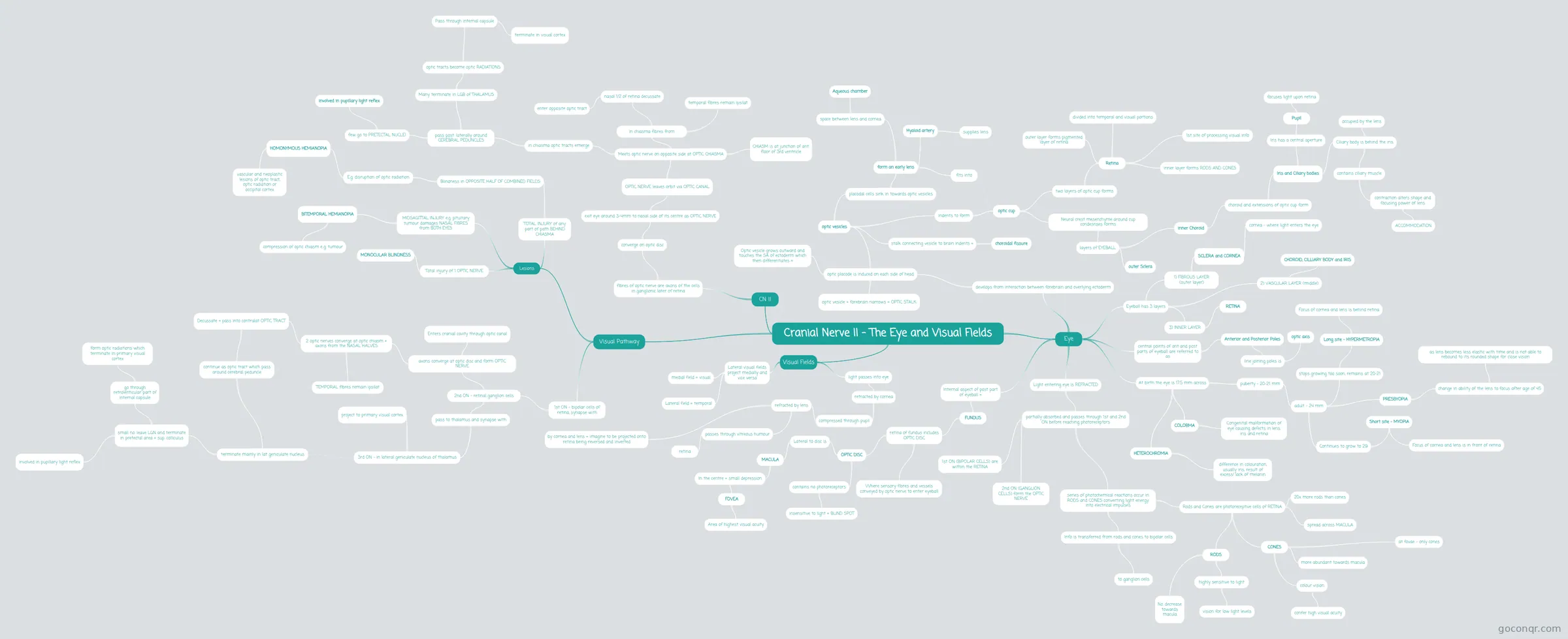
Mind map: Cranial Nerve II - The Eye and Visual Fields
Map content
Cranial Nerve II - The Eye and Visual Fields
CN !!
- fibres of optic nerve are axons of the cells in ganglionic later of retina
- converge on optic disc
- exit eye around 3-4mm to nasal side of its centre as OPTIC NERVE
- OPTIC NERVE leaves orbit via OPTIC CANAL
- Meets optic nerve on opposite side at OPTIC CHIASMA
- CHIASM is at junction of ant. floor of 3rd ventricle
- in chiasma fibres from
- nasal 1/2 of retina decussate
- enter opposite optic tract
- temporal fibres remain ipsilat
- nasal 1/2 of retina decussate
- in chiasma optic tracts emerge
- pass post. laterally around CEREBRAL PEDUNCLES
- Many terminate in LGB of THALAMUS
- optic tracts become optic RADIATIONS
- Pass through internal capsule
- terminate in visual cortex
- Pass through internal capsule
- optic tracts become optic RADIATIONS
- few go to PRETECTAL NUCLEI
- involved in pupillary light reflex
- Many terminate in LGB of THALAMUS
- pass post. laterally around CEREBRAL PEDUNCLES
- Meets optic nerve on opposite side at OPTIC CHIASMA
- OPTIC NERVE leaves orbit via OPTIC CANAL
- exit eye around 3-4mm to nasal side of its centre as OPTIC NERVE
- converge on optic disc
- fibres of optic nerve are axons of the cells in ganglionic later of retina
Eye
- develops from interaction between forebrain and overlying ectoderm
- optic placode is induced on each side of head
- optic vesicles
- placodal cells sink in towards optic vesicles
- form an early lens
- fits into
- Hyaloid artery
- supplies lens
- space between lens and cornea
- Aqueous chamber
- form an early lens
- indents to form
- optic cup
- two layers of optic cup forms
- Retina
- outer layer forms pigmented layer of retina
- inner layer forms RODS AND CONES
- 1st site of processing visual info
- divided into temporal and visual portions
- Retina
- Neural crest mesenchyme around cup condesnses forms
- layers of EYEBALL
- inner Choroid
- choroid and extensions of optic cup form
- Iris and Ciliary bodies
- Ciliary body is behind the iris
- contains ciliary muscle
- contraction alters shape and focusing power of lens
- ACCOMMODATION
- contraction alters shape and focusing power of lens
- occupied by the lens
- contains ciliary muscle
- Iris has a central aperture
- Pupil
- focuses light upon retina
- Pupil
- Ciliary body is behind the iris
- Iris and Ciliary bodies
- choroid and extensions of optic cup form
- outer Sclera
- inner Choroid
- layers of EYEBALL
- two layers of optic cup forms
- optic cup
- stalk connecting vesicle to brain indents =
- choroidal fissure
- placodal cells sink in towards optic vesicles
- Optic vesicle grows outward and touches the SA of ectoderm which then differentiates =
- optic vesicle + forebrain narrows = OPTIC STALK
- optic vesicles
- optic placode is induced on each side of head
- central points of ant and post parts of eyeball are referred to as
- Anterior and Posterior Poles
- line joining poles is
- optic axis
- line joining poles is
- Anterior and Posterior Poles
- At birth the eye is 17.5 mm across
- puberty - 20-21 mm
- adult - 24 mm
- stops growing too soon, remains at 20-21
- Long site - HYPERMETROPIA
- Focus of cornea and lens is behind retina
- Long site - HYPERMETROPIA
- Continues to grow to 29
- Short site - MYOPIA
- Focus of cornea and lens is in front of retina
- Short site - MYOPIA
- PRESBYOPIA
- change in ability of the lens to focus after age of 45
- as lens becomes less elastic with time and is not able to rebound to its rounded shape for close vision
- change in ability of the lens to focus after age of 45
- stops growing too soon, remains at 20-21
- adult - 24 mm
- COLOBMA
- Congenital malformation of eye causing defects in lens, iris and retina
- HETEROCHROMIA
- difference in colouration, usually iris, result of excess/ lack of melanin
- puberty - 20-21 mm
- Light entering eye is REFRACTED
- partially absorbed and passes through 1st and 2nd ON before reaching photoreceptors
- series of photochemical reactions occur in RODS and CONES converting light energy into electrical impulses
- Info is transferred from rods and cones to bipolar cells
- to ganglion cells
- Rods and Cones are photoreceptive cells of RETINA
- 20x more rods than cones
- RODS
- highly sensitive to light
- vision for low light levels
- No. decrease towards macula
- highly sensitive to light
- CONES
- colour vision
- confer high visual acuity
- more abundant towards macula
- at fovae - only cones
- colour vision
- spread across MACULA
- Info is transferred from rods and cones to bipolar cells
- 1st ON (BIPOLAR CELLS) are within the RETINA
- 2nd ON (GANGLION CELLS) form the OPTIC NERVE
- series of photochemical reactions occur in RODS and CONES converting light energy into electrical impulses
- partially absorbed and passes through 1st and 2nd ON before reaching photoreceptors
- Internal aspect of post part of eyeball =
- FUNDUS
- retina of fundus includes OPTIC DISC
- Where sensory fibres and vessels conveyed by optic nerve to enter eyeball
- OPTIC DISC
- contains no photoreceptors
- insensitive to light = BLIND SPOT
- Lateral to disc is
- MACULA
- In the centre = small depression
- FOVEA
- Area of highest visual acuity
- FOVEA
- In the centre = small depression
- MACULA
- contains no photoreceptors
- retina of fundus includes OPTIC DISC
- FUNDUS
- Eyeball has 3 layers
- 1) FIBROUS LAYER (outer layer)
- SCLERA and CORNEA
- cornea - where light enters the eye
- SCLERA and CORNEA
- 2) VASCULAR LAYER (middle)
- CHOROID, CILLIARY BODY and IRIS
- 3) INNER LAYER
- RETINA
- 1) FIBROUS LAYER (outer layer)
- develops from interaction between forebrain and overlying ectoderm
Visual Fields
- light passes into eye
- retracted by cornea
- compressed through pupil
- refracted by lens
- passes through vitreous humour
- retina
- by cornea and lens = imagine to be projected onto retina being reversed and inverted
- passes through vitreous humour
- refracted by lens
- compressed through pupil
- retracted by cornea
- Lateral visual fields project medially and vice versa
- Lateral field = temporal
- medial field = visual
- light passes into eye
Visual Pathway
- 1st ON - bipolar cells of retina, synapse with
- 2nd ON - retinal ganglion cells
- pass to thalamus and synapse with
- 3rd ON - in lateral geniculate nucleus of thalamus
- project to primary visual cortex
- 3rd ON - in lateral geniculate nucleus of thalamus
- axons converge at optic disc and form OPTIC NERVE
- Enters cranial cavity through optic canal
- 2 optic nerves converge at optic chiasm + axons from the NASAL HALVES
- Decussate + pass into contralat OPTIC TRACT
- continue as optic tract which pass around cerebral peduncle
- terminate mainly in lat geniculate nucleus
- small no. leave LGN and terminate in pretectal area + sup. colliculus
- involved in pupillary light reflex
- go through retrolenticular part of internal capsule
- form optic radiations which terminate in primary visual cortex
- small no. leave LGN and terminate in pretectal area + sup. colliculus
- terminate mainly in lat geniculate nucleus
- continue as optic tract which pass around cerebral peduncle
- TEMPORAL fibres remain ipsilat
- Decussate + pass into contralat OPTIC TRACT
- pass to thalamus and synapse with
- 2nd ON - retinal ganglion cells
- Lesions
- Total injury of 1 OPTIC NERVE
- MONOCULAR BLINDNESS
- MIDSAGITTAL INJURY e.g. pituitary tumour damages NASAL FIBRES from BOTH EYES
- BITEMPORAL HEMIANOPIA
- compression of optic chiasm e..g. tumour
- BITEMPORAL HEMIANOPIA
- TOTAL INJURY of any part of path BEHIND CHIASMA
- Blindness in OPPOSITE HALF OF COMBINED FIELDS
- E.g. disruption of optic radiation
- HOMONYMOUS HEMIANOPIA
- vascular and neoplastic lesions of optic tract, optic radiation or occipital cortex
- HOMONYMOUS HEMIANOPIA
- E.g. disruption of optic radiation
- Blindness in OPPOSITE HALF OF COMBINED FIELDS
- Total injury of 1 OPTIC NERVE
- 1st ON - bipolar cells of retina, synapse with
Information
- Description
- Anatomy Mind Map on Cranial Nerve II - The Eye and Visual Fields, created by Harriet Colquhoun on 26/03/2018.
- Created by
- Harriet Colquhoun
- Views
- 0
- Rating