
Mind map
Osteoporosis
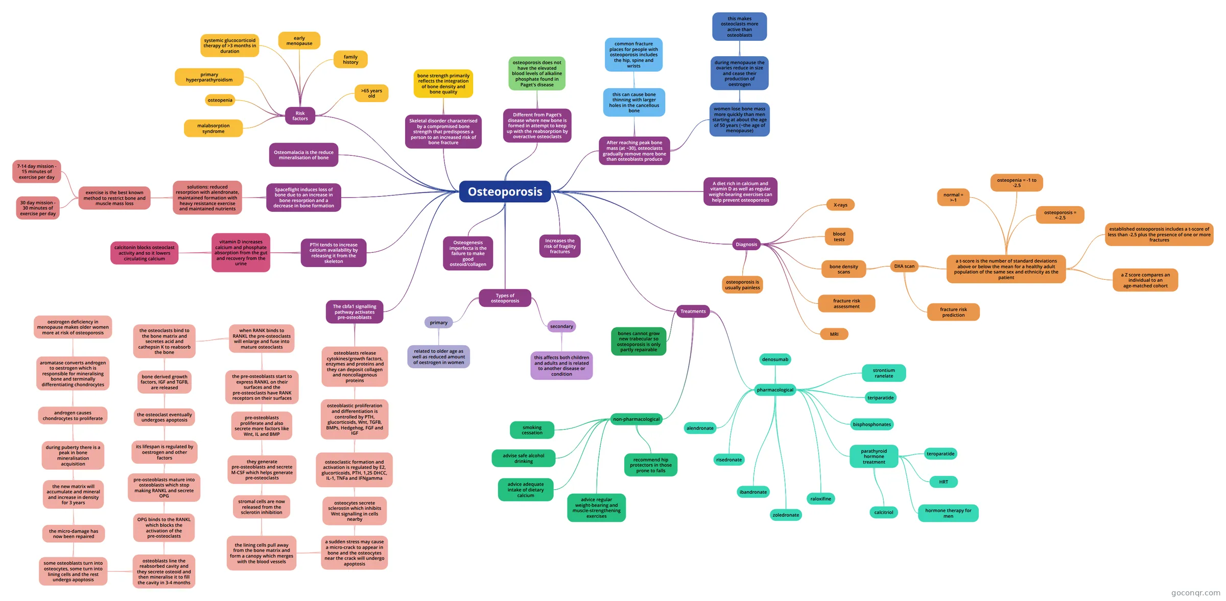
Mind map: Osteoporosis
Map content
Osteoporosis
- osteopertrosis is a failure of osteoclastic and chondroclastic resorption and failure of bone remodelling
- fluorosis is an abnormal matrix mineralisation where fluoride replaces calcium in the matrix
Skeletal disorder characterised by a compromised bone strength that predisposes a person to an increased risk of bone fracture
- bone strength primarily reflects the integration of bone density and bone quality
Different from Paget's disease where new bone is formed in attempt to keep up with the reabsorption by overactive osteoclasts
- new bone is produced haphazardly
- osteoporosis does not have the elevated blood levels of alkaline phosphate found in Paget's disease
After reaching peak bone mass (at ~30), osteoclasts gradually remove more bone than osteoblasts produce
- this can cause bone thinning with larger holes in the cancellous bone
- common fracture places for people with osteoporosis includes the hip, spine and wrists
- women lose bone mass more quickly than men starting at about the age of 50 years (~the age of menopause)
- during menopause the ovaries reduce in size and cease their production of oestrogen
- this makes osteoclasts more active than osteoblasts
- during menopause the ovaries reduce in size and cease their production of oestrogen
- this can cause bone thinning with larger holes in the cancellous bone
A diet rich in calcium and vitamin D as well as regular weight-bearing exercises can help prevent osteoporosis
Types of osteoporosis
- primary
- related to older age as well as reduced amount of oestrogen in women
- secondary
- this affects both children and adults and is related to another disease or condition
- primary
Risk factors
- >65 years old
- family history
- systemic glucocorticoid therapy of >3 months in duration
- primary hyperparathyroidism
- osteopenia
- early menopause
- malabsorption syndrome
Diagnosis
- there is no screening programme for osteoporosis in the UK
- X-rays
- blood tests
- bone density scans
- DXA scan
- a t-score is the number of standard deviations above or below the mean for a healthy adult population of the same sex and ethnicity as the patient
- normal = >-1
- osteopenia = -1 to -2.5
- osteoporosis = <-2.5
- established osteoporosis includes a t-score of less than -2.5 plus the presence of one or more fractures
- a Z score compares an individual to an age-matched cohort
- fracture risk prediction
- a t-score is the number of standard deviations above or below the mean for a healthy adult population of the same sex and ethnicity as the patient
- DXA scan
- fracture risk assessment
- MRI
- osteoporosis is usually painless
Treatments
- corticosteroids can induce osteoporosis as they can increase osteoclastic activity, decrease osteoblastic activity, impair collagen formation, increase bone turnover and result in poor bone formation and healing
- non-pharmacological
- smoking cessation
- advise safe alcohol drinking
- advice adequate intake of dietary calcium
- advice regular weight-bearing and muscle-strengthening exercises
- recommend hip protectors in those prone to falls
- pharmacological
- alendronate
- risedronate
- ibandronate
- zoledronate
- raloxifine
- selective oestrogen receptor modulator
- parathyroid hormone treatment
- teroparatide
- HRT
- hormone therapy for men
- calcitriol
- bisphosphonates
- the is the drug that is used the most
- teriparatide
- PTH decreases the amount of vertebral fractures in wormen and it decreases the risk of having ant other non-vertebral fracture
- PTH, 7-part receptor
- strontium ranelate
- reduces the risk of vertebral, hip and other fractures
- denosumab
- bones cannot grow new trabecular so osteoporosis is only partly repairable
Osteomalacia is the reduce mineralisation of bone
The cbfa1 signalling pathway activates pre-osteoblasts
- osteoblasts release cytokines/growth factors, enzymes and proteins and they can deposit collagen and noncollagenous proteins
- osteoblastic proliferation and differentiation is controlled by PTH, glucorticoids, Wnt, TGFB, BMPs, Hedgehog, FGF and IGF
- osteoclastic formation and activation is regulated by E2, glucorticoids, PTH, 1,25 DHCC, IL-1, TNFa and IFNgamma
- osteocytes secrete sclerostin which inhibits Wnt signalling in cells nearby
- a sudden stress may cause a micro-crack to appear in bone and the osteocytes near the crack will undergo apoptosis
- the lining cells pull away from the bone matrix and form a canopy which merges with the blood vessels
- stromal cells are now released from the sclerotin inhibition
- they generate pre-osteoblasts and secrete M-CSF which helps generate pre-osteoclasts
- pre-osteoblasts proliferate and also secrete more factors like Wnt, IL and BMP
- the pre-osteoblasts start to express RANKL on their surfaces and the pre-osteoclasts have RANK receptors on their surfaces
- when RANK binds to RANKL the pre-osteoclasts will enlarge and fuse into mature osteoclasts
- the osteoclasts bind to the bone matrix and secretes acid and cathepsin K to reabsorb the bone
- 2 weeks
- bone derived growth factors, IGF and TGFB, are released
- the osteoclast eventually undergoes apoptosis
- its lifespan is regulated by oestrogen and other factors
- pre-osteoblasts mature into osteoblasts which stop making RANKL and secrete OPG
- OPG binds to the RANKL which blocks the activation of the pre-osteoclasts
- osteoblasts line the reabsorbed cavity and they secrete osteoid and then mineralise it to fill the cavity in 3-4 months
- some osteoblasts turn into osteocytes, some turn into lining cells and the rest undergo apoptosis
- the micro-damage has now been repaired
- the new matrix will accumulate and mineral and increase in density for 3 years
- primary mineralisation takes less than 100 days and secondary mineralisation can take up to a year
- during puberty there is a peak in bone mineralisation acquisition
- BMD = bone mineral density
- androgen causes chondrocytes to proliferate
- aromatase converts androgen to oestrogen which is responsible for mineralising bone and terminally differentiating chondrocytes
- oestrogen deficiency in menopause makes older women more at risk of osteoporosis
- oestrogen normally inhibits bone resorption so a lack of oestrogen, like in menopause, means that bone resorption is not inhibited
- OPG is stimulated by oestrogen
- oestrogen deficiency in menopause makes older women more at risk of osteoporosis
- aromatase converts androgen to oestrogen which is responsible for mineralising bone and terminally differentiating chondrocytes
- the new matrix will accumulate and mineral and increase in density for 3 years
- the micro-damage has now been repaired
- some osteoblasts turn into osteocytes, some turn into lining cells and the rest undergo apoptosis
- osteoblasts line the reabsorbed cavity and they secrete osteoid and then mineralise it to fill the cavity in 3-4 months
- OPG binds to the RANKL which blocks the activation of the pre-osteoclasts
- pre-osteoblasts mature into osteoblasts which stop making RANKL and secrete OPG
- its lifespan is regulated by oestrogen and other factors
- the osteoclast eventually undergoes apoptosis
- the osteoclasts bind to the bone matrix and secretes acid and cathepsin K to reabsorb the bone
- when RANK binds to RANKL the pre-osteoclasts will enlarge and fuse into mature osteoclasts
- the pre-osteoblasts start to express RANKL on their surfaces and the pre-osteoclasts have RANK receptors on their surfaces
- pre-osteoblasts proliferate and also secrete more factors like Wnt, IL and BMP
- they generate pre-osteoblasts and secrete M-CSF which helps generate pre-osteoclasts
- stromal cells are now released from the sclerotin inhibition
- the lining cells pull away from the bone matrix and form a canopy which merges with the blood vessels
- a sudden stress may cause a micro-crack to appear in bone and the osteocytes near the crack will undergo apoptosis
- osteocytes secrete sclerostin which inhibits Wnt signalling in cells nearby
- osteoclastic formation and activation is regulated by E2, glucorticoids, PTH, 1,25 DHCC, IL-1, TNFa and IFNgamma
- osteoblastic proliferation and differentiation is controlled by PTH, glucorticoids, Wnt, TGFB, BMPs, Hedgehog, FGF and IGF
- osteoblasts release cytokines/growth factors, enzymes and proteins and they can deposit collagen and noncollagenous proteins
PTH tends to increase calcium availability by releasing it from the skeleton
- vitamin D increases calcium and phosphate absorption from the gut and recovery from the urine
- calcitonin blocks osteoclast activity and so it lowers circulating calcium
- vitamin D increases calcium and phosphate absorption from the gut and recovery from the urine
Spaceflight induces loss of bone due to an increase in bone resorption and a decrease in bone formation
- a lot of calcium salts are taken from the bone and excreted in urine, when first in space
- solutions: reduced resorption with alendronate, maintained formation with heavy resistance exercise and maintained nutrients
- exercise is the best known method to restrict bone and muscle mass loss
- 7-14 day mission - 15 minutes of exercise per day
- 30 day mission - 30 minutes of exercise per day
- exercise is the best known method to restrict bone and muscle mass loss
Increases the risk of fragility fractures
Osteogenesis imperfecta is the failure to make good osteoid/collagen
- there is a defect in collagen type I, the condition can cause recurrent childhood fractures, deformities, ligamentous laxity, low muscle tone and bluish sclerae
Information
- Description
- Year 1 Mind Map on Osteoporosis, created by Rana Al-Khayat on 29/04/2018.
- Created by
- Rana Al-Khayat
- Views
- 11
- Rating