
20697665
Description

Mind Map by Zainab Patel, updated more than 1 year ago
|
|
Created by Zainab Patel
almost 6 years ago
|
|
Skull views
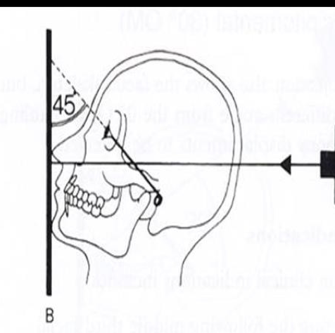
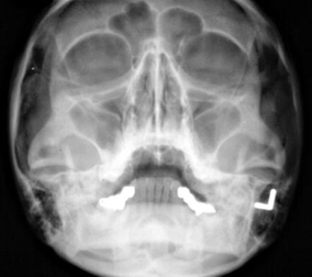
- Occipitomental view
- Investigate
- Coronoid process fractures
- Mid-face facial fractures
- Sinuses
- Coronoid process fractures
- Positioning
- Pt seated facing the film
with head tipped back so
aga tragal line at 45° to film.
Tubehead positioned so
beam centred through
occiput.
- Pt seated facing the film
with head tipped back so
aga tragal line at 45° to film.
Tubehead positioned so
beam centred through
occiput.
- Investigate
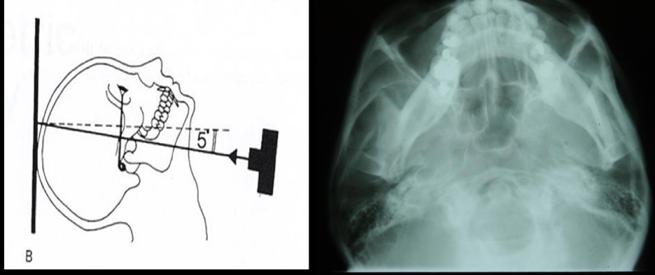
- Submento-vortex
- Investigate
- Base of skull & sphenoidal
sinus from below
- Fractures of
zygomatic arches
- Base of skull & sphenoidal
sinus from below
- Investigate
- Oblique lateral
- Investigate
- Not commonly used- now use DPT
- When not able to take
intra-oral e.g. gag
reflex
- Not commonly used- now use DPT
- Investigate
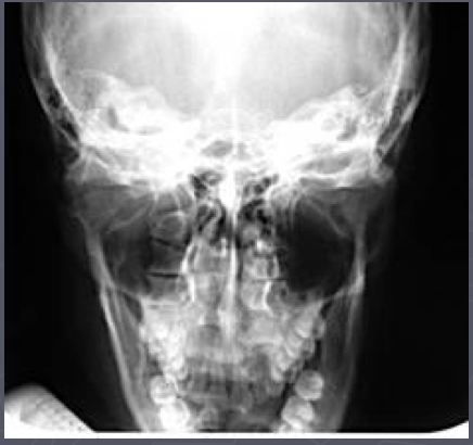
- PA
mandible
- AKA
- Posterior anterior mandible
- Posterior anterior mandible
- Investigate
- Fracture on
mandible
- At
- Body
- Angle
- Ramus
- Body
- At
- Lesions of
bone
- At
- Body of
mandible
- Body of
mandible
- At
- Fracture on
mandible
- Positioning
- Same patient position as PA
skull
- Tubehead with central
beam at through
cervical spine at level
of rami of mandible.
- Same patient position as PA
skull
- AKA
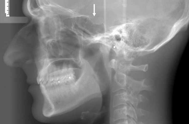
- True lateral skull
- Investigate
- Conditions of the cranium e.g
pagets, hyperparathyroidism
- Acromegaly
- Image sinuses
- Rarely taken
- Conditions of the cranium e.g
pagets, hyperparathyroidism
- Investigate
- Lateral Ceph
- Beam is set further forwards
compared to true lateral skull
- Standardised and
reproducible compared
to lateral skull
- Standardised and
reproducible compared
to lateral skull
- Investigate
- Ortho
- Orthognathic surgery
- Ortho
- Beam is set further forwards
compared to true lateral skull
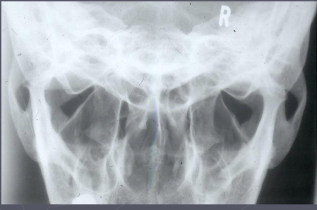
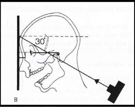
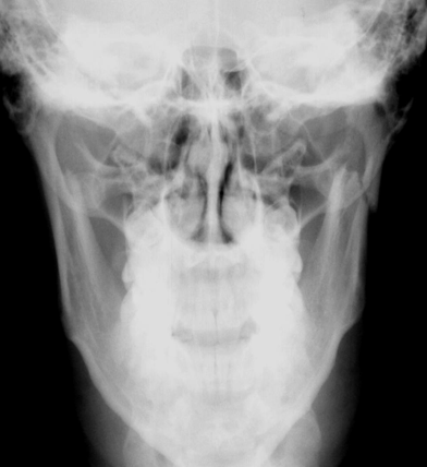
- Reverse Townes
- AKA
- Occipito frontal
- Occipito frontal
- Investigate
- Neck of condyle
- Condylar hypo/hyperplasia
- Neck of condyle
- AKA
Media attachments
Want to create your own Mind Maps for free with GoConqr? Learn more.