3728912

Tooth Development
- At the 6th week the oral epithelium
thickens and invaginates into the
surrounding mesenchyme
- The PRIMARY EPITHELIAL BAND
- Dental lamina
(lingually
positioned)
- Involved in the
development of the teeth /
tooth germs
- Bud -> Cap -> Bell stages
- Bud -> Cap -> Bell stages
- Local proliferation of the dental lamina
at positions of future deciduous teeth
causes production of epithelial
swellings into ectomesenchyme (future
tooth germs)
- Involved in the
development of the teeth /
tooth germs
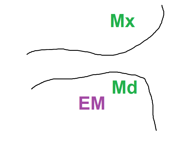
- Vestibular
lamina (buccally
positioned)
- Forms the vestibule of the mouth
- VL cell proliferation, then
degeneration of central epithelial cells -
creates the SULCUS of the vestibule
(gap between the cheek and
tooth-bearing area)
- Forms the vestibule of the mouth
- (Division)
- Dental lamina
(lingually
positioned)
- The PRIMARY EPITHELIAL BAND
- Key words
- Histo-differentiation
- In the context of tooth
development,
histo-differentiation is the
idea of cells differentiation
into morphologically and
functionally distinct groups
of cells e.g. odontoblasts
- Structure + Shape related to FUNCTION
- Structure + Shape related to FUNCTION
- In the context of tooth
development,
histo-differentiation is the
idea of cells differentiation
into morphologically and
functionally distinct groups
of cells e.g. odontoblasts
- Morpho-differentiation
- In the context of tooth
development,
morpho-differentiation is the
idea of the determination of
certain shapes (morpho) such
as the shape of the crown of
the tooth.
- In the context of tooth
development,
morpho-differentiation is the
idea of the determination of
certain shapes (morpho) such
as the shape of the crown of
the tooth.
- Histo-differentiation
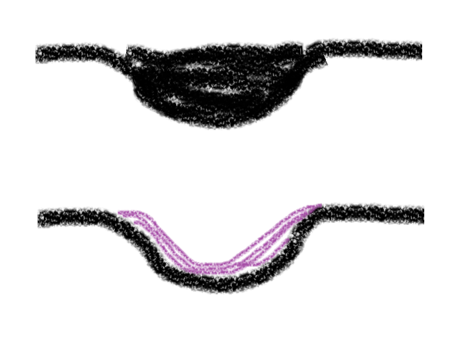
- Bud -> Cap -> Bell stages
- Bud
- Poor morphodifferentiation and
histodifferentiation of tissues at
this stage
- Ectomesenchymal
cells are closely
packed around the
bud
- An "epithelial incursion" into the
ectomesenchyme by the epithelial "bud" - the
early enamel organ
- Poor morphodifferentiation and
histodifferentiation of tissues at
this stage
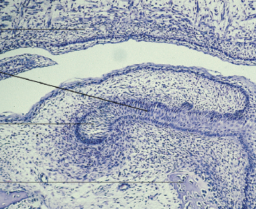
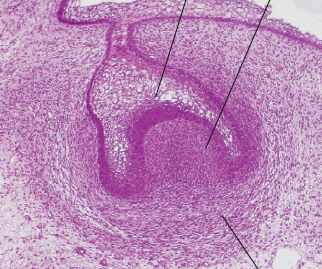
- Cap stage
- The epithelial bud
continues to proliferate
within the
ectomesenchyme that
it is embedded in
- Condensation of the
ectomesenchymal cells
that surround the bud
(forming the DENTAL
PAPILLA)
- Dentin
- Pulp
- Precursor to..
- Dentin
- We can clearly see the epithelial enamel
organ sitting upon a section of condensed
ectomesenchyme (the dental papilla)
- Dental follicle limiting dental papilla
and surrounding the enamel organ
- Periodontium
- Precursor to..
- Periodontium
- Dental follicle limiting dental papilla
and surrounding the enamel organ
- Invasion of vascular
supply to DP
- The epithelial bud
continues to proliferate
within the
ectomesenchyme that
it is embedded in
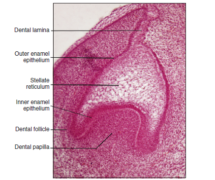
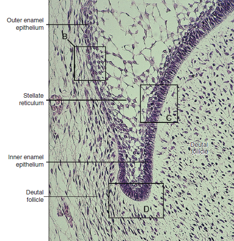
- Bell
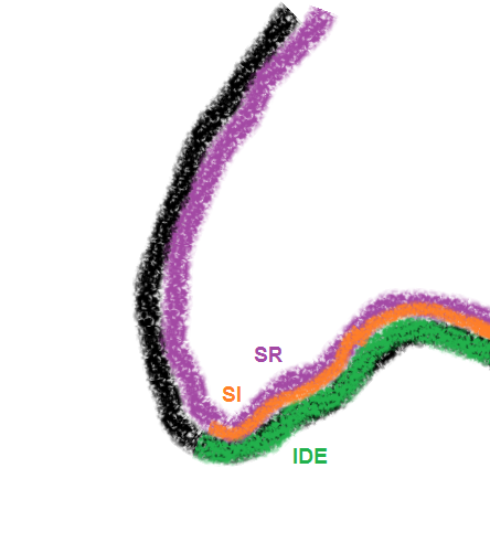
- Early bell stage (4 cell layers)
- Cells on the periphery of the enamel
organ assume a low cuboidal shape. This
creates the outer enamel epithelium
- Cells lining the dental papilla assume
a short columnar shape. This creates
the inner enamel epithelium (which
later differentiate into ameloblasts)
- The stratum intermedium forms via
differentation of epithelial cells
located between the IDE and SR. The
cells have a characteristically high
amount of alkaline phosphatase
- Works with the IDE as a single
functional unit in the production of
enamel, but its actual role is unclear.
- Works with the IDE as a single
functional unit in the production of
enamel, but its actual role is unclear.
- Stellate reticulum
- Cells on the periphery of the enamel
organ assume a low cuboidal shape. This
creates the outer enamel epithelium
- Late bell stage
- 1. Dental lamina breaks down 2. Tooth germ
loses attachment to the oral epithelium and
becomes encased in the bone of the jaw 3. Some
dental lamina remains in the jaw as epithelial
pearls (involved in future cyst formation)
- Differential rates of mitotic division of
IEE = epithelial folding = crown shape /
cuspal outline formed
- Dentin + Enamel formation occurs at the crest
of this folding (future cusp tips)
- Dentin + Enamel formation occurs at the crest
of this folding (future cusp tips)
- 1. Dental lamina breaks down 2. Tooth germ
loses attachment to the oral epithelium and
becomes encased in the bone of the jaw 3. Some
dental lamina remains in the jaw as epithelial
pearls (involved in future cyst formation)
- DF = >collagen fibrils in
extracellular space than DP
- Dental basement membrane separates IDE (or the enamel
organ for that matter) from the DP. Its role is in mediating
interactions between epith and mesen compartments during
odontoblast differentiation before dentine secretion - when
the DBM breaks down, the pre-dentine matrix will induce IEE
terminal differentiation into ameloblasts
- Dental papilla = progenitor cell
population for future dentine-pulp
complex, e.g. odontoblasts differentiate
from cells adjacent to the enamel organ
- Early bell stage (4 cell layers)
- Cap -> Bell transition
- The cells in the centre of the enamel organ secrete lots
of GAG into the extracel. compartment. These are
hydrophilic theref. water is drawn in. This increases
fluid volume in extracel. compartment and thus the
central cells are forced apart. Since desmosomal
contacts are still in place, this distorts the cells. This
forms star shaped cells termed the STELLATE
RETICULUM
- Bud
- Transitory structures
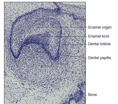
- Enamel knot
- Clusters of non-dividing epithelial
cells in molar cap stage tooth
germs
- Produces many signalling
molecules including BMP's etc
- Possible function is in the
organisation of cusp morphogenesis
- Precursor cells noted by
expression of p21
- Disappears by bell stage
- Clusters of non-dividing epithelial
cells in molar cap stage tooth
germs
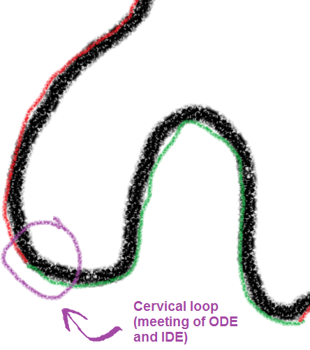
- Cervical loop
- Where the IDE meets the ODE
- After crown formation, gives
rise to the epithelial
component of root formation (HERS)
- This is the region where cells
continue dividing until the tooth
crown reaches its full size
- Where the IDE meets the ODE
- Enamel cord
- Unknown function - may be
involved in cap to bell
transition
- A strand of cells
from the stratum
intermedium to the
ODE which divides
the SR
- Unknown function - may be
involved in cap to bell
transition
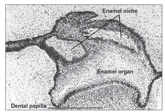
- Enamel niche
- Pockets of
ectomesenchyme within
the enamel organ
- Possible
sectioning
artefact
- Pockets of
ectomesenchyme within
the enamel organ
- Enamel knot
- Dental Organ / Tooth Germ =
- EO + DP + DF
- EO + DP + DF
- Supplies to the tooth
- Nerve supply
- Nerves penetrate
dental papilla with
onset of
dentinogenesis
- Nerves important for hypersensitivity
- Nerves important for hypersensitivity
- Nerves penetrate
dental papilla with
onset of
dentinogenesis
- Vascular supply
- Small vessels
invade the DP/DF in
the early bell
stage
- Increases a lot during the
bell stage during hard
tissue formation
- NEVER invades SR - EO = avascular
- Small vessels
invade the DP/DF in
the early bell
stage
- Nerve supply
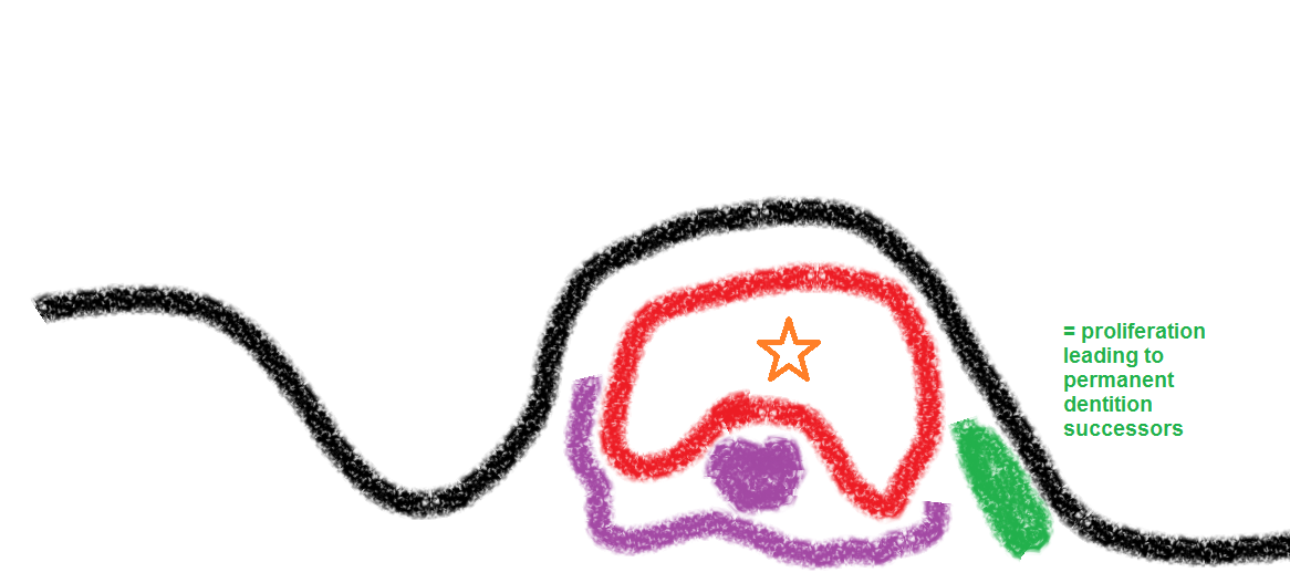
- Formation of the permanent dentition
- Permanent incisor, canine, premolar germs form as a
result of proliferation on the lingual aspect of the DL
next to deciduous predecessors
- Permanent molars do not have deciduous predecessors
(only 20 teeth in deciduous dentition). There is backwards
extension of the dental lamina which gives off epithelial
ingrowths - these will produce the 1st -> 3rd molars.
- Permanent incisor, canine, premolar germs form as a
result of proliferation on the lingual aspect of the DL
next to deciduous predecessors
- Chronology
- Deciduous = 6-8 weeks i.u
- Permanent successional teeth (i.e. 1 -> 5) = 20 weeks
i.u. to 10 months after
birth
- Permanent molars = 20 months i.u.
to 5 years after birth
- EXAM - GIVE ESTIMATES OF
CALCIFICATION/ERUPTION
DATES FOR DIFFERENT TYPES
OF TEETH
- Deciduous = 6-8 weeks i.u
Media attachments
Want to create your own Mind Maps for free with GoConqr? Learn more.