
Mind map
Spinal Cord
Map content
Spinal Cord
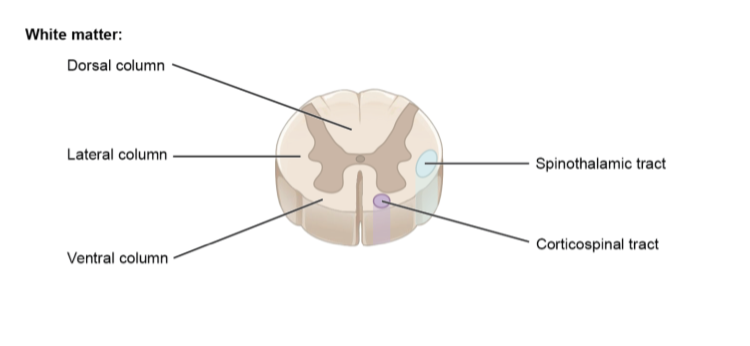
White Matter: myleinated axons
- Dorsal column
- posterior
- Ventral Column
- Anterior
- Lateral column
- Dorsal column
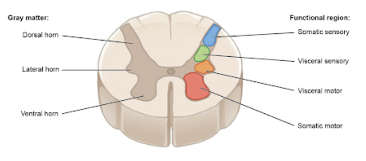
Grey matter: unmyelinated cell bodies/soma
- Horns: Bring information in/out of spinal cord
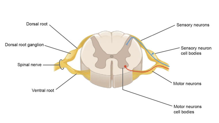
- Dorsal Horn
- Sensory (visceral and somatic)
- Lateral Horn
- Visceral motor
- Ventral Horn
- Somatic motor
- distinguished by type of information they carry
- Dorsal Horn
- 2. Choose a horn based on type (sensory v motor) and classification (somatic v visceral)
- Horns: Bring information in/out of spinal cord
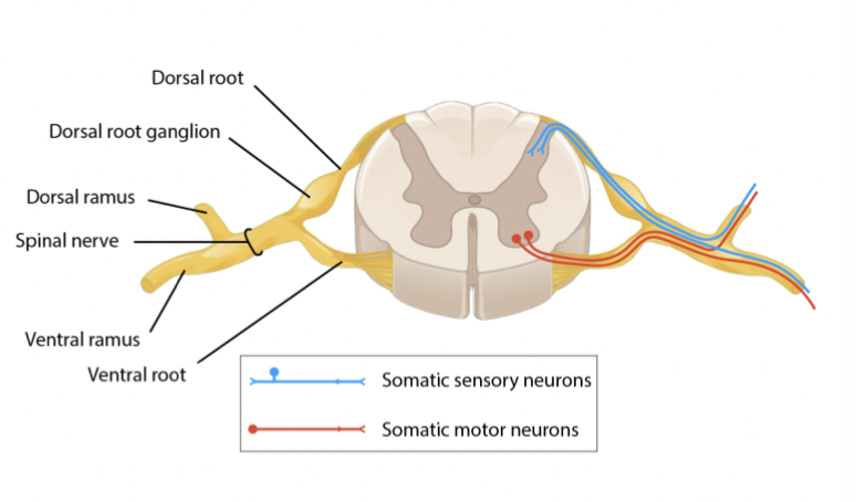
Roots: Connect spinal nerves to horns
- Dorsal Root
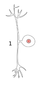
- Dorsal root ganglion
- Contains cell bodies
- Psuedounipolar neurons
- Dorsal root ganglion
- Ventral Root
- Contains cell bodies
- Distinguished by type of information (sensory or motor)
- 1. Choose a root based on type of information
- Dorsal Root
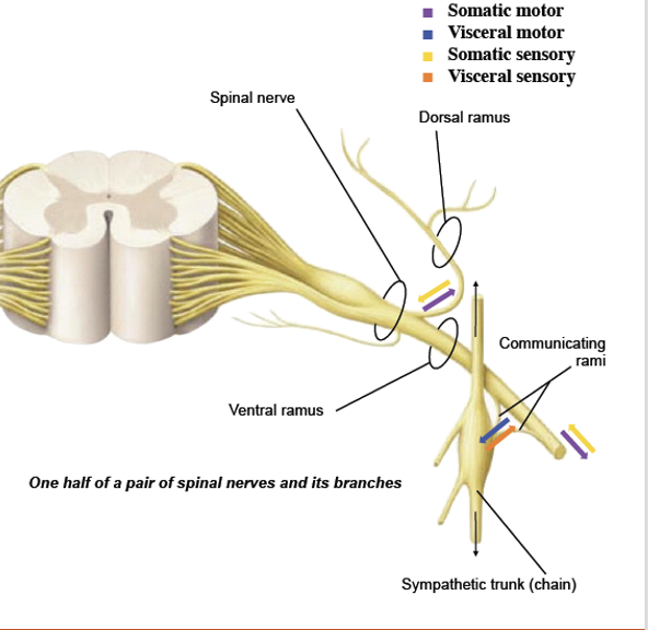
Rami: Travel from the body to the spinal nerve
- Classified by location of destination or origin
- Ventral Ramus
- Front sides of body
- Dorsal Ramus
- Back side of the body
- Communicating Rami
- Smooth/cardiac muscle, glands
- Cell bodies located in sympathetic ganglion
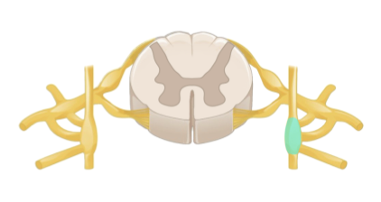
- Sympathetic chain
- Runs parallel and anterior to spinal cord
- Sympathetic chain
- Visceral motor and sensory
- Carried to sympathetic trunk
- 3. Choose a ramus based on destination
Pathways
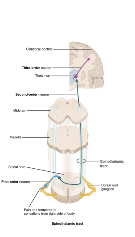
- Ascending: Send sensory information to the brain
- Neurons
- 1st order neuron
- Peripheral sensor neuron
- Body to dorsal horn
- 2nd order neuron
- Interneuron
- Travels from dorsal horn up lateral column to thalamus
- 3rd order neuron
- Interneuron
- Travels from thalamus to cerebral cortex
- Decussates at pyramids of medulla
- Cell bodies
- Located in dorsal horn and thalamus
- 1st order neuron
- 2 synapses
- Types
- Dorsal column-medial leminscus
- Spinothalamic
- Spinocerebellar
- Neurons
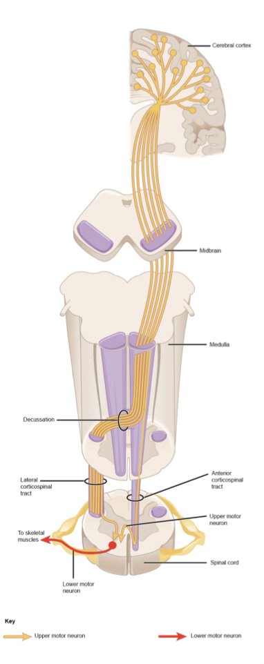
- Descending: Send instructions to muscles for motor function
- Neurons
- Upper motor neuron
- Interneuron
- Travels from primary motor cortex to ventral horn through the ventral column
- Lower motor neuron
- Motor neuron
- Travels from the ventral horn to the muscle
- Cell Bodies
- Located in ventral horn
- Upper motor neuron
- 1 synapse
- Neurons
- Ascending: Send sensory information to the brain
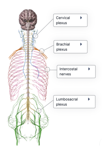
Plexuses: Groups of nerves from the ventral rami
- Intercostal nerves
- Runs along ribs
- Innervates muscles and skin of abdominal wall, bones
- Cervical plexus
- Innvervates neck
- Brachial Plexus
- Innervates arm
- C5-T1
- Lumbosacral plexus
- Innervates legs
- L2-S4
- Intercostal nerves
Reflexes
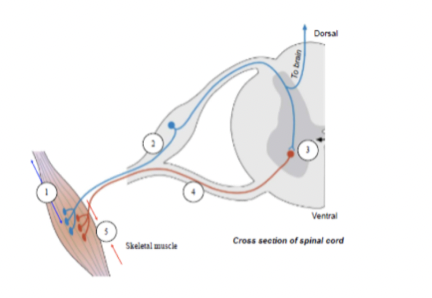
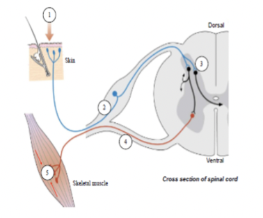
- Polysynaptic Reflex Arc
- Sensory Neuron (travels along rami to spinal nerve)
- Dorsal Horn
- Interneuron (gray matter)
- Ventral horn
- Ventral Root
- Muscle
- Where interneuron synapses
- Ventral Root
- Can also go outside of spinal column
- Ventral horn
- Where sensory neuron synapses with interneuron
- Interneuron (gray matter)
- Dorsal Horn
- Sensory Neuron (travels along rami to spinal nerve)
- Monosynaptic Reflex Arc
- Sensory neuron (travels along ramus)
- Ventral horn
- Muscle
- Ventral horn
- Sensory neuron (travels along ramus)
- Polysynaptic Reflex Arc
Anatomy
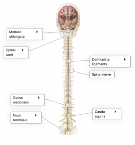
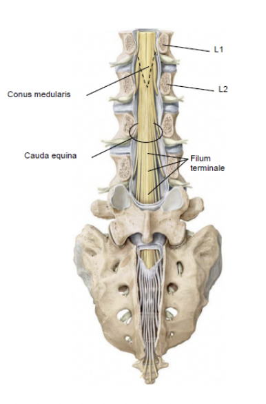
- Conus Medullaris
- Termination of spinal cord at L1/L2
- Cauda Equina
- Bundle of spinal nerve roots that lumbar, sacral, and coccygeal nerves pass through
- Filum Terminale
- Holds spinal cord down to sacrum and coccyx
- Continuation of pia mater
- Denticulate ligaments
- Extention of pia mater that stabilize spinal cord in vertebral canal
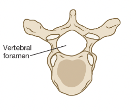
- Vertebral foramen
- Where spinal nerves emerge through foramina in vertebrae, forms vertebral canal
- Location of spinal cord
- Where spinal nerves emerge through foramina in vertebrae, forms vertebral canal
- Vertebral canal
- Where the spinal cord runs through
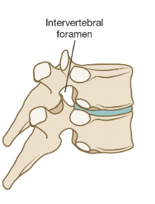
- Intervertebral foramen
- Spaces between vertebrae where spinal nerves exit
- Between vertebrae
- Conus Medullaris
Meninges
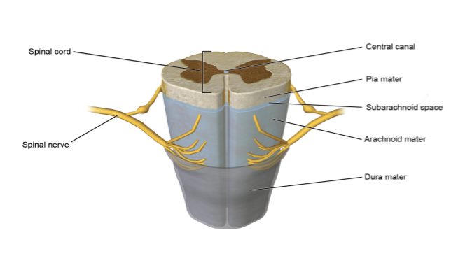
- Dura Mater
- Arachnoid Mater
- Subarachnoid Space
- Location of CSF
- Pia Mater
- Central Canal
- Fluid-filled
- Continuity with ventricles of brain
- Anterior median fissure
- Epidural Space
- Filled with fat and blood vessels
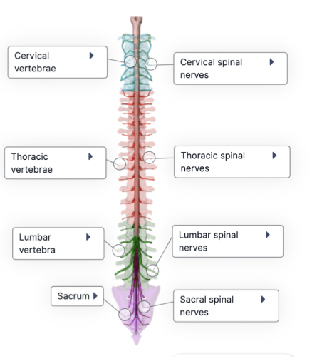
Vertebrae
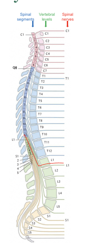
- Cervical
- 8
- Thoracic
- 12
- Lumbar
- 5
- Sacral
- 5
- Coccygeal
- 1
- Cervical
Images and files in the map
Information
- Description
- Mind Map on Spinal Cord, created by Julia Morneau on 14/09/2022.
- Created by
- Julia Morneau
- Views
- 0
- Rating