
Mind map
Week 2: Control of movement
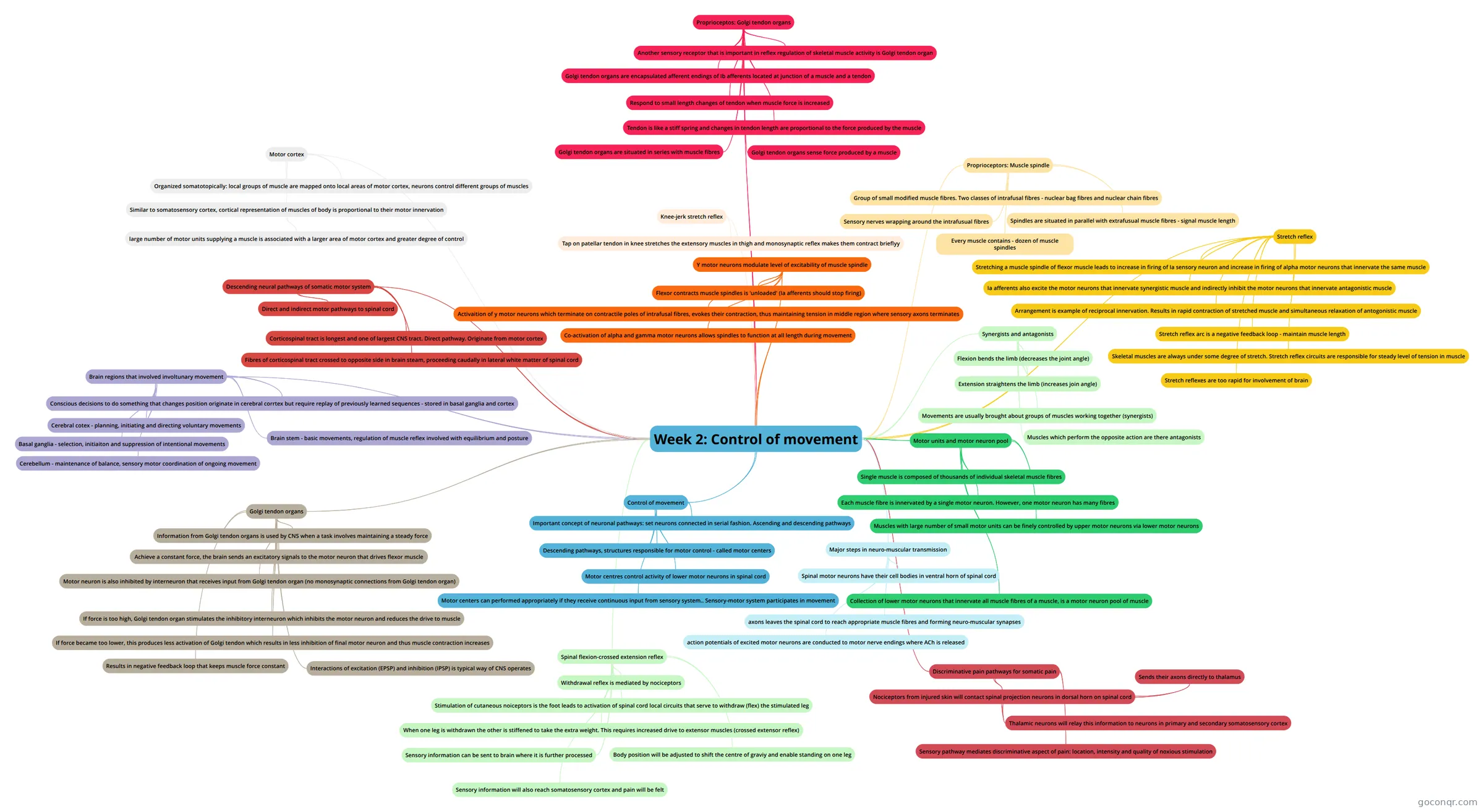
Mind map: Control of movement
Map content
Week 2: Control of movement
Control of movement
- Important concept of neuronal pathways: set neurons connected in serial fashion. Ascending and descending pathways
- Descending pathways, structures responsible for motor control - called motor centers
- Motor centres control activity of lower motor neurons in spinal cord
- Motor centers can performed appropriately if they receive continuous input from sensory system.. Sensory-motor system participates in movement
Major steps in neuro-muscular transmission
- Spinal motor neurons have their cell bodies in ventral horn of spinal cord
- axons leaves the spinal cord to reach appropriate muscle fibres and forming neuro-muscular synapses
- action potentials of excited motor neurons are conducted to motor nerve endings where ACh is released
Motor units and motor neuron pool
- Single muscle is composed of thousands of individual skeletal muscle fibres
- Each muscle fibre is innervated by a single motor neuron. However, one motor neuron has many fibres
- Muscles with large number of small motor units can be finely controlled by upper motor neurons via lower motor neurons
- Collection of lower motor neurons that innervate all muscle fibres of a muscle, is a motor neuron pool of muscle
Synergists and antagonists
- Flexion bends the limb (decreases the joint angle)
- Extension straightens the limb (increases join angle)
- Movements are usually brought about groups of muscles working together (synergists)
- Muscles which perform the opposite action are there antagonists
Proprioceptors: Muscle spindle
- Group of small modified muscle fibres. Two classes of intrafusal fibres - nuclear bag fibres and nuclear chain fibres
- Sensory nerves wrapping around the intrafusual fibres
- Every muscle contains - dozen of muscle spindles
- Spindles are situated in parallel with extrafusual muscle fibres - signal muscle length
Stretch reflex
- Stretching a muscle spindle of flexor muscle leads to increase in firing of Ia sensory neuron and increase in firing of alpha motor neurons that innervate the same muscle
- Ia afferents also excite the motor neurons that innervate synergistic muscle and indirectly inhibit the motor neurons that innervate antagonistic muscle
- Arrangement is example of reciprocal innervation. Results in rapid contraction of stretched muscle and simultaneous relaxation of antogonistic muscle
- Stretch reflex arc is a negative feedback loop - maintain muscle length
- Skeletal muscles are always under some degree of stretch. Stretch reflex circuits are responsible for steady level of tension in muscle
- Stretch reflexes are too rapid for involvement of brain
Y motor neurons modulate level of excitability of muscle spindle
- Flexor contracts muscle spindles is 'unloaded' (Ia afferents should stop firing)
- Activaition of y motor neurons which terminate on contractile poles of intrafusal fibres, evokes their contraction, thus maintaining tension in middle region where sensory axons terminates
- Co-activation of alpha and gamma motor neurons allows spindles to function at all length during movement
Knee-jerk stretch reflex
- Tap on patellar tendon in knee stretches the extensory muscles in thigh and monosynaptic reflex makes them contract brieflyy
Proprioceptos: Golgi tendon organs
- Another sensory receptor that is important in reflex regulation of skeletal muscle activity is Golgi tendon organ
- Golgi tendon organs are encapsulated afferent endings of Ib afferents located at junction of a muscle and a tendon
- Respond to small length changes of tendon when muscle force is increased
- Tendon is like a stiff spring and changes in tendon length are proportional to the force produced by the muscle
- Golgi tendon organs are situated in series with muscle fibres
- Golgi tendon organs sense force produced by a muscle
Golgi tendon organs
- Information from Golgi tendon organs is used by CNS when a task involves maintaining a steady force
- Achieve a constant force, the brain sends an excitatory signals to the motor neuron that drives flexor muscle
- Motor neuron is also inhibited by interneuron that receives input from Golgi tendon organ (no monosynaptic connections from Golgi tendon organ)
- If force is too high, Golgi tendon organ stimulates the inhibitory interneuron which inhibits the motor neuron and reduces the drive to muscle
- If force became too lower, this produces less activation of Golgi tendon which results in less inhibition of final motor neuron and thus muscle contraction increases
- Results in negative feedback loop that keeps muscle force constant
- Interactions of excitation (EPSP) and inhibition (IPSP) is typical way of CNS operates
Spinal flexion-crossed extension reflex
- Withdrawal reflex is mediated by nociceptors
- Stimulation of cutaneous noiceptors is the foot leads to activation of spinal cord local circuits that serve to withdraw (flex) the stimulated leg
- When one leg is withdrawn the other is stiffened to take the extra weight. This requires increased drive to extensor muscles (crossed extensor reflex)
- Sensory information can be sent to brain where it is further processed
- Body position will be adjusted to shift the centre of graviy and enable standing on one leg
- Sensory information will also reach somatosensory cortex and pain will be felt
Discriminative pain pathways for somatic pain
- Nociceptors from injured skin will contact spinal projection neurons in dorsal horn on spinal cord
- Sends their axons directly to thalamus
- Thalamic neurons will relay this information to neurons in primary and secondary somatosensory cortex
- Sensory pathway mediates discriminative aspect of pain: location, intensity and quality of noxious stimulation
- Nociceptors from injured skin will contact spinal projection neurons in dorsal horn on spinal cord
Motor cortex
- Organized somatotopically: local groups of muscle are mapped onto local areas of motor cortex, neurons control different groups of muscles
- Similar to somatosensory cortex, cortical representation of muscles of body is proportional to their motor innervation
- large number of motor units supplying a muscle is associated with a larger area of motor cortex and greater degree of control
Descending neural pathways of somatic motor system
- Direct and indirect motor pathways to spinal cord
- Corticospinal tract is longest and one of largest CNS tract. Direct pathway. Originate from motor cortex
- Fibres of corticospinal tract crossed to opposite side in brain steam, proceeding caudally in lateral white matter of spinal cord
Brain regions that involved involtunary movement
- Conscious decisions to do something that changes position originate in cerebral corrtex but require replay of previously learned sequences - stored in basal ganglia and cortex
- Cerebral cotex - planning, initiating and directing voluntary movements
- Basal ganglia - selection, initiaiton and suppression of intentional movements
- Cerebellum - maintenance of balance, sensory motor coordination of ongoing movement
- Brain stem - basic movements, regulation of muscle reflex involved with equilibrium and posture
Information
- Description
- Universität Physiology Mind Map on Week 2: Control of movement, created by Paras Chrysostomou on 13/03/2016.
- Created by
- Paras Chrysostomou
- Views
- 0
- Rating