
Mind map
Haemostasis (part 2: secondary haemostasis)
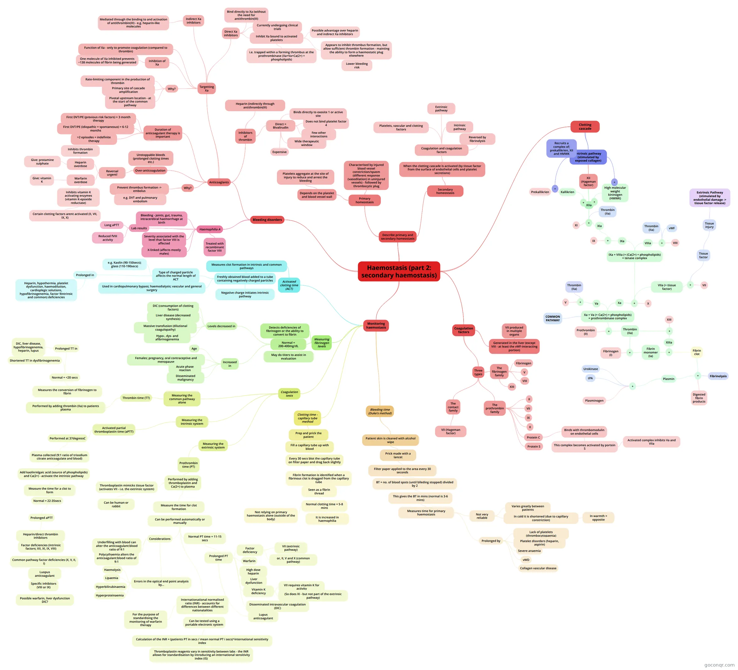
Mind map: secondary haemostasis
Map content
Haemostasis (part 2: secondary haemostasis)
Describe primary and secondary homeostasis
- Primary homeostasis
- Characterised by injured blood vessel constriction/spasm (different response (vasodilation) in uninjured vessels) - followed by thrombocytic plug
- Depends on the platelet and blood vessel wall
- Platelets aggregate at the site of injury to reduce and arrest the bleeding
- Secondary homeostasis
- When the clotting cascade is activated (by tissue factor from the surface of endothelial cells and platelet secretions)
- Coagulation and coagulation factors
- Platelets, vascular and clotting factors
- Extrinsic pathway
- Intrinsic pathway
- Reversed by fibrinolysis
- Coagulation and coagulation factors
- When the clotting cascade is activated (by tissue factor from the surface of endothelial cells and platelet secretions)
- Primary homeostasis
Clotting cascade
- Intrinsic pathway (stimulated by exposed collagen)
- XII (Hageman factor)
- XIIa
- +
- Kallikrien
- Prekallikrien
- Kallikrien
- +
- High molecular weight kininogen (HMWK)
- +
- XI
- XIa
- +
- IX
- IXa
- IXa + VIIIa (+ (Ca(2+) + phospholipids) = tenase complex
- +
- Xa
- Xa + Va (+ Ca(2+) + phospholipids) = prothrombinase complex
- Va
- +
- Thrombin (IIa)
- V
- +
- +
- Prothrombin (II)
- Thrombin (IIa)
- +
- Fibrinogen (I)
- Fibrin monomer (Ia)
- +
- Fibrin clot
- +
- Digested fibrin products
- Fibrinolysis
- Plasmin
- +
- Plasminogen
- Urokinase
- tPA
- +
- +
- Fibrin clot
- +
- +
- XIII
- XIIIa
- +
- COMMON PATHWAY
- Va
- Xa + Va (+ Ca(2+) + phospholipids) = prothrombinase complex
- X
- VIIa (+ tissue factor)
- +
- VII
- Tissue factor
- Tissue injury
- Extrinsic Pathway (stimulated by endothelial damage -> tissue factor release)
- Tissue injury
- +
- Xa
- VIIIa
- +
- VIII
- Thrombin (IIa)
- vWF
- +
- +
- IXa + VIIIa (+ (Ca(2+) + phospholipids) = tenase complex
- +
- Thrombin (IIa)
- +
- XIIa
- +
- +
- Recruits a complex of; prekallikrien, XII and HMWK
- XII (Hageman factor)
- Intrinsic pathway (stimulated by exposed collagen)
Coagulation factors
- Generated in the liver (except VIII - at least the vWF-interacting portion)
- VII produced in multiple organs
- Three types
- The fibrinogen family
- Fibrinogen
- V
- VIII
- XIII
- The prothrombin family
- II
- VII
- IX
- X
- Protein C
- Binds with thrombomodulin on endothelial cells
- This complex becomes activated by portein S
- Activated complex inhibits Va and VIIa
- This complex becomes activated by portein S
- Binds with thrombomodulin on endothelial cells
- Protein S
- The contact family
- VII (Hageman factor)
- The fibrinogen family
- Generated in the liver (except VIII - at least the vWF-interacting portion)
Monitoring haemostasis
- Bleeding time (Duke's method)
- Patient skin is cleaned with alcohol wipe
- Prick made with a lancet
- Filter paper applied to the area every 30 seconds
- BT = no. of blood spots (until blleding stopped) divided by 2
- This gives the BT in mins (normal is 3-6 mins)
- Measures time for primary haemostasis
- Not very reliable
- Varies greatly between patients
- In cold it is shortened (due to capillary constriction)
- In warmth = opposite
- Prolonged by
- Lack of platelets (thrombocytopaenia)
- Platelet disorders (heparin, aspirin)
- Severe anaemia
- vWD
- Collagen vascular disease
- Not very reliable
- Measures time for primary haemostasis
- This gives the BT in mins (normal is 3-6 mins)
- BT = no. of blood spots (until blleding stopped) divided by 2
- Filter paper applied to the area every 30 seconds
- Prick made with a lancet
- Patient skin is cleaned with alcohol wipe
- Clotting time - capillary tube method
- Prep and prick the patient
- Fill a capillary tube up with blood
- Every 30 secs blot the capillary tube on filter paper and drag back slightly
- Fibrin formation is identified when a fibrinous clot is dragged from the capillary tube
- Seen as a fibrin thread
- Normal clotting time = 5-8 mins
- It is increased in haemophilia
- Not relying on primary haemostasis alone (outside of the body)
- Normal clotting time = 5-8 mins
- Seen as a fibrin thread
- Fibrin formation is identified when a fibrinous clot is dragged from the capillary tube
- Every 30 secs blot the capillary tube on filter paper and drag back slightly
- Fill a capillary tube up with blood
- Prep and prick the patient
- Coagulation tests
- Measuring the intrinsic system
- Activated partial thromboplastin time (aPTT)
- Performed at 37degressC
- Plasma collected (9:1 ratio of trisodium citrate anticoagulate and blood)
- Add kaolin/elgaic acid (source of phospholipids) and Ca(2+) - activate the intrinsic pathway
- Measure the time for a clot to form
- Normal = 22-35secs
- Prolonged aPTT
- Heparin/direct thrombin inhibitors
- Factor deficiencies (intrinsic factors; XII, XI, IX, VIII)
- Common pathway factor deficiencies (X, V, II, I)
- Luopus anticoagulant
- Specific inhibitors (VIII or IX)
- Possible warfarin, liver dysfunction DIC?
- Specific inhibitors (VIII or IX)
- Luopus anticoagulant
- Common pathway factor deficiencies (X, V, II, I)
- Factor deficiencies (intrinsic factors; XII, XI, IX, VIII)
- Heparin/direct thrombin inhibitors
- Prolonged aPTT
- Normal = 22-35secs
- Measure the time for a clot to form
- Add kaolin/elgaic acid (source of phospholipids) and Ca(2+) - activate the intrinsic pathway
- Plasma collected (9:1 ratio of trisodium citrate anticoagulate and blood)
- Performed at 37degressC
- Activated partial thromboplastin time (aPTT)
- Measuring the extrinsic system
- Prothrombin time (PT)
- Performed by adding thromboplastin and Ca(2+) to plasma
- Thromboplastin mimicks tissue factor (activates VII - i.e. the extrinsic system)
- Can be human or rabbit
- Measure the time for clot formation
- Can be performed automatically or manually
- Normal PT time = 11-15 secs
- Prolonged PT time
- Factor deficiency
- VII (extrinsic pathway)
- or, II, V and X (common pathway)
- Warfarin
- High dose heparin
- Liver dysfunction
- Vitamin K deficiency
- VII requires vitamin K for activity
- (So does XI - but not part of the extrinsic pathway)
- Disseminated intravascular coagulation (DIC)
- Lupus anticoagulant
- Factor deficiency
- Prolonged PT time
- Considerations
- Underfilling with blood can alter the anticoagulant:blood ratio of 9:1
- Polycythaemia alters the anticoagulant:blood ratio of 9:1
- Errors in the optical end point analysis by...
- Lipaemia
- Haemolysis
- Hyperbilirubinaemia
- Hyperproteinaemia
- Internationational normalised ratio (INR) - accounts for differences between different nationalalities
- Can be tested using a portable electronic system
- Calculation of the INR = (patients PT in secs / mean normal PT i secs)^international sensitivity index
- Thromboplastin reagents vary in sensitivity between labs - the INR allows for standardisation by introducing an international sensitivity index (IS)
- Calculation of the INR = (patients PT in secs / mean normal PT i secs)^international sensitivity index
- For the purpose of standardising the monitoring of warfarin therapy
- Can be tested using a portable electronic system
- Normal PT time = 11-15 secs
- Can be performed automatically or manually
- Thromboplastin mimicks tissue factor (activates VII - i.e. the extrinsic system)
- Performed by adding thromboplastin and Ca(2+) to plasma
- Prothrombin time (PT)
- Measuring the common pathway alone
- Thrombin time (TT)
- Performed by adding thrombin (IIa) to patients plasma
- Measures the conversion of fibrinogen to fibrin
- Normal = <20 secs
- Shortened TT in dysfibrinogenemia
- Prolonged TT in
- DIC, liver disease, hypofibrinogenemia, heparin, lupus
- Normal = <20 secs
- Measures the conversion of fibrinogen to fibrin
- Performed by adding thrombin (IIa) to patients plasma
- Thrombin time (TT)
- Measuring the intrinsic system
- Measuring fibrinogen levels
- Detects deficiencies of fibrinogen or the ability to convert to fibrin
- Normal = 200-400mg/dL
- May do titers to assist in evaluation
- Levels decreased in
- DIC (consumption of clotting factors)
- Liver disease (decreased synthesis)
- Massive transfusion (dilutional coagulopathy)
- Hypo-, dys- and afibrinogenemia
- Increased in
- Age
- Females; pregnancy, oral contraceptive and menopause
- Acute phase reaction
- Disseminated malignancy
- Activated clotting time (ACT)
- Measures clot formation in intrinsic and common pathways
- Freshly obtained blood added to a tube containing negatively charged particles
- Negative charge initiates intrinsic pathway
- Type of charged particle affects the normal length of ACT
- e.g. Kaolin (90-150secs); glass (110-190secs)
- Prolonged in
- Heparin, hypothermia, platelet dysfunction, haemodilution, cardioplegic solutions, hypofibrinogenemia, factor 9intrinsic and common) deficiencies
- Used in cardiopulmonary bypass; haemodialysis; vascular and general surgery
- Freshly obtained blood added to a tube containing negatively charged particles
- Measures clot formation in intrinsic and common pathways
- Bleeding time (Duke's method)
Bleeding disorders
- Haemophilia A
- X-linked (affects mostly males)
- Severity associated with the level that factor VIII is affected
- Bleeding - joints, gut, trauma, intracerebral haemorrhage at birth
- Treated with recombinant factor VIII
- Lab results
- Long aPTT
- Reduced fVIII activity
- Anticoaglants
- Why?
- Prevent thrombus formation -> embolus
- e.g. DVT and pulmonary embolism
- Prevent thrombus formation -> embolus
- Over-anticoagulation
- Reversal urgent!
- Heparin overdose
- Give: protamine sulphate
- Inhibits thrombin formation
- Warfarin overdose
- Give: vitamin K
- Inhibits vitamin K activating enzymes (vitamin K-epoxide reductase)
- Certain clotting factors arent activated (II, VII, IX, X)
- Heparin overdose
- Unstoppable bleeds (prolonged clotting times etc.)
- Reversal urgent!
- Duration of anticoagulant therapy is important
- First DVT/PE (previous risk factors) = 3 month therapy
- First DVT/PE (idiopathic = spontaneous) = 6-12 months
- >2 episodes = indefinite therapy
- Targetting Xa
- Why?
- Pivotal upstream location - at the start of the common pathway
- Primary site of cascade amplification
- Rate-limiting component in the production of thrombin
- Inhibtion of Xa
- One molecule of Xa inhibited prevents ~138 molecules of fibrin being generated
- Function of Xa - only to promote coagulation (compared to thrombin)
- Indirect Xa inhibitors
- Mediated through the binding to and activation of antithrombin(III) - e.g. heparin-like molecules
- Direct Xa inhibitors
- Bind directly to Xa (without the need for antithrombin(III)
- Currently undergoing clinical trials
- Inhibit Xa bound to activated platelets
- i.e. trapped within a forming thrombus at the prothrombinase (Xa+Va+Ca(2+) + phospholipids)
- Possible advantage over heparin and indirect Xa inhibitors
- Appears to inhibit thrombus formation, but allow sufficient thrombin formation - mainting the ability to form a haemostatic plug elsewhere
- Lower bleeding risk
- Why?
- Inhibitors of thrombin
- Heparin (indirectly through antithrombin(III)
- Direct = Bivalirudin
- Binds directly to exosite 1 or active site
- Does not bind platelet factor 4
- Few other interactions
- Wide therapeutic window
- Expensive
- Why?
- Haemophilia A
Information
- Description
- Blood Science Mind Map on Haemostasis (part 2: secondary haemostasis), created by maisie_oj on 30/04/2013.
- Created by
- maisie_oj
- Views
- 138
- Tags
- Rating