
Mind map
Flavivirus
Map content
Flavivirus
Replication
- 1. Virus attachment to plasma membrane
- Adhesion
- 2. Entry to cytoplasm/nucleus
- Endocytosis (clathrin)
- Low pH/ fusion nucleocapsid release
- 3. Un-coating of virus particle
- Synthesis of polyprotein
- Golgi associated protein cleavage (host and virus proteases)
- Long protein formed, which is then cut up into smaller parts
- 4. Synthesis of mRNA and protein
- Genome replication via RNA intermediate
- 5. Replication of nucleic acid
- 6. Virus assembly
- Budding from ER (peplomers)
- 7. Exit of virus from cell
- Maturation and exocytosis, need not be cytopathic
- Louping ill virus replication
- Receptors: sialic acid on glycolipids /gangliosides, found on a wide variety of cells including nerves
- Hence can cause encephalitis
- Encephalitis is a sudden onset inflammation of the brain
- Hence can cause encephalitis
- Virus enters and replicates well in a variety of mammalian and avian cell types.
- Therefore disease not species restricted.
- Pig = intermediate host
- Double stranded RNA intermediates, potentially good interferon inducers.
- Virus does not usually shut of host cell protein synthesis or only does so very late in replication cycle.
- Virus does not lyse the host cell: exit by exocytosis.
- Variably cytopathic to mammalian or avian cells
- Non cytopathic in insect cells.
- Receptors: sialic acid on glycolipids /gangliosides, found on a wide variety of cells including nerves
- Bovine Viral Diarrhoea replication
- Receptor CD46 (complement control protein)
- Located on surface of most cells
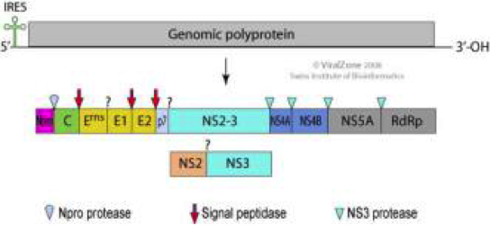
- Virus Polyprotein cleaved by Host protease and virus NSP2/3 protease
- Does not shut down host protein synthesis
- Blocks Type 1 interferon production
- Non lytic infection with exocytosis
- Most strains non cytopathic
- Persistent infection of cell lines in vitro, cattle in vivo
- Disease associated with conversion to cytopathic strain with NSP3 mutations
- Will start to destroy other cells inside the cow
- The N-terminal protease Npro catalyses its own separation (grey arrow)
- Npro also binds to Interferon regulatory factor-3 (IRF3) leading to proteasomal degradation thereby inhibiting the transcription of IFN.
- Mechanism inside cell blocking interferon production
- Npro also binds to Interferon regulatory factor-3 (IRF3) leading to proteasomal degradation thereby inhibiting the transcription of IFN.
- Structural glycoprotein protein Erns (rns = RNAse) is released from infected cells
- Intrinsic RNase activity - degrades extracellular viral ss-and dsRNA
- Prevents activation of interferon responses by TLR3 in the endosome of plasmacytoid dendritic cells.
- Suppress monocytes response to anything else – blocks interferon response and causes animal to be susceptible to other diseases/viruses
- Prevents activation of interferon responses by TLR3 in the endosome of plasmacytoid dendritic cells.
- Intrinsic RNase activity - degrades extracellular viral ss-and dsRNA
- Receptor CD46 (complement control protein)
- 1. Virus attachment to plasma membrane
Characteristics
- Genome
- Flaviviridae small +ve RNA virus
- Structure
- Spherical capsid (icosahedral symmetry)
- Surrounded by a tight lipid envelope which has indistinct peplomeres
- Double layer – has an envelope
- Spherical capsid (icosahedral symmetry)
- Genus: Flavivirus
- (Arboviruses – mosquitoes in the tropics and ticks in temperate areas)
- Genome
Louping ill
- Pathogenesis - infection from bite of carrier tick
- Replication at site of entry in leucocytes/macrophages DCs
- Spread to local lymph node where more cells are infected
- 1o Fever associated with interferon response and viraemia spread to other lymphoid organs
- Further replication and continuing prolonged viraemia (not a true persistent infection).
- Opportunity for infection of ticks (extended duration of feeding)
- In some animals prolonged viraemia allows spread to CNS and onset of neurological signs.
- 2o fever (ataxia, tremors, louping, paralysis, death)
- 1. Blood: viraemia and infected lymphoid cells Virus attached to RBCs
- 2. Infection of endothelium
- 3. Infection of neurones microglia astrocytes: Death of nerve cells Pass across endothelial cells + BBB → binds to gangliocytes (high levels in neurones)
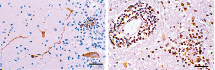
- Perivascular cuffing: infiltration of lymphocytes, monocytes and dendritic cells across inflamed endothelium
- Stained for macrophage /dendritic cells
- Virus encephalitis – leukocytes clumping around the edge of blood vessels
- Cerebellum: Purkinje cells infected with Louping Ill
- Stained for virus antigen
- Epidemiology
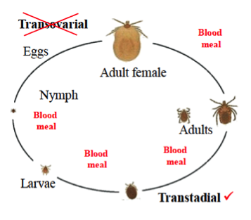
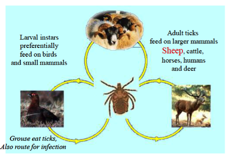
- Tick borne disease: Ixodes ricinus
- Reservoir hosts many birds and small mammals
- Red grouse, Voles, Hares etc. with sporadic epidemics in livestock and humans
- Moorland – can be quite pathogenic in grouse
- Can infect sheep, goats, cattle, deer, horses
- Louping ill virus in Ticks (non pathogenic)
- Virus first acquired by blood feeding on viraemic host
- Immunology
- More severe disease associated with Immunosuppression by Ehrlichia Phagocytophillia (cow infection)
- Rise in Antibody titre prior to 2o vireamia is protective
- Lambs are protected by maternal antibodies; most disease seen in sheep from 6 months -2yrs of age
- Born in spring – high tick burden, protected by maternal antibodies
- By next spring – turned back out on the moor – then develop disease
- Pathogenesis - infection from bite of carrier tick
Pestivirus: Bovine Viral Diarrhoea/mucosal disease
- Infection of pregnant cows
- (by non-cytopathic strain)
- Trans-placental infection of foetus
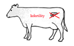
- 1st 30 days early embryonic death
- Cow mounts an immune response and clears the virus
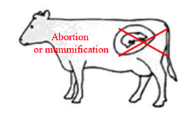
- 30-90 days embryonic/ foetal death
- Cow mounts an immune response and clears the virus
- Trans placental infection > 125 days
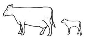
- Cow and calf mount immune response and calf reaches term
- Some Foetal defects may occur, retinal dysplasia, cerebellar hypoplasia, cerebral cavitation low birth weight, neonatal death.
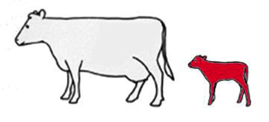
- 90-120 days gestation
- Infection during a critical period of calf’s immune system development
- Learning what is self and what is non self
- Cow mounts immune response and clears virus
- Calf survives, but is immuno-tolerant to virus
- Lifelong persistent infection with no antibodies or immune response
- Infection during a critical period of calf’s immune system development
- Mucosal disease
- Animal that have become persistently infected (tolerant) with a non cytopathic strain may go on to develop mucosal disease
- This is associated with the appearance of cytopathic variants of the virus in the blood of infected animals
- These arise from virus mutants of the original strain (deletions, duplications, rearrangements, re-combinations or insertion of cellular sequences)
- All lead to production of an 80kd. NS3 that mediates the cytopathic effect
- Virus acquires a tropism for the mucosal immune system e.g. peyers patch, lamina propria and MLN
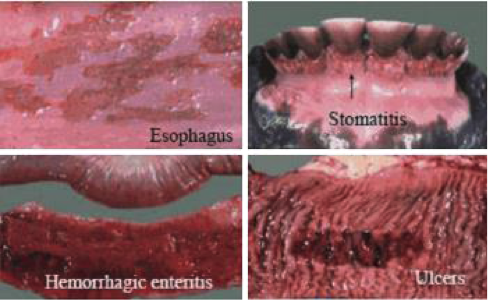
- Onset may be acute or gradual, pyrexia, anorexia, profuse watery diarrhoea, nasal discharge erosive stomatitis, dehydration emaciation and death
- Pathology
- Erosions in mouth, oesophagus, forestomachs
- Inflammation, hyperemia, intestines
- Necrosis of peyer’s patches and lymphoid tissues
- Animal that have become persistently infected (tolerant) with a non cytopathic strain may go on to develop mucosal disease
- Endemic herds
- High level of immunity (80% seropositive) virus infection of susceptible adults is rare
- Newborn calves are protected by maternal antibody
- Heifers are most likely to become infected when they join the main herd (i.e. during second pregnancy)
- Persistent infection of cows at around 1% of animals
- Common virus infection of cattle throughout the world
- Doesn’t often cause diarrhoea – rota and corona viruses are more common causes of diarrhoea
- Only a proportion of infected animals show any signs of disease
- Many different isolates mainly non cytopathic
- Can be divided into 2 serologically distinct biotypes.
- Biotype 1 – most common in UK, conversion to cytopathic form causes “typical” mucosal disease.
- Biotype 2 – less common in UK also non cytopathic
- But conversion to cytopathic form causes thrombocytopaenia and haemorrhagic disease
- Can be divided into 2 serologically distinct biotypes.
- Conversion to Cytopathic strains arise de novo by mutations to NSP3 (protease) and other virus genes
- Including point mutations, duplication of virus genome segments and incorporation of host RNA (many cases 80kD NSP3)
- One virus, many diseases
- BVD: acute infection of susceptible cattle.
- Infection via nasal route replication in epithelium, viraemia associated with T and B cells leukopenia (decrease in number of WBCs)
- Disease usually mild (non cytopathic virus): high morbidity, few clinical signs (if any), transient fever and recover, antibody response to virus.
- May be off colour for a bit, may have fever
- Immuno-suppressive: blocks interferons, leukopaenia, alters monocyte function, promotes T-helper 2 response (exacerbation of concurrent respiratory disease)
- Will worsen any other concurrent diseases
- Occasionally moderate/severe outbreaks
- Fever depression, diarrhoea, erosions in mouth some deaths.
- + immuno-suppression may exacerbate respiratory disease.
- Recovered animals seroconvert to BPV
- Infection of pregnant cows
Control
- Louping ill
- Inactivated cell culture vaccines given at 1 yr. annual boosters given to breeding flock
- Bird populations moving around and very high tick burdens can give rise to peaks in disease
- Tend to be well vaccinated
- Inactivated cell culture vaccines given at 1 yr. annual boosters given to breeding flock
- BVD Control Programme
- Drawbacks
- Testing and removal to generate BVD free herd
- Risk of heard breakdown
- Vaccine not all totally effective, may not protect from all strains of virus and are not certain to prevent foetal infection
- Some evidence that attenuated vaccines may contribute to disease onset in tolerant animals (BVDV can be spread by dirty needles)
- 1. Testing and removal of persistently infected animals
- Virus capture assay +ve/antibody –ve
- Difference between inactivated vaccine and non-cytotoxic disease? Probably not much
- 2. Vaccination of remaining animals
- 1st dose inactivated vaccine (no response to NSP3)
- 2nd booster dose (live attenuated vaccine)
- 3. Continue to monitor and remove of virus +ve animals
- 4. Vaccination of replacements @ 6 months
- 5. Maintain bio-security
- Drawbacks
- Louping ill
Diagnosis
- Louping ill
- Virus isolation, serology, (IgM), immunohistology
- IgM – response at the present – first immunoglobulin produced by antibodies
- IgG – may have been a historical infection, and is just antibody left over
- Virus isolation, serology, (IgM), immunohistology
- BVD
- Tissue culture (plus Immunofluoresence, non cytopathic).
- Detection of viral RNA by RTPCR. (ear punch test)
- Serology by ELISA
- Remember no antibody in tolerant carriers
- Milk antibody for herd test
- NS3 antigen commonly used highly conserved
- Not induced by inactivated vaccine.
- Antigen capture ELISA for persistent infection.
- Louping ill
Images and files in the map
Information
- Description
- BVSc3 AD2 (Viruses) Mind Map on Flavivirus, created by Jess Pope on 21/12/2016.
- Created by
- Jess Pope
- Views
- 0
- History
-
- Created by Jess Pope · Dec 21, 2016
- Copied by Jess Pope · Dec 21, 2016
- Copied by Jess Pope · Dec 21, 2016
- Copied by Jess Pope · Dec 21, 2016
- Copied by Jess Pope · Dec 21, 2016
- Copied by Jess Pope · Dec 21, 2016
- Copied by Jess Pope · Dec 21, 2016
- Rating