Description
|
|
Created by Jonathan Williams
over 8 years ago
|
|
Page 1
Summary of Morse et al. 2008 - SCI induced bone loss in growing rats
Method Overview SCI model - T10 severe contusion (BBB to confirm injury severity) Rats - 7-week old male Sprague-Dawley (200-225 grams) Controls - body weight (age) and gender matched (no surgery -not sham) Sacrificed on Day 10 post-SCI MicroCT bone imaging TRAP-staining for mature osteoclast identification dynamic histomorphometry for bone apposition rate determination. Here the contusion SCI model is used to determine its impact on bone density, microarchitecture and microenvironment at the distal femoral metaphyses.
intro/justification SCI causes osteoporosis leads to increased risk of low trauma fracture There is evidence that the bone loss is not due just to lack of mobility. CNS is a major regulator of bone metabolism (bone is highly innervated) This study suggests an important role of the neural system in regulating bone cell formation and influencing bone homeostasis. No studies before this on bone loss following contusion SCI.
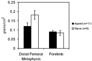
In-vivo BMD Assessment Femoral metaphysis chosen for analysis. (clincally relevant) Distal forelimb (radius/ulna) chosen as supralesional control in-vivo BMD assessment w/ PIXImus scanner Assessment areas - (16 x 13 pixels) @ distal femoral metaphysis and (11 x 13) @ distal radius/ulna. BMD reproducibility (RMS-coefficient of variation) -4% @ dfm and 5% @ dr/u.
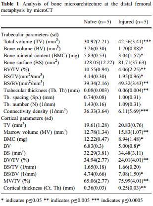
MicroCt microarchitecture analysis µCT scanner - µCT 40 by Scanco. Indices assessed: TV, BV, MV, BMC, BS, Th, BV/TV. Tb.Th, TB.N, Tb.Sp Indices reproducibility (precession of error) 0.06 - 2.16% for cortical and 0.59-5.24% for trabecular compartment.[33]
Results - BMD 34%* decrease in BMD in T10 group at dfm. (n = 11) compared to CTR (n = 9) @ 10 days post-injury. No significant difference in forelimb BMD between groups.
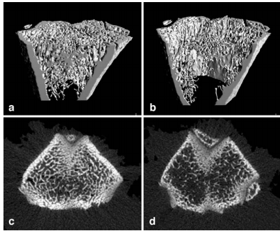
results - microarchitecture significant alterations due to SCI, as shown in Figure. 48% decrease in trabecular BMC, with an associated decrease in Tb.Th and Conn.D (n = 5) compared to control (n = 5). mean cortical bone content decreased by 35%, with a significant decrease in Ct.Th. BS.BC increased in both compartments indicating increased trabecular and cortical bone resoption.
Notes To obtain bone (apposition) formation rates need biological/chemical tests e.g. a form of histomorphometry. Also for bone mineralisation, osteoclast/blast activity etc.
Discussion Before study it was assumed rapid bone loss following acute SCI was due to enhanced bone resorption, with normal bone formation. Here report suppressed osteoblast activity for the first time in the development of osteoporosis.
Want to create your own Notes for free with GoConqr? Learn more.