
Mapa mental
Object Recognition
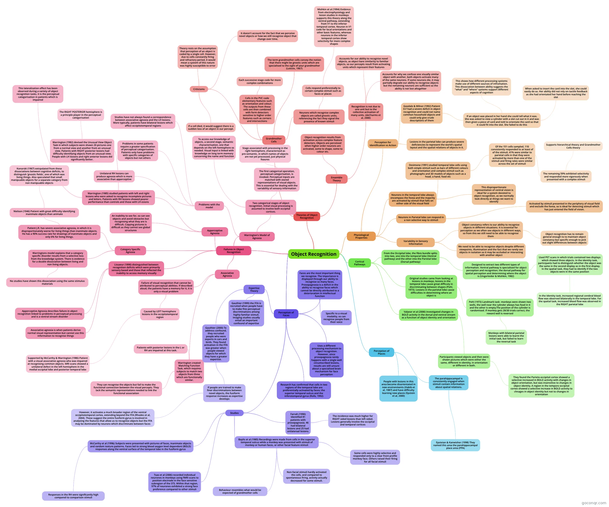
Mind map: Object recognition
Conteúdo do mapa
Object Recognition
Cortical Pathways
- From the Occipital lobe, the fibre bundle splits into two, one into the temporal lobe (Ventral pathway) and the other into the Parietal lobe (Dorsal pathway)
- Designed to extract two different types of information. Ventral pathway is specialised for object perception and recognition, the dorsal pathway for spatial perception and determining where the object is (Ungerleider & Mishkin, 1982)
- Used PET scans in which trials contained two displays which showed three objects. In the identity task, participants had to distinguish whether the object was the same in the second display as in the first display. In the spatial task, they had to identify if the two objects were in the same position
- In the Identity task, increased regional cerebral blood flow was observed bilaterally in the temporal lobe. For the spatial task, increased blood flow was observed in the RIGHT parietal lobe
- Used PET scans in which trials contained two displays which showed three objects. In the identity task, participants had to distinguish whether the object was the same in the second display as in the first display. In the spatial task, they had to identify if the two objects were in the same position
- Designed to extract two different types of information. Ventral pathway is specialised for object perception and recognition, the dorsal pathway for spatial perception and determining where the object is (Ungerleider & Mishkin, 1982)
- Original studies came from looking at lesions in monkeys, lesions in the temporal lobe cause great difficuty in discriminating between shapes (Pohl, 1973). Lesions in the parietal lobe cause difficulties in determining where an object is
- Pohl (1973) Landmark task- monkeys were shown two wells, the well near the cylinder always has food in it and the other is empty.The position of the cylinder is randomised. If monkey gets 28/30 trials correct, the reward well is reversed
- Monkeys with bilateral parietal lesions were able to learnt the initial task, but failed to learn the reversal task
- Pohl (1973) Landmark task- monkeys were shown two wells, the well near the cylinder always has food in it and the other is empty.The position of the cylinder is randomised. If monkey gets 28/30 trials correct, the reward well is reversed
- Valyear et al (2006) investigated changes in BOLD activity in the dorsal and ventral stream as a function of object identity and orientation
- Participants viewed objects and then were shown pictures which were either the same, different in identity, in orientation or different in both.
- They found the Parieto-occipital cortex showed a selective increased in BOLD activity with changes in object orientation, but was insensitive to changes in object identity. A region in the temporo occipital cortex showed a selective increase in BOLD activity to chnages in object identity but not to changes in orientation
- Participants viewed objects and then were shown pictures which were either the same, different in identity, in orientation or different in both.
- From the Occipital lobe, the fibre bundle splits into two, one into the temporal lobe (Ventral pathway) and the other into the Parietal lobe (Dorsal pathway)
Physiological Properties
- Variability in Sensory Information
- Object constancy refers to our ability to recognise objects in different situations. It is essential for perception as we often see objects in different ways, so from this we still need to be able to recognise them
- Object recognition has to remain general enough to to maintain object constancy but specific enough to pick out slight differences between objects
- We need to be able to recognise objects despite different viewpoints, illumination and the fact that we rarely see objects in isolation so it may be occluded or interacting with another object
- Object constancy refers to our ability to recognise objects in different situations. It is essential for perception as we often see objects in different ways, so from this we still need to be able to recognise them
- Perception for Identification vs Action
- Lesions in the parietal lobe can produce severe deficiencies to represent the world's spatial layout and the spatial relations of objects in it.
- Goodale & Milner (1992) Patient D.F had a severe deficit in object recognition and could not name common household objects and could only give crude descriptions of them
- If an object was placed in her hand she could tell what it was. She was asked to view a cylinder with a slot cut out in it and was then given a piece of card and told to orientate the card so that it could fit into the slot. She failed to do this
- When asked to insert the card into the slot, she could easily do so. Her ability did not rely on tactile feedback as she had orientated her hand before reaching the slot
- This shows how different processing systems make use of different sources of infromation. This dissociation between ability suggests the "what" and "where" systems support different aspects of cognition
- If an object was placed in her hand she could tell what it was. She was asked to view a cylinder with a slot cut out in it and was then given a piece of card and told to orientate the card so that it could fit into the slot. She failed to do this
- Neurons in Parietal lobe can respond in a non selective way to stimuli
- Activated by stimuli presented in the periphery of visual field and exclude the fovea, so is ideal for detecting stimuli which has just entered the field of vision.
- Neurons in the temporal lobe always encompass the fovea and the majority are activated by stimuli that falls on either side of the visual field
- This disproportionate representation of central vision is ideal for a system devoted to object recognition, as we normally look directly at things we want to identify
- Desimone (1991) studied temporal lobe cells using both simple stimuli such as bars of different colours and orientation and complex stimuli such as photographs and 3D models of objects such as a head, a hand, food etc
- Of the 151 cells sampled, 110 consistently responded to at least of of the stimuli. 41% were similar to parietal cells in that they were activated by more than one of the stimuli and firing rates were similar across the set of stimuli
- The remaining 59% exhibited selectivity and responded more vigorously when presented with a complex stimuli
- Supports hierarchical theory and Grandmother Cells theory
- The remaining 59% exhibited selectivity and responded more vigorously when presented with a complex stimuli
- Of the 151 cells sampled, 110 consistently responded to at least of of the stimuli. 41% were similar to parietal cells in that they were activated by more than one of the stimuli and firing rates were similar across the set of stimuli
- Variability in Sensory Information
Theories of Object Recognition
- Grandmother Cells
- Cells in the PVC code elementary features such as orientation and colour. The outputs from these cells are then combined with form detectors sensitive to higher order features such as corners and intersections
- Each successive stage code for more complex combinations
- Neurons which recognise complex objects are called gnostic units, referencing the fact they signal the presence of known stimuli
- Cells respond preferentially to certain complex stimuli such as faces or places
- Mishkin et al (1994) Evidence from electrophysiology and lesion studies in monkeys supports this theory along the ventral pathway, extending from V1 to the inferior temporal cortex. Neuron in V1 code for local orientations and other basic features, whereas neurons in the inferior temporal cortex show selectivity for more complex shapes
- Cells respond preferentially to certain complex stimuli such as faces or places
- The term grandmother cells convey the notion that there might be gnostic units which are specialised to the sight of your grandmother (Lettvin, 1967)
- Criticisms
- Theory rests on the assumption that perception of an object is coded by a single cell. However, due to cells constantly firing and refractory period, it would mean a system of this nature was highly susceptible to error
- If a cell died, it would suggest there is a sudden loss of an object in our percept.
- It doesn't account for the fact that we perceive novel objects or how we still recognise object that change over time.
- Cells in the PVC code elementary features such as orientation and colour. The outputs from these cells are then combined with form detectors sensitive to higher order features such as corners and intersections
- Ensemble Theory
- Object recognition results from activation across complex feature detectors. Objects are perceived when higher order neurons are activated, some to shape, some to colour etc.
- Recognition is not due to one unit but to the collective activation of many units. (deCharms et al, 2000)
- Accounts for why we confuse one visually similar object with another, both objects activate many of the same neurons. If some neurons die, it may partially degrade our ability to recognise objects but the remaining neurons are sufficient so the ability is not lost altogehter
- Accounts for our ability to recognise novel objects, as object bare similarity to familiar objects, so our percepts result from activating units which represent their features
- Grandmother Cells
Failures in Object Recognition
- Apperceptive Agnosia
- An inability to see for, so can see objects and avoid obstacles but recognising what they are is difficult. Copying pictures is difficult as they cannot see global structures
- Warrington (1985) studied patients with left and right lesions who were asked to recognise incomplete pictures and letters. Patients with RH lesions showed poorer performance than controls and those with LH esions
- This lateralization effect has been observed during a variety of object recognition tasks, it is the perceptual categorisation in patients which is impaired
- Warrington (1982) devised the Unusual View Object Task in which subjects were shown 20 pictures one from a normal view and another from an unusual view. Patients with RIGHT posterior lesions had difficulty identifying objects from an unusual view. People with LH lesions and right anterior lesions did significantly better.
- The RIGHT POSTERIOR hemisphere is a principle player in the perceptual categorisation
- Associative Agnosia
- Failure of visual recognition that cannot be attributed to perceptual abilities. If described aloud, the patients have a memory for it, it is only a visual problem
- Caused by LEFT hemisphere lesions in the occipitotemporal region
- Supported by McCarthy & Warrington (1986) Patient with a visual associative agnosia who was impaired at recognising common objects. MRI scans showed a unilateral defect in the left hemisphere in the medial occipital lobe and posterior temporal lobe
- Caused by LEFT hemisphere lesions in the occipitotemporal region
- Warrington created Matching Function Task, which requires subjects to match two objects from three which are functionally similar.
- Patients with posterior lesions in the L or RH are impaired at this task.
- They can recognise the objects but fail to make the functional connection between the visual percepts. They lack the semantic representations needed to link the functional association
- Failure of visual recognition that cannot be attributed to perceptual abilities. If described aloud, the patients have a memory for it, it is only a visual problem
- Category Specific Agnosia
- Patient J.R. has severe associative agnosia, in which it is disproportionately worse for living things than inanimate objects. He has a 90% success rate for drawings of inanimate objects and only 6% for living things.
- Nielson (1946) Patient with great difficulty identifying inanimate objects than animate
- Konorski (1967) extrpolated from these dissociations between cognitive deficits, to distinguish 'gnostic fields', one of which was living things. Also speculated that small manipuable objects for a separate category from non manipuable objects
- Nielson (1946) Patient with great difficulty identifying inanimate objects than animate
- Warringtons model explains that a category specific disorder results from a selective loss from the knowledge system. There is evidence for a double dissociation between living and non living objects.
- No studies have shown this dissociation using the same stimulus materials
- Patient J.R. has severe associative agnosia, in which it is disproportionately worse for living things than inanimate objects. He has a 90% success rate for drawings of inanimate objects and only 6% for living things.
- Warrington's Model of Agnosia
- Two categorical stages of object recognition. Initial visual processing is assumed to involve both occipital cortices.
- The first categorical operation, perceptual categorisation, is invoked. Perceptual inputs are matched with stored representations of visual objects. This is essential for dealing with the variability of sensory information
- Stage associated with processing in the right hemisphere, characterised as pre-semantic, in which names of objects are not yet processed, just physical feaures
- To access our knowledge of objects, a second stage, semantic characterisation, one that depends on the left hemisphere os needed. Visual input is linked with knowledge on long term memory concerning the name and function
- The first categorical operation, perceptual categorisation, is invoked. Perceptual inputs are matched with stored representations of visual objects. This is essential for dealing with the variability of sensory information
- Problems with the model
- Studies have not always found a correspondence between associative agnosia and the LH lesions. More typically, patients have bilateral lesions which affect occipitotemporal regions
- Unilateral RH lesions can produce agnosia which is more associative than apperceptive
- Problems in some patients require a greater specification of perceptual categorisation. Patients only have problems with specific categories of objects but not others
- Two categorical stages of object recognition. Initial visual processing is assumed to involve both occipital cortices.
- Lissaeur (1890) distinguished between recognition deficits which were primarily sensory based and those that reflected the inability to access memory visually
- Apperceptive Agnosia describes failure in object recognition linked to problems in perceptual processing and is a ventral stream disorder
- Associative agnosia is when patients derive normal visual representation but cannot use this information to recognise things
- Apperceptive Agnosia
Perception of Faces
- Expertise Theory
- Gauthier (1999) the FFA is recruited when people have to make fine perceptual discriminations among highly familiar stimuli. Imaging studies usually entail an important confound of expertise
- Gauthier (2000) To address confound, they recruited people who were experts in cars and birds. They found activation in the FFA was greater when people viewed objects for which they have a greater expertise.
- If people are trained to make finer discriminations between novel objects, the fusiform response increases as expertise develops
- However, it activates a much broader region of the ventral occipitotemporal cortex, extending beyond the FFA (Rhodes et al, 2004). These suggest the entire fusiform gyrus is involved in analysing the features that allow us to recognise objects but the FFA may be dominated by neurons which discriminate between faces
- If people are trained to make finer discriminations between novel objects, the fusiform response increases as expertise develops
- Faces are the most important thing we recognise. The importance is displayed through our ability to recognise so many faces. Prosopagnosia is a deficit in the ability to recognise faces which cannot be directly attributed to a deterioration in intellectual function
- Specific to a visual modality, so can recognise people from their voice
- Uses a different processing mechanism to object recognition. However, since prosopagnosia rarely happens with a single well circumscribed lesions, results are still unsure about a specialised brain mechanism for face perception
- Studies
- Farrah (1990) Identified 81 patients with prosopagnosia. 46 had bilateral lesions and 25 had unilateraal lesions.
- The incidence was much higher for RIGHT sided lesions than left sided. Lesions generally involve the occipital and temporal cortices
- Baylis et al (1985) Recordings were made from cells in the superior temporal sulcus while a monkey was presented with stimuli of monkey or human faces, or other facial feature stimuli
- Some cells were highly selective and responded only to a clear front profile monkey face. Others raised their firing for all facial stimuli
- Non-facial stimuli hardly activated the cells, and compared to spontaneous firing, activity actually decreased for some stimuli.
- Behaviour resembles what would be expected of grandmother cells
- Tsao et al (2006) recorded individual neurones in monkeys using fMRI scans to position electrode in the face sensitive subregion of the STS. Within that region, 97% of neurones exhibited a strong face preference compared to other stimuli
- McCarthy et al (199&) Subjects were presented with pictures of faces, inanimate objects and random texture patterns. Faces led to strong blood oxygen level dependent (BOLD) responses along the ventral surface of the temporal lobe in the fusiform gyrus
- Responses in the RH were significantly high compared to comparison stimuli
- Farrah (1990) Identified 81 patients with prosopagnosia. 46 had bilateral lesions and 25 had unilateraal lesions.
- Research has confirmed that cells in two regions of the temporal lobe are preferentially activated by faces; the superior temporal sulcus and the inferotemporal gyrus (Rolls, 1992)
- Expertise Theory
Perception of Places
- The parahippocampal is consistently engaged when stimuli contain information about spatial relations.
- Epsteian & Kanwisher (1998) They named this area the parahippocampal place area (PPA)
- People with lesions in this area become disorientated in new environments (Habib et al, 1987) and have difficulty learning new places (Epstein et al, 2000)
- The parahippocampal is consistently engaged when stimuli contain information about spatial relations.
Informações
- Descrição
- Biological Mapa Mental sobre Object Recognition, criado por eloisefeeney27 em 05-05-2014.
- Criado por
- eloisefeeney27
- Visualizações
- 37
- Avaliação
Mais sobre Object and Face Recognition