
Mapa mental
Microscopes
Conteúdo do mapa
Microscopes
Compound Light microscope
- Lower resolution - 0.2 um
- Maximum magnification is 1500x
- Monocular one eyepiece
- Binocular two eyepiece lens
- What limits the microscope is the wavelength of light
- Image created on retina
- Preparing a specimen
- Always put on a glass slide
- Light must be able to travel through the specimen so it needs to be very thin
- Water can be used for temporary specimens - to last longer you have to use something other than water
- A method of making something thin enough is squashing it
- Another way is cutting it
- Making a section slide
- Fixation
- Put in chemical called fixative which preserves it
- Dehydration
- Transfer specimen to dehydration chemical such as alcohol
- Clearing
- Put it in another chemical to drive out the alcohol
- Embedding
- Put the specimen in molten wax and let it solidify so you can cut it
- Sectioning
- Cut the specimen using a microtome
- Staining
- Coloured chemical which will bind to specific chemicals in the cell
- Mounting
- Put it in the slide, put clear liquid and coverslip
- Fat Dudes Can Eat Subway So Much
- Fixation
- Artifacts are parts seen down the microscope which were not there before
Magnification = image size / object size
Confocal laser scanning microscope
- Called optical sectioning
- Uses laser to focus at different levels at tissue
- Uses fluorescent stains
- Image is 3D
- Clearer than light microscope
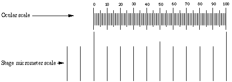
Calibrating light microscope
- There is a different eyepiece with a scale that has 100 small measurements of epu
- Stage micrometer - 1cm - 10mm - 10 000 um
- To calibrate you need to find out how many small measurements of the stage micrometer are in 100 epu
- Then work out the um it would equal
- Then divide the um by 100 to get the size of 1 epu
Electron microscope
- Transmission electron microscope (TEM)
- Electrons pass through specimen
- Works like X-ray
- Electrons have bad penetration
- Even air molecules will block electrons so a vacuum is needed
- An electromagnetic lens is used to focus the beam of electrons
- Specimen has to go through double door air lock to prevent a loss of vacuum
- Image is called electron micrograph
- Electron hits electron sensitive fluorescent sheet - under is another sheet that will create a permanent image
- Has to be bolted to concrete base to prevent vibrations - also has to be on bottom floor of building
- Magnification is from 500 000x to 2 000 000x
- Resolution is 0.2-05nm
- Specimen has to be dead
- Preparing a section
- First 3 steps same as light - Fixation Dehydration and Clearing - Fat Dudes Can
- Embedding
- Resin instead
- Sectioning
- Glass or diamond blade because they have to be nm thick
- Staining
- Heavy metal stains to block electrons - mercury lead silver
- Mounting
- Copper grid
- More artifacts
- Uses an electron beam made by electron gun instead of light
- Scanning electron microscope (SEM)
- Creates 3D image of surface of specimen
- Not using sections
- Beam of electrons scans the specimen
- Needs to be coated in gold to make it more reflective
- The specimen is surrounded in electron detectors which create an image on a computer
- The specimen has to be frozen in liquid nitrogen and is carefully broken and will fracture through the weakest point in the cell - allowing you to look inside
- Transmission electron microscope (TEM)
Imagens e arquivos do mapa
Informações
- Descrição
- Mapa Mental sobre Microscopes, criado por Fin Carson em 12-10-2017.
- Criado por
- Fin Carson
- Visualizações
- 7
- Avaliação