
Mapa mental
Immunology
Conteúdo do mapa
Immunology
Inflammation
- Cellular response to injury
- 4 main reactions
- Vascular changes
- Vasodilation
- Decreases blood flow
- Blood normally flows centrally within a vessel
- Results white blood cell margination
- As cells are able to move peripherally
- Especially larger WBCs
- As cells are able to move peripherally
- Occurs first in arterioles
- Then in capillary beds
- May be mediated by histamine and nitric oxide
- Results in increased HEAT
- HEAT =
- CALOR
- HEAT =
- Results in increased REDNESS/ERYTHMEA
- REDNESS/ERYTHMEA =
- RUBOR
- REDNESS/ERYTHMEA =
- Decreases blood flow
- Vascular permeability changes
- Leaky blood vessels
- Causes loss of proteins
- Occurs due to:
- Endothelical contraction
- Results in histamine, bradykinin, substance P and leukotrienes entering surrounding tissues
- Direct injury
- For ecample:
- Toxins
- Burns
- Cuts
- May cause damage DIRECTLY to blood vessel itself
- For ecample:
- White blood cells
- May destroy "self" cells as well as foreign cells
- Damage WEAKENS vessel
- May destroy "self" cells as well as foreign cells
- New vessel formation
- VEGF makes new vessels
- This is brought into the cell via transcytosis.
- TRANSCYTOSIS: Process by which various macromolecules are transported across the interior of a cell Vesicles are employed to take the macromolecules on one side of the cell draw them across the cell and then eject them on the other side. Most commonly observed in cells of an epithelium , the process is also present elsewhere e.g. neuron, osteoclasts and intestinal cells
- Relatively fragile structures
- Thus increase leakiness
- VEGF makes new vessels
- Endothelical contraction
- Change in osmotic pressure also happens
- Water flow out of vessel after proteins
- Swelling occurs
- SWELLING =
- TUMOUR
- SWELLING =
- Leaky blood vessels
- Vasodilation
- Cellular changes
- Stasis
- Diminuation of blood flow
- Occurs in a blood vessel
- Results in white blood cell margination
- Adhesion occurs after white blood cell margination
- White cells drift towards the edge of the blood vessel
- Adhesion
- Blood vessels express various receptors on the luminal surface that have a matching protein/ligand on the WBCs surface
- Ligands on WBCs attach to selectins and or integrins on endothelial cells
- Selectins
- Are a family of CAMS
- Cell Adhesion Molecules
- VCAMS
- Vascular Cell Adhesion Molecules
- ICAMS
- Intercellular adhesion molecule
- Endothelial expression of ICAM and VCAM increased by TNF and IL-1
- TNF
- Tumour Necrosis Factor
- Tyoe of CYTOKINE
- Type of CYTOKINE
- Tumour Necrosis Factor
- IL-1
- Interleukin 1
- TNF
- PROTEOGYCANS increase affinits of ICAMs and VCAMs for integrins
- VCAMS
- Cell Adhesion Molecules
- Expression increased by histamine and thrombin
- Are a family of CAMS
- Integrins
- Transmembrane receptors
- Fascillitate cell extracellular matrix adhesion
- Over 30 types
- Selectins
- Ligands on WBCs attach to selectins and or integrins on endothelial cells
- Rolling Adhesion
- Leukocyte binding to selectin/integrin molecules
- Causes Leukocytes to slow down
- It beings "rolling" along inner surface of blood vessel wall
- Transitory bonds between selectin and integrins and their ligands
- Easily formed and broken
- Causes Leukocytes to slow down
- Leukocyte binding to selectin/integrin molecules
- Blood vessels express various receptors on the luminal surface that have a matching protein/ligand on the WBCs surface
- Migration
- Accumilation and shesion of leukocytes to the epithelical cells ofblood vessel walls at the site of injury in the early stages of inflammation Occurs as a result of dilation of capillaries and slowing of the blood flow in blood vessels
- WBCs migrate out of the vessel into affected tissues
- Occurs due to openings
- WBCs follow a bacteria gradient
- Chemotaxis
- Somatic cells direct their movements according to certain chemicals in their environment
- Cells follow a chemical gradient made of bacterial components and move along this
- Chemotaxis
- Pavementing
- Term used to describe the margination of leukocytes on the endothelium neat a site of damage
- Stasis
- Chemical Mediators
- Morphologic Patterns
- Vascular changes
- 4 main reactions
- Necrosis
- Results in inflammation
- Site of injury
- Chemokines
- Bind to PROTEOGLYCANS on endothelial cells
- PROTEOGLYCANS increase the affinity of VCAMs and ICAMs for integrins
- Bind to PROTEOGLYCANS on endothelial cells
- Chemokines
- Cell activated by antigen
- Leukotriene
- A lipid, C20 H30 O3 produced by white blood cells in an immune response to antigens that contributes to allergic asthma and inflammatory reactions
- Cytokines
- Protein molecules, released by cells when activated by an antigen.
- They are involved in cell-to-cell conmmunications, acting as enhancing mediators for immune responses through interaction with specific cell-surface receptors in leukocytes
- Kinds include:
- Interleukin
- Produced by leukocytes
- Lymphokine
- Interferon
- Tumor Necrosis factor
- Interleukin
- Leukotriene
- Phagocytosis
- 3 phases
- Recognition and attachment
- Engulfment
- Pseudopods surround foreign cell and form a vesicle around it
- Vesicle known as a PHAGOSOME
- A membrane bound vesicle that contains the ingested matter of a phagocytic cell.
- This then fuses with a lysosome
- To from a PHAGOLYSOSOME
- Vesicle known as a PHAGOSOME
- Pseudopods surround foreign cell and form a vesicle around it
- Killing and degradation
- REACTIVE OXYGEN SERIES
- NADPH oxidase
- Enzyme
- Causes oxygen to gain an electron from NADPH
- Oxygen becomes
- SUPEROXIDE
- Oxygen becomes
- NADPH oxidase
- REACTIVE NITROGEN SERIES
- Nitric oxide synthase
- Enzyme
- Combines NO with SUPEROXIDE
- Produces ONOO
- Nitric oxide synthase
- REACTIVE OXYGEN SERIES
- Carried out by various molecules
- Neutrophils
- Group of leukocyts that have granules that only stain with neutral dyes.
- Inflammatory cell that characterises acute inflammation
- CHARACTERISISES acute inflammation
- Polymorphs
- Granuloytes
- Only survive for a couple of hours outside the safety of a blood vessel
- Macrophages
- A large phagocytic cell present in connective tissue Slower to arrive to injury site than neutropils
- Mannose receptors
- Mediates the endocytosis of glycoproteins by macrophages
- Binds to high-mannose structures
- Examples include potentially pathogenic viruses, bacteria and fungi
- Neutralises the for phagocytic engulfment
- Examples include potentially pathogenic viruses, bacteria and fungi
- High mannose structures on found in human cells
- Mannose residures
- Terminal residues contained by bacterial surface glycoproteins and glycolipids
- Phagocytes
- Some can be professional presenting cells
- Contain Pattern Recognition Receptors
- Example is a SCAVENGER RECEPTOR
- A group of receptors that recognise modified low density lipoprotein (LDL) by oxidation or acetylation
- It is though that they particiate in the removal of foreign substances and waste materials in the living body by extensive ligand specificity and a variety of receptor molecules
- Mainly found on professional phagocytes
- Similar mechanism to used by phagocytes to recognise foreign cells is employed to recognise LDL.
- There are 3 classes
- Example is a SCAVENGER RECEPTOR
- Contain Pattern Recognition Receptors
- Is an example of a polymorph
- A polymorph is any group of white blood cells that have lobed nuclei and granular cytoplasm and function and phagocytes. They include neutrophils, basophils and eosinophils
- AKA "POLYMORPHONUCLEAR LEUKOCYTE
- Most are also granulocytes
- Any group of white blood cells having granules in the cytoplasm e.g. neutrophils, basophils and eosinophils
- Some can be professional presenting cells
- Neutrophils
- 3 phases
- 4 clinical characteristics of inflamaation
- Swelling
- Tumour
- Caused by increased perfusion of tissue, slow blood flow and increased permeability of vessels
- Heat
- Calor
- Redness
- Rubor
- Pain
- Dolar
- 5th feature of inlammation
- Loss of function
- Swelling
- VERY GOOD AT DAMAGING TISSUES
- Thus is mediated
- 2 common characteristics of mediators are:
- Short lived (Short-half life)
- Only produced as long as the stimulus persists
- 2 common characteristics of mediators are:
- Thus is mediated
- MYOCARDIAL INFARCTION
- Infarction
- Death of tissue after loss of oxygen
- Hypoxia
- Hypoxic injury that is pathological results in NECROSIS
- Causes cell injury and acute inflammation
- No OXYGEN =
- No ATP
- Causes sodium and potassium pump to fail
- Increased levels of extracellular Potassium causes swelling
- Calcium pump FAILS
- Increased levels of calcium stimulates
- ATPase (makes things worse)
- Phospholipase (membrane damage)
- Proteases (membrane and cytoskeleton damage)
- Endonuclease (DNA damage and breakdown)
- Mitochondrial permeability (release pro death factors)
- Increased levels of calcium stimulates
- Causes sodium and potassium pump to fail
- No ATP
- No OXYGEN =
- Short time window for drug delivery
- 20 minutes
- After 30 minutes the use of drugs is less effective (point less) because cells are going to die anyway.
- 20 minutes
- Cell death
- 2 main causes
- Apoptosis
- Necrosis
- 3 types
- Caseous
- Liquefactive
- Coagulative
- Cell death with some structure of cells left as "ghost outline" before complete phagocytosis of materials.
- Ghost outline of cells
- 3 types
- 2 main causes
- 8 terms vascular changes may occur in a blood vessel
- Vasodilation
- Slowing of blood flow
- White blood cell margination
- Rolling
- Pavementing
- Diapedesis
- The passage of the corpuscular elements of the blood from the blood vessel to the surrounding tissues without rupture of the walls of the blood vessels.
- Chemtaxis
- Phagocytosis
- Risk of cardiac rupture
- Greatest risk 3 to 7 days after MI
- At this point the wall of the heat is at it's weakest. Necrosis and neutrophils may be all that is holding it together if the full thickness of the wall is affected
- Greatest risk 3 to 7 days after MI
- After acute inflammation
- 4 things can happen
- Resolution
- Occurs if there is no suppuration or persistence of injury occurs in the heart when cells begin to die
- Restitution
- AKA Repair, organisation and fibrosis
- Occurs if there is no suppuration or persistence of injury occurs in the heart when cells begin to die
- Gradual process where PROGRESSIVE scarring occurs
- Scarring in the heart is an issue
- The heart muscle can't pump as well as the scarred muscle has loss of function (won't contract). This results in a weak heart and the picture of heart failure. The problems caused depend on the amount of damage.
- Loss of function
- Heart failure
- Nerve damage
- The nerve bundles in the area of the heart that is damaged may also be damaged.
- Pacemaker may consequently not be heard properly
- Suppuration
- Chronic inflammation
- Resolution
- 4 things can happen
- Macrophages
- As time fades away (e.g. 10 days after injury was inflicted), neutrophils fade away from injury site and are replaced by macrophages
- Presence of these in autopsy
- Imparts yellow appearance
- An MI that occurred more than 6 weeks prior to an autopsy cannot be dated
- Down a microscope all that would be seen is scarring as collagen fibre would have been laid down by fibroblasts.
- Replaced by fibroblasts at injury site
- These lay gradually down collagen
- This process occurs progressively after 2 weeks and is complete at 6 weeks.
- These lay gradually down collagen
- Infarction
- Cellular response to injury
AFTER ACUTE INFLAMMATION
- 4 stages
- These stages need not be mutually exclusive, they can occur simultaneously. Suppuration can progress to fibrosis as can chronic inflammation
- Resolution
- Complete restoration of the tissue to the normal after removal of inflammatory components.
- Type depends upon:
- 3 factors
- SITE of injury
- Affects resolution because different organs have different capacity for repair. Different organs have different vascular supplies.
- TYPE of injury
- Affects resolution because the severity of the injury/ pathogenicity of organism will determine the type of response required.
- Duration of injury
- SITE of injury
- 3 factors
- For this to occur there MUST be:
- MINIMAL cell death
- The tissue itself must have the capacity to repair
- Good vascular supply present for the delivery of WBCs and rapid removal of injurious agents
- Injurious agent easily removed
- Requires a "scaffold" to work around
- Hence necessity of minimal cell damage
- Abrasion
- Describe injury with the basement membrane still attached
- These heal RAPIDLY with COMPLETE resolution
- Erosion
- Describe injury with the basement membrane still attached
- Suppuration
- Pus Contains living, dying and dead cells e.g. neutrophils, bacteria, inflammatory debris (fibrin)
- May be termed abscess
- A collection of pus or purulent matter in any tissue or organ of the body
- May be termed empyema
- Accumilation of pus in body cavity
- Antibiotics fails to work on pus/abscess
- There is no blood supply to the pus/abscess. This means that there is no direct route of entry for the antibiotics into the centre of the pus/abscess. Since the antibiotics cannot be administered into the middle to the pus/abscess it must be removed.
- Repair, organisation and fibrosis
- Scarring of tissue which occurs if:
- Injury produces lots of necrosis
- Lots of fibrin that isn't easily cleared accuminlates
- There is poor blood supply, so there is difficulty removing debris
- The tissue type cannot regenerate
- Favoured from of healing if there is damage to MUCOSA which goes BEYOND the basement membrane
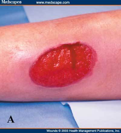
- Ulcer
- Open sore on the body caused by a break in the skin or mucous membrane which fails to heal
- Ulcer
- Common response to all tissue damage in an attempt to reolace it
- Granulation tissue formation
- Defect is slowly infiltrated by capillaries and then by myofibroblasts from the edges
- Collagen and smooth muscle cells are deposited also
- Given constituents look very red
- This tissue is SLOW to form
- Granulation tissue formation
- Scarring and fibrosis
- Bad scars cause loss of sensation
- Areas of fibrous tissue (fibrosis) that replace normal skin after injury
- Results in loss of function e.g. ability to contract or tightness in skin for example
- Scarring of tissue which occurs if:
- Chronic inflammation
- NOT RELATED TO TIME
- Favoured if
- Suppuration occurs e.g. empyema or scarring
- Injury persists e.g. foreign material present that cannot be easily broken down
- Foreign bodies may be found in a granuloma
- Granuloma
- Form of a localised nodular inflammation found in tissues
- Exogenous examples
- Talc
- Asbestos
- Suture material
- Oil
- Endogenous examples
- Keratin
- Bone
- Crystals
- Exogenous examples
- Tuberculous granulomas
- CASEOUS NECROSIS
- Latin name for tuberculous granulomas
- Tuberculosis
- Caused by a myobacterium
- Myobacterium idenfitied useding Zieh-Neelsen stain
- Ziehl Neelsen stain: a special bacteriological stain used to identify acid-fast organisms, mainly mycobacteria. Myobacterium tuberculosis is the most important of this group because it is responsible for causing tuberculosis
- Also known as the acid-fast bacilli. Will be bright red after staining.
- Myobacterium idenfitied useding Zieh-Neelsen stain
- Caused by a myobacterium
- CASEOUS NECROSIS
- Form of a localised nodular inflammation found in tissues
- Granuloma
- Foreign bodies may be found in a granuloma
- Infection persists e.g. mycobacterium or Infectious agent persists e.g. virus
- Due to injury type e.g. autoimmune or transplant rejection
- CHARACTERISED by the presence of the LYMPHCYTE (small round blue cells) and other cells such as the macrophage (monocyte within tissue)
- 4 stages
Imagens e arquivos do mapa
Informações
- Descrição
- Mapa Mental sobre Immunology, criado por Theresa Namaalwa em 14-10-2017.
- Criado por
- Theresa Namaalwa
- Visualizações
- 7
- Avaliação